A subscription to JoVE is required to view this content. Sign in or start your free trial.
Method Article
Microdissection من الأنسجة الجنينية العين اسماك الزرد
In This Article
Summary
توضح هذه المقالة نهج لشبكية العين microdissect الزرد مع وبدون صباغ الشبكية ظهارة المرفقة ، 1-3 أجنة postfertilization يوما.
Abstract
الزرد هو نموذج حيواني شعبية للبحث حول التنمية بسبب العين السريعة لها
Protocol
الجزء 1 : التحضيرات قبل microdissection
- الحلول
- E3 المتوسطة 4 (5 ملي مول كلوريد الصوديوم ، 0.17 ملي بوكل ، 0.33 ملي CaCl 2 و 0.33 ملي MgSO 4).
- حل رينغر 1 ، 5 (116 ملي مول كلوريد الصوديوم ، و 2.9 ملي بوكل ، 1.8 ملي CaCl 2 ، 5 HEPES مم ، pH7.2) ، تصفية تعقيمها.
- 5N هيدروكسيد الصوديوم.
- النقش الكيماوية من الإبر التنغستن
- آمن كوب صغير يحتوي 5N هيدروكسيد الصوديوم على طبق بيتري بواسطة الطين.
- إرفاق مشبك الورق إلى جانب الكأس ، بحيث يأتي في اتصال مع محلول هيدروكسيد الصوديوم.
- توصيل القطب السالب من العاصمة امدادات الطاقة الى مشبك الورق والقطب الموجب إلى واحدة من نهاية قطعة قصيرة (~ 2cm) من سلك التنجستن.
- التفاف على الطرف الآخر من سلك التنغستن مع كرة صغيرة من الطين وتراجع هذه الغاية في محلول هيدروكسيد الصوديوم.
- زيادة الجهد ل3V تقريبا وتراجع السلك صعودا وهبوطا في محلول هيدروكسيد الصوديوم حتى يتم التوصل إلى سعر النقش جيدة. يمكن عادة إبرة في نوعية جيدة يكون محفورا في 10 دقيقة.
- وسلك التنغستن عرضة لتصبح أرق حل تدريجيا. عندما يسقط الكرة من الطين ، فإن الجانب الآخر من الأسلاك التي لا تزال تعلق على القطب الموجب لها شكل إبرة حادة.
- شطف إبرة حادة مع حل رينغر E3 أو لإزالة رواسب الملح.
- إرفاق الإبرة إلى قضيب خشبي بواسطة الشريط أو حامل إبرة لسهولة التعامل.
- الثقافة لوحات
- وفالكون 60 مم X البوليسترين لوحات ثقافة 15 جاهزة للاستخدام لmicrodissection الشبكية.
- لتشريح RPE المرفقة شبكية العين ، سحق two أجنة تماما في لوحة الثقافة التي تحتوي على حل رينغر للتخلص من صوقية من السطح قبل 30 دقيقة من تشريح. قبل وقت قصير من التشريح ، وغسل لوحة على نطاق واسع مع الحل رينغر الطازج.
- أجنة جمع والتدريج
- تمسك الزرد الكبار كما هو موضح 4 ، 5.
- فصل الأسماك في أحواض تربية الوالدين ثابتة البشرى بواسطة مقسم ليلة قبل التزاوج.
- إزالة الفاصل بعد تشغيل الضوء على غرفة في صباح اليوم التالي. تسمح للوالدين لعبور 10 دقيقة في فترات كل نصف ساعة. فصل الأسماك بعد كل معبر.
- الحفاظ على الأجنة التي تم جمعها من كل عبور في E3 المتوسطة بشكل منفصل في 28 درجة مئوية.
- الأجنة في مرحلة 10-12 التسميد آخر ساعة (HPF) وعلى الفور restage قبل التشريح في نقطة زمنية محددة ، وفقا للمعايير المقررة 1 ، 6.
الجزء 2 : تشريح -- إزالة المخ والتعرض العين 1
- الإجراءات المذكورة في هذا المقطع هي مشتركة لتشريح شبكية العين (الجزء 3A) وRPE المرفقة شبكية العين (الجزء 3B).
- تتم جميع تشريح غرامة من قبل غيض من ملقط دومون ويعاونه زوج آخر من ملقط دومون لتحديد المواقع الأنسجة. ويمكن استخدام إبرة التنجستن كيميائيا محفورا عن التلاعب أدق إذا لزم الأمر.
- يتم تنفيذ كافة تشريح غرامة على التكبير ~ 5 - 8X تحت المجهر SZX16 أوليمبوس مجهزة هدفا 1X أو ما يعادلها.
- الحفاظ على الأجنة في E3 المتوسطة في مقعد الحاضنة الأولى في 28 درجة مئوية بجانب المجهر للوصول السهل الجنين أثناء تشريح.
- وتحسنت المرحلة المجهر إلى 28 درجة مئوية بحلول لوحة الحرارية للحد من تأثير تقلبات درجة الحرارة على التعبير الجيني.
- أعد نقل الجنين في مرحلة تطويرية معينة إلى حل رينغر في 60 X 15 مم فالكون لوحة الثقافة كما هو موضح في الجزء 1 # 3.
- قطع الرأس مع جزء من الجذع الأمامي بسرعة من الجسم.
- دبوس نهاية الخلفي من هذا النسيج على لوحة الثقافة مع ملقط مساعدة.
- فتح الرأس على الجانب الظهري بدءا من الجبهة الأمامية.
- إزالة المخ بحيث يتعرض الجانب الإنسي للعيون وتواجه التصاعدي لمزيد من التلاعب.
3A الجزء : 1 تشريح الشبكية
- تحضير الجنين كما هو موضح في الجزء 2.
- فرشاة بعناية RPE تعرض على الجانب الإنسي للكرة العين التي تواجه صعودا من قبل غيض من الملقط حتى ينظر إلى فتحة صغيرة في شبكية العين.
- الاستمرار في تنظيف وتقشير العمل حتى في الجانب الإنسي للشبكية يكاد يتعرض جميع. تجنب خدش واللكم على شبكية العين تتعرض لها.
- لإزالة RPE الوحشي على الشبكية التي تواجه الآن نحو الانخفاض ، والنهج الذي الفرشاة بزاوية 45 درجة تقريبا من سطح لوحة الثقافة.
- إزالة RPE التي ترتبط بقوة نسبيا في مشرشر ORA التي تعلق جزء من فصل إلى RPE جلوحة ulture التشريح ، ومن ثم رفع شبكية العين برفق.
- لفة الشبكية في الجزء السفلي من الطبق الثقافة لتنظيف RPE المتبقية.
- لاحظنا أن هذا البوليسترين فالكون لوحة خاصة لديه ثقافة الالتزام التفضيلية للRPE لحوالي 20 دقيقة مرة واحدة سحقت جنين. وتستخدم هذه الخاصية لإزالة مخلفات RPE. ومع ذلك ، فإن خلايا الشبكية لا عصا على السطح إلى حد أقل ، والتي يمكن تفتيشها بشكل وثيق تحت التكبير عالية. على المرء أن توازن بين اكتمال إزالة RPE والنزاهة في شبكية العين.
- العدسة تتمسك كثير من الأحيان إلى لوحة الثقافة ويفصل من شبكية العين خلال عملية المتداول. أحيانا ، فمن الضروري فصل العدسة عن طريق إبرة التنغستن محفورا مع دوامات العمل على سطح العدسة.
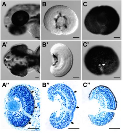
- ويمكن إزالة هذه الإجراءات بنجاح RPE من الشبكية دون المساومة على سلامة الأنسجة. ويدل على هذا التشكل شامل جيدة (الشكل 1B و "B) والأنسجة (الشكل 1B") ، السهم وعلى وجه الخصوص ، والحفاظ على المصفوفة خارج الخلية في مبصرة بين طبقة وRPE هو جيد بشكل استثنائي (1B الشكل "؛ مقارنة مع الأنسجة من الجنين كله (الشكل 1A ")).
3B الجزء : RPE المرفقة تشريح الشبكية 3
- تحضير الجنين كما هو موضح في الجزء 2.
- رأس دبوس إلى لوحة الثقافة التي ملقط مساعد. رفع العين بلطف من الجانب الخلفي والجانبي لفة على الجانب الأمامي.
- ويعلق المشيمية الظنية والأنسجة الصلبة على السطح الخارجي للطبقة RPE نسبيا بإحكام على الجلد ، ويمكن مقشر معظمها من قبل المتداول خارج العين بعناية إلى الجانب الأمامي.
- إزالة العدسة بإبرة التنغستن بعد تعرض الجانب الوحشي من العين ، وعندما لا تزال تعلق على العين على الجلد.
- يمكن لهذه الإجراءات المحافظة على طبقة RPE بنجاح كامل مع شبكية العين. يمكن choroids الافتراضي وإزالة الأنسجة الصلبة إلى حد كبير (الشكل 1C ، 'C و C").
الجزء 4 : الأنسجة جمع العينات لعمل الحمض النووي الريبي
- ويمكن جمع عينات تشريح في TRIzol في أنبوب microfuge ريبونوكلياز خالية من الحمض النووي الريبي لتوصيف المصب صفه قبل 1.
ممثل النتائج

الشكل 1 (أ) والجانبي (A ') عرض الظهرية من رأس اليرقات الزرد في 52 HPF قبل تشريح. (A ") والفرع المقابل النسيجي للرئيس اليرقات في الفقرة (أ) و (A') ، (ب) والجانبي (B ') عرض الظهرية لتشريح شبكية العين في 54 HPF. سطح الشبكية كان سليما من كل الاطراف وجهات النظر الظهرية (B ") والفرع المقابل النسيجي للتشريح في شبكية العين (ب) و (B'). وكان هيكل في شبكية العين وتصفيح الشبكية سليمة. على وجه الخصوص ، "كانت محفوظة بشكل جيد (والسهام في شبكية العين تشريح (B المصفوفة خارج الخلية بين طبقة مبصرة وRPE A)" والسهام). (C) والجانبي (C ') عرض الإنسية لشبكية العين RPE المرفقة تشريح في 52 HPF. وكان RPE طبقة سليمة ومستمرة ، وهو ما تدل أيضا المقطع النسيجي للتشريح الأنسجة (C "). وتبلغ مساحة بيضاء في' C هو العصب البصري (السهم) للحصول على الأنسجة ، تم جمع عينات من الأنسجة وإصلاحها في 4 ٪ أجريت بارافورمالدهيد. بلاستيك وsectioning تضمين هذه العينات كما هو موضح 3. شريط مقياس = 50 ميكرومتر.
Discussion
ويمكن للأنسجة العين Microdissection الزرد الحصول على شبكية العين سليمة وفعالة شبكية العين RPE المرفقة. هذا يساعد كثيرا الدراسات المتعلقة التعبير نسيج العين محددة (شبكية العين أو أي RPE). في الواقع ، لقد نجحنا في استغلال هذه الإجراءات للحصول على الحمض النووي الريبي ملامح التعبي...
Disclosures
Acknowledgements
ويؤيد هذا العمل من قبل صندوق بدء التشغيل من قسم العلوم البيولوجية في جامعة بوردو.
Materials
| Name | Company | Catalog Number | Comments |
| Cordless pestle motor | VWR international | 47747-370 | |
| DC power supply | Lascar | PSU130 | Any DC supply would work. The specific voltage of a different machine will need further optimization. |
| Disposable pestle & microtube, 1.5 mL (DNase, RNase and pyrogen-free) | VWR international | 47747-366 | These are used for tissue collection in TRIzol for expression analysis. |
| Dumont #5 forceps, Tips: 0.05 x 0.01mm, Inox | World Precision Instruments, Inc. | 500341 | Fine tip dimension is desirable but is not inflexible, as one may need to sharpen the tip from time to time. |
| Dumont #5SF forceps, Tips: 0.025 x 0.005mm, Inox | Fine Science Tools | 11252-00 | Fine tip dimension is desirable but is not inflexible, as one may need to sharpen the tip from time to time. |
| Falcon polystyrene culture plates, 60 X 15 mm | BD Biosciences | 351007 | These plates are used as dissection plates. |
| Olympus SZX16 Stereomicroscope | Olympus Corporation | SZX16 | Any stereomicroscope would work. We used Leica stereomicroscope in previous studies1-3 without any issues. We also use the 1X objective exclusively for the dissection even though we have a 2X objective installed. |
| Sharpening stone | Fine Science Tools | 29008-01 | Use this to sharpen the tip of the forceps if necessary |
| Thermo plate | Tokai Hit | MATS-U55SZX2B | This is used to maintain the temperature of the tissue throughout dissection and minimize the influence of temperature fluctuation on gene expression. We also put the whole microscope in an environmentally controlled room at 28°C during dissection in previous studies1-3 with good success. |
| Trizol, 100 mL | Invitrogen | 15596-026 | |
| tungsten wire, 0.015 inch diameter | World Precision Instruments, Inc. | TGW1510 | |
| Wooden Applicator | Puritan | 807 | This is used for holding the chemically-etched tungsten needle. |
References
- Leung, Y. F., Dowling, J. E. Gene expression profiling of zebrafish embryonic retina. Zebrafish. 2, 269-283 (2005).
- Leung, Y. F., Ma, P., Link, B. A., Dowling, J. E. Factorial microarray analysis of zebrafish retinal development. Proc Natl Acad Sci U S A. 105, 12909-12914 (2008).
- Leung, Y. F., Ma, P., Dowling, J. E. Gene expression profiling of zebrafish embryonic retinal pigment epithelium in vivo. Invest Ophthalmol Vis Sci. 48, 881-890 (2007).
- Nusslein-Volhard, C., Dahm, R. . Zebrafish : a practical approach. , (2002).
- Westerfield, M. The zebrafish book : a guide for the laboratory use of zebrafish (Danio rerio). , (2000).
- Kimmel, C. B., Ballard, W. W., Kimmel, S. R., Ullmann, B., Schilling, T. F. Stages of embryonic development of the zebrafish. Dev Dyn. 203, 253-310 (1995).
- Fadool, J. M., Dowling, J. E. Zebrafish: a model system for the study of eye genetics. Prog Retin Eye Res. 27, 89-110 (2008).
Reprints and Permissions
Request permission to reuse the text or figures of this JoVE article
Request PermissionExplore More Articles
This article has been published
Video Coming Soon
Copyright © 2025 MyJoVE Corporation. All rights reserved