
A subscription to JoVE is required to view this content. Sign in or start your free trial.
Method Article
فيفو تصوير لايف الرئة ورم خبيث والمكروية بهم
In This Article
Summary
نحن تصف طريقة بسيطة نسبيا لخارج الجسم الحي التصوير الحي للورم التفاعلات خلية سدى داخل ورم خبيث في الرئة، وذلك باستخدام صحفيين الفلورسنت في الفئران. باستخدام الغزل القرص مبائر المجهر، هذه التقنية تمكن التصور من الخلايا الحية لمدة 4 على الأقل ساعة ويمكن تكييفها لدراسة الأمراض الرئوية الالتهاب الأخرى.
Abstract
ورم خبيث يشكل سببا رئيسيا للمراضة والوفيات المرتبطة بالسرطان. الانبثاث هي عملية متعددة الخطوات، ونظرا لتعقيدها، والعمليات الخلوية والجزيئية الدقيقة التي تحكم نشر المنتشر والنمو لا تزال بعيدة المنال. تصوير مباشر يسمح التصور من التفاعلات الديناميكية والمكانية للخلايا والمكروية بهم. الأورام الصلبة metastasize عادة إلى الرئتين. ومع ذلك، فإن الموقع التشريحي للرئتين يشكل تحديا لتصوير حيوي داخلي. يوفر هذا البروتوكول طريقة بسيطة نسبيا وسريعة لخارج الجسم الحي التصوير الحي للتفاعلات الدينامية بين الخلايا السرطانية وسدى المحيط بهم داخل ورم خبيث في الرئة. باستخدام هذا الأسلوب، والقدرة على الحركة من الخلايا السرطانية فضلا عن التفاعل بين الخلايا السرطانية والخلايا اللحمية في المكروية التي يمكن تصور في الوقت الحقيقي لعدة ساعات. باستخدام المعدلة وراثيا الفئران مراسل الفلورسنت، خط خلية فلوري، عن طريق الحقن fluorescently المسمىجزيئات و / أو الأجسام المضادة، مكونات متعددة من المكروية الرئة يمكن تصور مثل الأوعية الدموية والخلايا المناعية. لصورة أنواع مختلفة من الخلايا، وقد استخدم قرص الغزل المجهر متحد البؤر التي تتيح التصوير المستمر على المدى الطويل مع الحصول على الصور السريع، أربعة ألوان. الوقت الفاصل بين الأفلام التي تم تجميعها من الصور التي تم جمعها خلال مواقف متعددة وطائرات التنسيق تظهر التفاعلات بين النقيلي الحية والخلايا المناعية لا يقل عن 4 ساعة. هذه التقنية يمكن زيادة استخدامها لاختبار العلاج الكيميائي أو العلاج الموجه. وعلاوة على ذلك، يمكن تكييف هذه الطريقة لدراسة الأمراض ذات الصلة الرئة الأخرى التي قد تؤثر على المكروية الرئة.
Introduction
The deadliest aspect of cancer is metastasis, which accounts for more than 90% of cancer-related morbidity and mortality1. Metastasis is a multistep process and due to its complexity, the exact cellular and molecular mechanisms that govern metastatic dissemination and growth are still elusive. To metastasize, tumor cells in the primary tumor must detach from their neighboring cells and basement membrane, cross through the extracellular matrix, intravasate, travel via blood or lymphatic vessels, extravasate at the secondary site, and finally, survive and establish secondary tumors. In addition to the properties of the tumor cells, the contribution from the microenvironment, which includes the adjacent stroma along with the normal counterparts of the cancer cells, is crucial for the seeding and establishment of metastatic lesions2.
Traditional methods to study metastatic seeding and growth examine static states, as tissues are excised and sectioned for histology. These data only generate a snapshot of this highly dynamic process. Although some useful information can be gained from these studies, the complicated process by which tumor and stromal cells interact during metastatic formation cannot be adequately assessed by these methods. Furthermore, it is not possible to gain insights into tumor or stromal cell migration patterns, which are important in establishing a colony at the distant site. In order to effectively study the metastatic process, it is essential to visualize various interactions between cancer cells and their microenvironment in a continuous manner and at real time.
The lung is a common site for metastases from solid tumors as breast, colorectal, pancreatic cancer, melanoma and sarcoma3. Intravital imaging was previously used to study cell-cell interaction in various primary tumor and metastatic models4,5. Methods of lung imaging in mice, including intravital imaging, lung section imaging, and an ex vivo pulmonary metastasis assay have been published6–9. Intravital imaging of mouse lungs utilizes a thoracic suction window to stabilize the lungs6. This method is used for time-lapse imaging of the lung microcirculation and alveolar spaces. The anatomical location of the lungs poses a challenge to intravital imaging. In order to access the lungs, the chest cavity must be opened which leads to loss of negative pressure and collapsed lungs. This method only allows the visualization of a small part of the lungs and is technically demanding; an unnecessary complication in studies that examine processes that are independent of blood flow. Moreover, this method also requires gating out movement caused by breathing. This is done either by collecting images between breaths or during post image acquisition analyses10. The alternative ex vivo lung section imaging provides stability and depth, and also prepares lung parenchyma for immunostaining7. However, the lengthy sectioning process leads to an extensive delay between the time of animal sacrifice and the start of the imaging session. Moreover, the process of sectioning a mouse lung causes considerable amount of cell death8, thus interfering with the quality and quantity of imaging samples and perhaps needlessly altering tumor-stroma interactions. In order to technically bridge between the methods of intravital imaging and lung section imaging, while exploiting the advantages of the two techniques, a relatively fast and easy method for ex vivo lung imaging was developed. This method was achieved by imaging of non-sectioned whole lung lobes. Using this method, the motility of cancer cells as well as interactions between cancer cells and stromal cells in their microenvironment can be visualized in real time for several hours.
Protocol
يجب أن يتم تنفيذ جميع الإجراءات المذكورة وفقا للمبادئ التوجيهية واللوائح لاستخدام الحيوانات الفقارية، بما في ذلك الحصول على موافقة مسبقة من قبل المؤسسات ورعاية الحيوان المحلية واللجنة الاستخدام (IACUC).
1. الجيل الرئة الانبثاث لخارج الجسم الحي تصوير لايف (المعدلة وراثيا أو الذيل الوريد حقن)
ملاحظة: يمكن أن تتولد الانبثاث الرئة عن طريق استخدام نماذج الماوس المعدلة وراثيا أو عن طريق الوريد (IV) حقن الخلايا السرطانية.
- توليد الانبثاث الرئة للتصوير عن طريق عبور نموذج الفأر الورم وراثيا في الماوس مراسل المعدلة وراثيا، على سبيل المثال، عبر سرطان الثدي نموذج الماوس، ماوس الثديية فيروس الورم محطة الطويلة تكرار-التورام منتصف تي مستضد (MMTV-PyMT) 11 في ACTB-ECFP نموذج الفأر 12.
ملاحظة: نموذج ACTB-ECFP تعرب عن تعزيز البروتين الفلوري سماوي (ECFP) تحت β-عملفي المروج بحيث كل الخلايا يتألق في الزرقاء، قناة CFP. ومع ذلك، فإن الخلايا السرطانية إلى حد بعيد أبرز وتظهر بوصفها الأكبر من الخلايا ECFP إيجابي تحت المجهر. نموذج الفأر MMTV-PyMT يتطور المرض تدريجيا، حيث يرتبط نمو الورم الثديية مع نشر الخلايا السرطانية إلى المحيط، وخاصة إلى الرئتين. في الفئران MMTV-PyMT على FVB / ن الخلفية، micrometastases يمكن ملاحظة حول 10-11 أسابيع من العمر. عموما، هذه التقدم إلى macrometastases في حوالي 14 أسبوعا من العمر 13 عاما.
أو - توليد الانبثاث التجريبية باستخدام الخلايا الأولية أو خطوط الخلايا مسانج. استخدام في المختبر التلاعب الخلايا السرطانية الأولية أو خطوط الخلايا (على سبيل المثال، التنبيغ) تليها حقن د 14.
- لفترة وجيزة، في هذا البروتوكول، حقن البروتين الفلوري الأخضر (GFP) -expressing (+) خط الخلية MMTV-PyMT في الفئران الفلورسنت مراسل (ACTB-ECFP) أو الفئران wildtype. ثم،تصور هذه الخلايا يشار إلى خلايا VO-PyMT 15 باستخدام الخضراء، قناة GFP.
ملاحظة: تم اشتقاق خط الخلية VO-PyMT الأصلي في جراحة العظام فاندربيلت في ناشفيل، تينيسي. VO لتقف على فاندربيلت العظام. - بعد حقن الخلايا 6 10 (في 200 ميكرولتر)، ومراقبة تسرب الخلايا السرطانية مباشرة وتصل إلى بضعة ساعات بعد الحقن. مراقبة micrometastases بين 1-3 أسابيع بعد الحقن وكشف macrometastases 3 أسابيع بعد الحقن 15.
ملاحظة: أقل الخلايا يمكن حقنها لإطالة وقت من الحقن لنمو النقيلي.
- لفترة وجيزة، في هذا البروتوكول، حقن البروتين الفلوري الأخضر (GFP) -expressing (+) خط الخلية MMTV-PyMT في الفئران الفلورسنت مراسل (ACTB-ECFP) أو الفئران wildtype. ثم،تصور هذه الخلايا يشار إلى خلايا VO-PyMT 15 باستخدام الخضراء، قناة GFP.
2. وضع العلامات من مكونات الاهتمام في الإنتقالية المكروية (المعدلة وراثيا و / أو الحقن)
ملاحظة: وصفها يمكن أن يتحقق عن طريق الفئران المعدلة وراثيا و / أو عن طريق الحقن المختلفة. تأكد من استخدام الألوان الفلورسنت مختلفة لوضع العلامات على مختلف أنواع الخلايا.
- مكونات تسمية المكروية المتنقل باستخدام الفئران المعدلة وراثيا. عبور نموذج ورم الماوس المذكورة سابقا (على سبيل المثال، MMTV-PyMT س ACTB-ECFP) في نموذج الفأر وراثيا التي وصفت الخلايا اللحمية من الفوائد بنسبة بروتين فلوري الذي لا ECFP، على سبيل المثال.، ج-FMS-EGFP 4،16.
ملاحظة: بالإضافة إلى التصور من الخلايا السرطانية في قناة CFP، وهذا يمكن التصور من خلايا الدم النخاعي في القناة GFP 4.
و / أو - تسمية مختلف مكونات المكروية المتنقل باستخدام الحقن في الفئران مراسل فلوري المعدلة وراثيا أو (غير الفلورسنت) الفئران wildtype.
ملاحظة: العديد من المركبات ويمكن حقن لتسمية مختلف مكونات المكروية المنتشر، على سبيل المثال، يتم استخدام AF647 مترافق GR-1 الضد هنا لتسمية العدلات وتستخدم بعض وحيدات 13 و مختلفة dextrans الوزن الجزيئيلتسمية الشعيرات الدموية في الرئة. لإعداد هذه الحقن راجع الخطوة 4.
3. إعداد المواد قبل تشريح
- 2٪ الاغاروز
- تزن 0.2 غرام من الاغاروز وإضافة إلى 10 مل 1 × برنامج تلفزيوني. الحرارة الحل بحل الاغاروز. Agarose سوف يصلب على RT، لذلك الحفاظ عليه في 37 ° C حمام الماء حتى تستخدم للتضخم.
- CO 2 وتحكم في درجة الحرارة
- تحقق ده 2 O في غرفة الترطيب. إعادة ملء عند الحاجة. إدراج لوحة التكوين في درجة حرارة حامل مرحلة لوحة (غرفة المناخ). تشغيل وحدة تحكم CO 2 ومجموعة CO 2 بنسبة 5٪. تأكد من تعيين معدل تدفق الهواء عند 0.4 نيكولا لانغ / دقيقة.
- فتح الهواء وثاني أكسيد الكربون 2 الصمامات. تشغيل وحدة تحكم في درجة الحرارة. تأكد من ضبط درجة حرارة الغرفة المناخ والغطاء عند 37 درجة مئوية.
- الافراج عن الضغط الجوي على CO 2 متر. تحقق CO 2 زيادة، هquilibration قد يستغرق ما يصل إلى 30 دقيقة.
- الغزل القرص مجهر متحد البؤر
ملاحظة: تفاصيل المجهر انشاء وقد وصفت سابقا 4،17.- بدوره على الليزر (ليزر الأرجون ل488 الإثارة نانومتر ونانومتر الحالة الصلبة 405، 561 نانومتر و 640 نانومتر ليزر). بدوره على المجهر، والكاميرا، وحدة الغزل تحكم القرص، AOTF، وحدة التحكم ليزر وجهاز تحكم الكاميرا.
- فتح مصراع المجهر، بدوره على جهاز كمبيوتر يستخدم المجهر وفتح البرنامج.
- إعداد الأدوات ومنصة التشريح.
- بدوره على حبة تعقيم الساخن والسماح لها تصل إلى 250 درجة مئوية. نظيفة 2 أزواج من مقص جراحي وملقط بالماء والصابون. تعقيم الأدوات لمدة 30 ثانية على الأقل. السماح للأدوات يبرد. استخدام غطاء البوليسترين كمنصة تشريح. تغطية ذلك مع قطعة من معتاد على الثمالة المختبر.
4. إعداد الحقن
ملاحظة: اعتمادا على عمر النصف والاستجابة المفضلة، حقن fluorescently المسمى الأجسام المضادة و / أو جزيئات الفلورسنت إما مباشرة قبل التضحية الحيوانية أو بضع ساعات إلى أيام قبل.
- لالعدلات صورة GR1 إيجابية وحيدات، وإعداد حقنة مع 7 ميكرولتر من الأسهم AF647 مترافق GR-1 الأجسام المضادة (1 ملغ / مل) في 100 ميكرولتر من برنامج تلفزيوني العقيمة تحت غطاء محرك السيارة. وضع G ½ إبرة 27 على حقنة.
- إلى الشعيرات الدموية في الرئة الصورة، وإعداد حقنة الثانية والثالثة مع 100 ميكرولتر من إما 70 KD-رودامين مترافق ديكستران (4 ملغ / مل) أو ديكستران 10 دينار كويتي AF647 مترافق (4 ملغ / مل). وضع G ½ إبرة 27 على الحقن.
- حقن محلول الأجسام المضادة الرابع 5 ساعة-AF647 مترافق قبل الختان الرئتين.
- حقن واحد أو كلا حلول ديكستران الرابع مباشرة قبل استئصال الرئتين.
5. إعداد الرئتين لخارج الجسم الحي تصوير لايف
ملاحظة: حاول أن تعمل عقيمة والحذر قدر الإمكان لتجنب التحديات التي لا داعي لها من الخلايا المناعية داخل الرئتين.
- حقن داخل الصفاق الماوس (الملكية الفكرية) مع جرعة زائدة قاتلة من مخدر يسمح به بروتوكول الحيوانية التي وافقت عليها IACUC، على سبيل المثال، 1 مل من 2.5٪ آفيرتين. انتظر الماوس لوقف التنفس ويكون تماما غير استجابة للمؤثرات الضارة (الخلفيتين مخلب قرصة).
ملاحظة: خلع عنق الرحم والقتل الرحيم ثاني أكسيد الكربون التي ينبغي تجنبها لأنها يمكن أن تؤثر تأثيرا ضارا على بقاء الخلية الرئة. - لشل حركة الماوس على لوحة التشريح وتعقيم الفأر مع الايثانول 70٪.
- استخدام مقص جراحي لإجراء أول شق شرسوفي عرضية من خلال الجلد، تليها شق مماثل من خلال الغشاء البريتوني. عقد مجلس تشريح في وضع عمودي وقطع الشريان الأورطي النازل، بحيث حمامات الدم باستمرار في البطن وليس في تجويف الصدر.
- Sارتشف فتحة صغيرة في الحجاب الحاجز للافراج عن فراغ. قطع على طول الضلع 10TH وال12 لاستئصال الحجاب الحاجز والحصول البصرية إلى الرئتين.
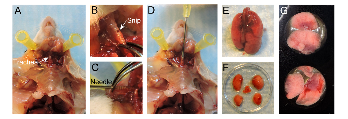
- استخدام مقص جراحي لقطع الجلد تصل إلى القصبة الهوائية فوق القفص الصدري ولكن ترك القفص الصدري سليمة. فصل الجلد من القفص الصدري. فضح القصبة الهوائية عن طريق إزالة الأنسجة الضامة المحيطة بها، والحرص على عدم الإضرار القصبة الهوائية نفسه (الشكل 1A).
- قص فتحة صغيرة حوالي 1 ملم في القطر في موازاة القصبة الهوائية تتعرض لحلقات غضروفية، وأقرب إلى الحنجرة ممكن (الشكل 1B). يجب الحرص على عدم قطع تماما من خلال القصبة الهوائية.
- خذ 20 G إبرة وإدراج بلطف ملم إبرة 4-5 في القصبة الهوائية دون أي قوة مضادة (1D الشكل). وينبغي أن تكون نهاية الإبرة وضوحا من خلال القصبة الهوائية (الشكل 1C). استخدام ملقط لتثبيت الإبرة في القصبة الهوائية. بدلا من ذلك، قد تكون مرتبطة خياطة آرواوند القصبة الهوائية لعقد إبرة في المكان.
ملاحظة: من خلال إدخال عميق جدا، وكارينا قد تكون صدمة أو جانب واحد فقط من الرئتين قد يكون مبالغا فيه. - حقنة ملء مع 400 ميكرولتر من 37 درجة مئوية 2٪ الاغاروز منخفضة ذوبان في درجة الحرارة (مأخوذة مباشرة من حمام درجة حرارة ثابتة). تأكد من أن لوحة تشريح واقفا وغرس ببطء الاغاروز الدافئ من خلال إبرة في الرئتين، واستخدام ~ 400 ميكرولتر لتضخيم الرئتين.
ملاحظة: ووتش الرئتين تضخيم داخل القفص الصدري. لا تبالغ في تضخيم الرئة لأنها سوف تمزق. - بمجرد تضخم الرئتين، وملء ~ ⅔ من القفص الصدري، فصل حقنة والحفاظ على إبرة داخل القصبة الهوائية لمنع أي الاغاروز من تسرب.
- صب حوالي 50 مل من 20 درجة مئوية في برنامج تلفزيوني على الرئتين مبالغ للسماح للالاغاروز داخل الرئتين لوضع وتوطيد. ببطء إزالة الإبرة وإغلاق القصبة الهوائية مع ملقط لمنع أي غير عزز الاغاروز من تسرب.
- فضح الرئتين عن طريق إجراء القص وبعد استئصال الرئة. لاستئصال الرئتين، الابقاء على القصبة الهوائية في حين قطع طريق القصبة الهوائية تماما. برفق القصبة الهوائية فوق، قطع النسيج الضام والمريء في حين سحب الرئتين من تجويف الصدر حتى يتم فصل الرئتين من الفأرة (الشكل 1E).
- تزج الرئتين في حارة RPMI-1640 ليغسل الدم المفرط وفصل بلطف الفص باستخدام مقص وملقط لقطع القصبات الهوائية الجذعية الرئيسية الفصوص 'في نقير (الشكل 1F).
- وضع فصوص، مع سطح مستو وصولا الى تحقيق أقصى قدر من سطح التصوير، في بئر من 24-جيدا لوحة التصوير (الشكل 1G). إضافة 100 ميكرولتر من 37 درجة مئوية RPMI-1640 على قمة الفصوص. وضع عدة 15 مم غطاء المجهر دائري الشرائح على قمة الفصوص لمنعها من العائمة.
- صب برنامج تلفزيوني الدافئ في الآبار المحيطة بها لمنع وسائل الإعلام RPMI-1640 من EVAPorating. إدراج 24 لوحة جيدا في غرفة المناخ معايرتها والحفاظ على فصوص الرئة عند 37 درجة مئوية مع الهواء و 5٪ CO 2. أدخل غرفة المناخ على مرحلة من مراحل المجهر متحد البؤر.
ملاحظة: خليط الغاز الأخرى (على سبيل المثال، 5٪ O 2 و 5٪ CO 2 في N 2 لدراسة سلوك الخلية تحت ظروف نقص الأكسجة / انخفاض الأكسجين) ويمكن أيضا أخذها بعين الاعتبار.

الشكل 1. بروتوكول لإعداد الرئتين للتصوير الحية. (A) تعرض القصبة الهوائية بعد إعداد الماوس. (ب) القصاصة الصغيرة المحرز في موازاة القصبة الهوائية تتعرض لحلقات غضروفية. (C) 20 G إبرة 4-5 ملم في القصبة الهوائية. (D) تقطير من 400 ميكرولتر 2٪ منخفضة ذوبان درجة الحرارة الاغاروز إلى الرئتين. (E) Inflالرئتين ATED فصل من الماوس. (F) فصوص فصل بعد التضخم. (G) الفصوص وضعت في بئر من لوحة التصوير 24-جيدا. الرجاء انقر هنا لعرض نسخة أكبر من هذا الرقم.
6. اكتساب وتحليل الصور
يمكن الحصول على الصور مع مجموعة متنوعة من الغزل القرص المجاهر مبائر بدعم من البرامج المختلفة: ملاحظة. في هذا البروتوكول، ويستخدم إما μManager مع قرص الغزل المجهر حسب الطلب متحد البؤر أو زن مع المتاحة تجاريا الغزل القرص مجهر متحد البؤر لاقتناء الصور، في حين يتم استخدام Imaris لتحرير الأفلام والتحليل.
- الحصول على صور باستخدام μManager. خطوة مفصلة من قبل بروتوكول خطوة لاقتناء الصور باستخدام برنامج μManager يوصف سابقا 18.
أو - الحصول على صوراستخدام صورة برامج التحليل مثل زن (انظر الشكل S1).
- انقر فوق علامة التبويب "تحديد موقع" و، واختيار الهدف (10X أو 20X) في 'مسار الضوء "أداة (الشكل S1A، مربع أحمر). وفي وقت لاحق، انقر على 'عيون - دابي "للنظر في قناة CFP من خلال العدسة (الشكل S1A، المربع الأزرق). إضفاء الطابع المحلي على عينة يدويا باستخدام المجهر. انقر على "جميع Off'after النسيج هو مركز مجال الرؤية.
- انقر على علامة التبويب "شراء" لتعيين كافة المعلمات من أجل الحصول على الصور.
- في أداة "قنوات"، انقر فوق الزر '+' (الشكل S1B، مربع أحمر). تظهر القائمة المنبثقة والبحث عن الصبغة (ق) في العينة في "قاعدة صبغ" (الشكل S1B). حدد صبغ وانقر على 'إضافة'.
ملاحظة: سيقوم البرنامج وضع جميع المرشحات على الوجه الأمثل. يمكن حذف صبغةعن طريق تحديده تليها النقر على زر سلة المهملات (الشكل S1B، المربع الأصفر). - في قائمة "الوضع اكتساب، مجموعة 'Binning" ل5X5. انقر مرتين على ECFP في قائمة القنوات لتحديده. خفض قوة الليزر إلى 20٪ وذلك حتى لا يكون ابيض العينة أثناء إعداد المعلمات لاقتناء الصور.
- ضع علامة في المربع "بلاط" في قسم "مدير تجربة" وتظهر أداة البلاط في "متعددة الأبعاد اقتناء" مجموعة أداة (الشكل S1C). انقر على زر "الإعداد المتقدم" لعرض صورة حية من كاميرا. انقر على زر "إضافة" في قسم "المراكز" لإضافة 4-6 مواقف للتجربة. لحذف الموقف، وتحديد هذا الموقف، وانقر على زر سلة المهملات.
- في المجموعة أداة "اكتساب المعلمة"، افتح الأداة "استراتيجية التركيز"، واختر "مطلقالبريد ثابت Z-موقف "من القائمة المنسدلة.
- ضع علامة في المربع Z-المكدس في قسم 'مدير تجربة "وأداة Z-المكدس يظهر في مجموعة أداة" متعددة الأبعاد اقتناء "(الشكل S1D). انقر مرتين على أحد المواقع في قسم "المراكز" واضغط على "لايف". يدويا تعيين أول وتحديد موقف الأخير من مجال التصوير. تعيين الفاصل الزمني في 4 ميكرون.
ملاحظة: سيقوم البرنامج تحديد عدد شرائح للمجموعة المختارة والفاصلة. من الناحية المثالية، 5-7 شرائح هي مريحة للسماح التصور الكافي والحصول على الصور سريعا. - تحقق مربع "السلاسل الزمنية" في قسم "مدير تجربة". مجموعة المرجوة "مدة وأوقات الاستراحة في أداة" السلاسل الزمنية "التي ظهرت في" متعددة الأبعاد اقتناء "مجموعة أداة (الشكل S1E).
- في 'Acquisiنشوئها وضع "القائمة، حدد" Binning "ل2X2. انقر مرتين على fluorophore في قائمة القنوات لتحديده، وزيادة قوة الليزر إلى 100٪. اضغط على "لايف" وضبط "الوقت التعرض. كرر ذلك كل fluorophore.
- تحقق "تمكين السيارات حفظ" مربع. تحديد مجلد واكتب في اسم الملف. سيتم حفظ جميع الصور المكتسبة تلقائيا في هذا المجلد.
- انقر على "تجربة ابدأ" في قسم "مدير تجربة" لبدء الحصول على الصور.
- بعد الحصول على الصور، تجميع البيانات الخام في برنامج Imaris. تحويل الصور إلى .ims الملفات ويمكن إجراء التعديلات. خطوة مفصلة من قبل بروتوكول خطوة لتحويل الملفات، إجراء تعديلات وتوفير الأفلام باستخدام Imaris يوصف سابقا 18.
- عند حفظ الفيلم، مجموعة 'معدل الإطار' إلى 5 إطار في الثانية (fps).
النتائج
باستخدام الغزل القرص مبائر المجهري، ومختلف النظم نموذج الفأر والحقن، المكروية المنتشر يمكن تصور وتتبع مع مرور الوقت. باستخدام MMTV-PyMT. ACTB-ECFP. وصفت ج-FMS-EGFP الثلاثي نموذج الفأر وراثيا، والمكونات الخلوية المختلفة fluorescently (الشكل 2A، فيلم 1). هيكل ن...
Discussion
توضح هذه المخطوطة طريقة مفصلة لخارج الجسم الحي التصوير الحي من ورم خبيث في الرئة في نماذج الفئران من ورم خبيث. يوفر هذا البروتوكول التصوير والتصوير المباشر للتفاعلات ورم الخلايا سدى ديناميكية والمكانية داخل المكروية الرئة. وهي طريقة سهلة وسريعة نسبيا التي تسمح...
Disclosures
The authors have no conflicts of interest to disclose. All animal experiments were conducted in accordance with IACUC approved protocols, UCSF.
Acknowledgements
We thank Nguyen H. Nguyen for her technical help and Audrey O’Neill for support with the Zeiss Cell Observer spinning-disk confocal microscope. This work was supported by a Department of Defense postdoctoral fellowship (W81XWH-11-01-0139) and the Weizmann Institute of Science-National Postdoctoral Award Program for Advancing Women in Science (to V.P.).
Materials
| Name | Company | Catalog Number | Comments |
| MMTV-PyMT/FVB mice | Jackson Laboratory | 2374 | Female mice |
| ACTB-ECFP/FVB mice | UCSF Werb lab | Female mice | |
| c-fms-EGFP/FVB mice | UCSF Werb lab | Female mice | |
| FVB mice | Jackson Laboratory | 1800 | Female mice |
| GFP+ VO-PyMT cells | UCSF Werb lab | ||
| 70,000 kDa Dextran, rhodamine-conjugated | Invitrogen | D1818 | Dilute to 4mg/ml in 1 x PBS and store at -20 °C. Use 0.4 mg per animal. |
| 10,000 kDa Dextran, Alexa Fluor 647 conjugated | Invitrogen | D22914 | Dilute to 4mg/ml in 1 x PBS and store at -20 °C. Use 0.4 mg per animal. |
| Anti-mouse Gr-1 antibody Alexa Fluor 647 | UCSF Monoclonal antibody core | Stock 1mg/ml. Use 7 ug per animal. | |
| Anesthetic | Anesthesia approved by IACUC, used for anesthesia and/or euthanesia | ||
| 1X PBS | UCSF cell culture facility | ||
| PBS, USP sterile | Amresco INC | K813-500ML | Ultra pure grade for i.v. injection |
| Styrofoam platform | Will be used as dissection board | ||
| Fine scissors sharp | Fine Science Tools | 14060-11 | |
| Forceps | Roboz Surgical Store | RS-5135 | |
| Hot bead sterilizer | Fine Science Tools | 18000-45 | Turn ON 30min before use |
| Air | UCSF | ||
| Oxygen | UCSF | ||
| Carbon dioxide | UCSF | ||
| 1 mL syringe without needle | BD | 309659 | |
| 27 G x 1/2 needle | BD | 305109 | for i.v. injection |
| 20 G x 1 needle, short bevel | BD | 305178 | |
| Low-melting-temperature agarose | Lonza | 50111 | To make 10 ml of solution, weigh 0.2 g of agarose, add to 10 ml 1 x PBS, and heat to dissolve. Agarose will solidify at room temperature, so maintain in a 37 °C water bath until used for inflation. |
| RPMI-1640 medium without phenol red | Life Technologies | 11835-030 | |
| 24 well Imaging plate | E&K scientific | EK-42892 | |
| Glass cover slides, 15 mm | Fisher Scientific | 22-031-144 | |
| Digital CO2 and temperature controller | Okolab | DGTCO2BX | http://www.oko-lab.com |
| Climate chamber | Okolab | http://www.oko-lab.com | |
| Cell Observer spinning disk confocal microscope | Zeiss | ||
| Zen software | Zeiss | ||
| Inverted microscope | Carl Zeiss Inc | Zeiss Axiovert 200M | |
| ICCD camera | Stanford Photonics | XR-Mega-10EX S-30 | |
| Spinning disk confocal scan-head | Yokogawa Corporation | CSU-10b | |
| Imaris | Bitplane | ||
| mManager | Vale lab, UCSF | Open-source software |
References
- Chaffer, C. L., Weinberg, R. A. A perspective on cancer cell metastasis. Science. 331 (6024), 1559-1564 (2011).
- Plaks, V., Kong, N., Werb, Z. The cancer stem cell niche: how essential is the niche in regulating stemness of tumor cells. Cell stem cell. 16 (3), 225-238 (2015).
- Nguyen, D. X., Bos, P. D., Massague, J. Metastasis: from dissemination to organ-specific colonization. Nat Rev Cancer. 9 (4), 274-284 (2009).
- Egeblad, M., Ewald, A. J., et al. Visualizing stromal cell dynamics in different tumor microenvironments by spinning disk confocal microscopy. Dis Model Mech. 1 (2-3), 155-167 (2008).
- Ellenbroek, S. I. J., van Rheenen, J. Imaging hallmarks of cancer in living mice. Nat Rev Cancer. 14 (6), 406-418 (2014).
- Looney, M. R., Thornton, E. E., Sen, D., Lamm, W. J., Glenny, R. W., Krummel, M. F. Stabilized imaging of immune surveillance in the mouse lung. Nat Methods. 8 (1), 91-96 (2011).
- Thornton, E. E., Krummel, M. F., Looney, M. R. Live imaging of the lung. Cur Protoc Cytom. , (2012).
- Thornton, E. E., Looney, M. R., et al. Spatiotemporally separated antigen uptake by alveolar dendritic cells and airway presentation to T cells in the lung. J Exp Med. 209 (6), 1183-1199 (2012).
- Mendoza, A., Hong, S. -. H., et al. Modeling metastasis biology and therapy in real time in the mouse lung. J Clin Invest. 120 (8), 2979-2988 (2010).
- Lelkes, E., Headley, M. B., Thornton, E. E., Looney, M. R., Krummel, M. F. The spatiotemporal cellular dynamics of lung immunity. Trends Immunol. 35 (8), 379-386 (2014).
- Guy, C. T., Cardiff, R. D., Muller, W. J. Induction of mammary tumors by expression of polyomavirus middle T oncogene: a transgenic mouse model for metastatic disease. Mol Cell Biol. 12 (3), 954-961 (1992).
- Hadjantonakis, A. -. K., Macmaster, S., Nagy, A. Embryonic stem cells and mice expressing different GFP variants for multiple non-invasive reporter usage within a single animal. BMC Biotechnol. 2, (2002).
- Casbon, A. -. J., Reynaud, D., et al. Invasive breast cancer reprograms early myeloid differentiation in the bone marrow to generate immunosuppressive neutrophils. Proc Natl Acad Sci USA. 112 (6), 566-575 (2015).
- Donovan, J., Brown, P. Parenteral injections. Curr Protoc Immunol. , (2006).
- Halpern, J., Lynch, C. C., et al. The application of a murine bone bioreactor as a model of tumor: bone interaction. Clin Exp Metastas. 23 (7-8), 345-356 (2006).
- Sasmono, R. T., Oceandy, D., et al. A macrophage colony-stimulating factor receptor-green fluorescent protein transgene is expressed throughout the mononuclear phagocyte system of the mouse. Blood. 101 (3), 1155-1163 (2003).
- Ewald, A. J., Werb, Z., Egeblad, M. Dynamic, long-term in vivo imaging of tumor-stroma interactions in mouse models of breast cancer using spinning-disk confocal microscopy. Cold Spring Harb Protoc. (2), (2011).
- Bonnans, C., Lohela, M., Werb, Z. Real-time imaging of myeloid cells dynamics in ApcMin/+ intestinal tumors by spinning disk confocal microscopy. J Vis Exp. (92), (2014).
- Nakasone, E. S., Askautrud, H. A., et al. Imaging tumor-stroma interactions during chemotherapy reveals contributions of the microenvironment to resistance. Cancer cell. 21 (4), (2012).
- Cheng, N., Lambert, D. L. Mammary transplantation of stromal cells and carcinoma cells in C57BL/6J mice. J Vis Exp. (54), (2011).
- Al-Mehdi, A. B., Tozawa, K., Fisher, A. B., Shientag, L., Lee, A., Muschel, R. J. Intravascular origin of metastasis from the proliferation of endothelium-attached tumor cells: a new model for metastasis. Nat Med. 6 (1), 100-102 (2000).
- Wong, C. W., Song, C., et al. Intravascular location of breast cancer cells after spontaneous metastasis to the lung. Am J Pathol. 161 (3), 749-753 (2002).
- Liang, C. -. C., Park, A. Y., Guan, J. -. L. In vitro scratch assay: a convenient and inexpensive method for analysis of cell migration in vitro. Nat protoc. 2 (2), 329-333 (2007).
- Nelson, K., Bobba, C., Ghadiali, S., Hayes, D. J., Black, S. M., Whitson, B. A. Animal models of ex vivo lung perfusion as a platform for transplantation research. World J Exp Med. 4 (2), (2014).
- Magness, S. T., Bataller, R., Yang, L., Brenner, D. A. A dual reporter gene transgenic mouse demonstrates heterogeneity in hepatic fibrogenic cell populations. Hepatology. 40 (5), 1151-1159 (2004).
- Motoike, T., Loughna, S., et al. Universal GFP reporter for the study of vascular development. Genesis. 28 (2), (2000).
- Srivastava, M. K., Andersson, A., et al. Myeloid suppressor cells and immune modulation in lung cancer. Immunotherapy. 4 (3), (2012).
- Craig, A., Mai, J., Cai, S., Jeyaseelan, S. Neutrophil recruitment to the lungs during bacterial pneumonia. Infect Immun. 77 (2), 568-575 (2009).
- Kreisel, D., Nava, R. G., et al. In vivo two-photon imaging reveals monocyte-dependent neutrophil extravasation during pulmonary inflammation. Proc Natl Acad Sci USA. 107 (42), 18073-18078 (2010).
Reprints and Permissions
Request permission to reuse the text or figures of this JoVE article
Request PermissionExplore More Articles
This article has been published
Video Coming Soon
Copyright © 2025 MyJoVE Corporation. All rights reserved