需要订阅 JoVE 才能查看此. 登录或开始免费试用。
Method Article
男性和女性盆腔器官脱垂的磁共振成像对左旋的定量研究
摘要
在这里, 我们提出了一个协议, 以标准化测量的提升机, 并通过磁共振成像。目的是通过比较男女盆腔脱垂患者的休息和紧张值, 使用一致的解剖骨性地标, 从图像分析中提取生物力学推断。
摘要
在这里, 我们提出了一个协议, 以检查提升和盆腔器官脱垂, 在 Valsalva 机动和同时疏散声学凝胶, 使用水平方向 1.5 T 磁共振 (MR) 扫描仪的男性和女性的悬浮液间隙。在中矢状图像上, 骨盆器官的垂直距离以毫米为单位, 相对于 hymen 平面 (女性) 和与共生 pubis (男性) 的下边界, 前面有-(上) 或 + (下) 的迹象。在轴向图像上, 利用三个关键图像的自由手追踪方法, 以平方厘米的形式计算悬浮物, 通过中突 (i 级), 切线到共生的下边界 (ii 级), 并在最大前直肠的边缘。墙体凸起 (三级)。对休息和紧张的地区进行比较, 以找到增加的百分比的证据。目的是在不受异物干扰或考官接近的情况下, 提供盆腔器官下降和分离最大程度的客观证据, 以克服盆腔检查和会阴检查的局限性(即主观性和性方面的局限性 [仅限女性])。
引言
当在悬浮物间隙内作用的力不再被外界对抗, 导致异常扩大和器官撞击时, 骨盆器官脱垂 (POP) 就会发展起来。有几个因素导致这种疾病, 包括韧带, 筋膜, 或肌肉强直活动。无论所涉及的机制如何, 增加的中断规模都被认为是评估无法关闭该机制的可靠指数。通常情况下, 盆腔支撑的状态是在盆腔检查1中通过观察子宫颈、阴道先端和阴道壁在 Valsalva 机动过程中的位置来确定的。然而, 这种方法的不准确, 加上未能识别所有相关的网站2,3和性相关的收缩 (仅限女性), 导致临床医生和研究人员寻求替代方法, 即诊断成像。
目前确定中断大小的方法包括会阴超声 (tpus)4,5和最近的磁共振成像 (mri)。不幸的是, 现有的方法进行检查和测量的个别参数在研究人员6,7,8,9, 10,11,12,1 3日, 对研究结果进行比较很困难。此外, 在最常见的盆腔下降过程的定义和术语方面, 以及在所采用的系统14、15 的分类和量化方面, 仍然存在重大差异。
这项研究强调了 MRI 相对于其他方法的优势, 并描述了定量的技术细节和诊断标准的 POP 在男性和男性。特别是, 描述的重点是在紧张的情况下, 在病人仰视的情况下, 对盆腔器官下降和提升体的扩大进行量化, 以证明缺乏垂直定向的 mr 系统16,17 (即, 重力不会对检测与 POP 相关的各种变化产生不利影响)。
研究方案
根据意大利放射学会的《国家准则》进行了涉及人体的程序
1. 患者准备
- 帮助患者填写一份表格, 提供有关其病史、当前症状、治疗 (医疗或手术) 以及先前的病历 (如果有的话) 的信息。
- 在开始检查之前, 请征得每位患者的书面同意。
- 事先清楚地解释了程序的特点和目的, 包括各种动作的表现, 如挤压、拉紧和直肠排空。
- 提供有关手术的持续时间 (平均时间: 25分钟) 的信息, 以及将一个小导管插入肛管进行对比管理 (声学凝胶) 的需要。
- 在开始检查之前, 要求病人把膀胱倒在马桶里。
注: 根据患者的病史和症状, 根据所管理的对比量和使用的扫描平面和脉冲序列的数量, 根据每个病例量身定制程序。
2. 诊断室和设施
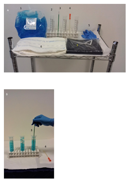
- 在诊断室区域内放置手推车, 配备所有必要的仪器和用品, 包括手套、注射器、导管、润滑剂果冻、声学凝胶等 (见材料表)。

图 1: 用品.这张图片显示 (a) 一个手推车, 带有 mr 检查用品, (b) 在给药前, 在诊断室附近的区域用水稀释声学凝胶 (每个注射器 50/30 毫升)。请点击这里查看此图的较大版本.
- 要求患者将 mr 扫描仪的诊断表放置在左侧位置 (Sims 的位置)。轻轻地将导管插入直肠, 并给药造影剂 (声学耦合凝胶), 直到患者体验到典型的撤离欲望 (平均剂量为250毫升)。之后把病人变成仰角。
- 调整臀部下方的吸水垫, 并将表面相控阵线圈包裹在患者的骨盆周围, 以获取图像。
注: 在预期的感觉, 紧急, 不适, 或非自愿泄漏的情况下, 中断注射, 并登记在泄漏之前注射的总体积, 以及体积泄漏。
3. 技术和图像采集
- 获取日冕、轴向和矢状平面 (TFE t1 脉冲序列、8毫秒的 TR、5毫秒的 TE、25°的翻转角度 (FA)、15毫米的翻转角度 (FA)、15毫米的图像数和图像数量: 5-11) 中的定位器侦察扫描, 以标记感兴趣区域的边界 (ROI)。
- 然后, 在中矢状面上得到三个后续动态序列 (见表 1, 系列 1:trp, 2.7/1.3 ms;在肛门直肠交界处中心的 FA, 与病人休息, 挤压他/她的肛门括约肌, 并紧张 (每个 10秒)。
- 此后, 指示患者立即开始直肠排空的运动, 并在开始时 (通过声学装置) 通知患者, 以便在58秒的整个时间周期内模拟采集图像 (见表 1, 系列 1:trcte, 2.7/1.3 ms;FA 45 °;厚度35毫米;获取时间为 58s)。
注: 如有必要, 请重复该序列, 直到获得足够的对比度流。 - 在日冕平面上重复后一个序列 (见表 1, 系列 2:trp, 2.8/1.3 ms;FA 45 °;厚度35毫米;当患者排出残余直肠对比时, 获得时间为58秒。
- 然后, 指示患者在不中断9个月的情况下进行稳态 Valsalva 机动。
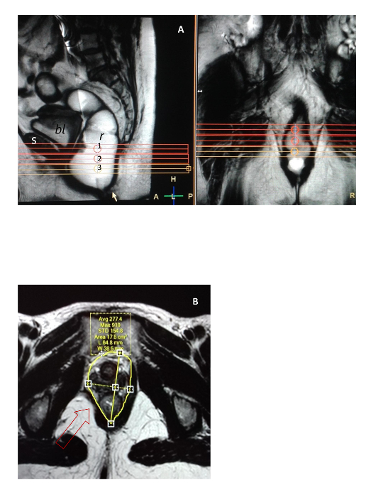
- 以直肠排空过程中获得的矢状图像作为参考, 选择轴向平面上的三个水平平面 (见图 3) 来成像提升体的间隙, 如下所示: 第一个在中部 (一级), 第二个切线到在直肠前壁 (iii 级) 最大鼓胀点的下边界 (二级), 第三边界。
注: 上述原因是在 ROI 中包括男女间隔边界内外最相关的解剖区域: 膀胱基地、尿道、阴道、子宫颈、会阴体、肛门直肠结合部、内侧筋膜和脂肪凹槽 (女性), 或膀胱基部、后间隙、前列腺、精囊、丹尼虫筋膜、肛门直肠结合部、肠系膜筋膜和前间隙 ( 男性 ) 。 - 在轴向平面上获得水平1厘米厚的截面 (见表 1, 系列 3:trp, 3/1.5 ms;FA 45 °;厚度为10毫米;在瓦尔萨尔瓦机动期间, 从每个级别获得9s 的时间, 使患者在随后的两次机动之间有60秒的间隔来放松。
- 最后, 在轴向、矢状和日冕平面下静止时获取静态 T2 加权图像 (见36499-46600100 系列4、5和6:trpe 表1;90° fa;厚度间隙为 4/0.4 mm;采集时间为 3: 00:3:44 分钟), 为盆腔解剖提供完整的评估。
注: 有关技术设置, 请参阅表 1 。

表 1: 使用 1.5 T 扫描仪和外部线圈进行 MR 排便成像的技术设置。
4. 图像分析和测量
- 要测量盆腔器官在静止时和紧张时的位置, 从分析软件中的中矢状动态 MR 图像, 转到位于屏幕顶部的工具栏选项列表, 并将鼠标悬停在 "注释工具" 上。
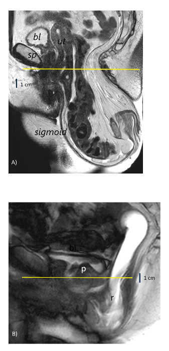
- 然后, 单击箭头并选择标尺, 从两条参考线获得膀胱颈、子宫颈、前列腺基、精囊和直肠地板垂直距离的线性测量 (以毫米为单位), 如下所示:相对于处女膜平面 (女性) 或与共生普比 (雄性) 的下边界相切的水平线所具有的负 (近端) 或正 (远端) 数字的距离。

图 2: 在中矢状 mr 图像上的盆腔器官下降参考线.(A) 一名61岁妇女, 直肠下降在 hymen 平面 (黄线) 和 sigmoidocele 下方 gt;10 厘米。(B) 一名 42 岁男子 , 直肠肠套叠 , 在联合 ( 黄线 ) 的下缘下降到 gt ; 3 厘米。bl = 膀胱;sp = 联合性 ;ut = 子宫;r = 直肠;p = 前列腺。请点击这里查看此图的较大版本.
- 要从轴向静态和动态图像中测量裂孔前直径和横向直径 ( 毫米 ) , 请重复步骤 4 . 1 - 4 . 2 中描述的相同线性测量选择 , 并计算从联合到拟波勒索的前缘和悬浮物肌肉内侧边界之间的距离。
- 要测量休息时和最大应变时的裂孔面积 (以平方厘米为单位), 请再次单击"注释工具" , 然后选择"自由 roi " 以选择自由轮廓跟踪技术 (参见图 3)。
- 说明提升体肌肉的内部区域, 并将静息测量和紧张测量表示为绝对值, 并表示确认骨性地标所确定的同一级别部分的百分比增加, 即共发和同性结节 ( 2 级 ) 。
注: 登记对悬浮液内器官的任何撞击, 并在对器官脱垂的定义和分级时, 参考国际盆腔器官脱垂委员会建议的标准 14、15和传统的盆腔器官脱垂 "HMO" mri 分类系统.9个。

图 3: 提升机的间隙成像和面积测量方法.(A) 从相对于中突状 ( 1 级 ) 拍摄的中矢状图像中选择三个轴向扫描段 , 与其下边界 ( 2 级 ) 相切 , 并在直肠排空期间直肠前壁 ( 第 3 级 ) 的最大凸起处进行选择。(B) 在一名 52 岁的妇女中 , 用左手轮廓追踪法测量的非对称区域的例子 , 该妇女的右球菌肌肉 ( 箭头 ) 有焦点缺陷。s = 联合 ;bl = 膀胱;r = 直肠。左侧面板 = 矢状视图; 右侧面板 = 日冕视图。面积值以平方厘米 1 = 第一级表示;2 = 第二层;3 = 三级。请点击这里查看此图的较大版本.
结果
2012年至2018年期间, 该协议已在意大利三个不同的诊断中心成功采用, 平均每月累计考试速度为 30±4, 使用相同的 1.5 Tmr 扫描仪型号和技术设置 (见表 1和材料)。在此期间, 对以下三个主要疾病类别的男女患者进行了 2 000多次检查: 盆腔器官脱垂和疏散障碍 (第1组)、肛周瘘 (第2组) 和慢性盆腔疼痛。已知或怀疑神经病变 ( 第 3 组 ) 。患者人口中最相关?...
讨论
这种方法比盆腔检查有明显的优势, 仅限于评估泌尿生殖道的中断, 只在女性。相反, 这里提出的方法检查整个提升机在男性和男性之间的间隙。此外, 虽然很容易通过检查的触诊的妇科医生, 女性裂隙只能近似地计算与统治者, 以产生一个椭圆形1的面积。同样, 与2和 3-d tpus4,5相比, 确实存在优势, 这两种优势不适合男性患者。然而, 该协?...
披露声明
作者没有什么可透露的。
致谢
提交人特别感谢护士保拉·加拉维洛和朱利亚·梅拉拉在检查期间提供的宝贵协助。
材料
| Name | Company | Catalog Number | Comments |
| MR scanner | Philips Medical Systems, High Tech Campus 37, 5656 AE, Eindhoven, The Netherlands | Description: 1.5 T horizontally oriented, Multiva model, SENSE XL Torso coil Procedure: Position the patient in the left lateral decubitus on the diagnostic table with the coil warpped around the pelvis | |
| Catheter | Convatek ltd, First avenue Deeside, Flintshire CH5 2NU UK | Description: Sterile vaginal catheter, 16 ch,180 mm long, 3 mm wide Procedure: Gently insert the lubricated tip inside the anal canal for contrast administration with patients in the left side position | |
| Holder | Kartell Plastilab, Artiglas Srl, Via Carrara Padua, Italy | Description: Universal test-tube holder with multiple 13-mm holes Procedure: Put 3 empty syringes vertically inside the holes with the outlet cone down | |
| Syringe | Pikdare Srl, Via Saldarini Catelli 10 , 22070 Casnate con Bernate (Como) Italy | Description: Sterile, latex free,60 mL graduated transparent cylinder, catheter cone Procedure: Fill with contrast, adjust the plunger and connect to the catheter | |
| Contrast | Ceracarta SpA, Via Secondo Casadei 14 47122 Forlì Italy | Description: Eco supergel not irritant, water soluble, salt free Procedure: Dilute the content of each syringe adding 30 mL of tap water to 50 mL of acustic gel | |
| Mixer device | Kaltek Srl, Via del Progresso 2 Padua Italy | Description: Kito-Brush for endovaginal sampling Procedure: Rotate one full turn 10-20 times until obtaining an homogeneous gel | |
| Pad | Fater SpA, Via A. Volta 10, 65129 Pescara Italy | Description: Pad for incontinent subjects Procedure: Wrap around patient's pelvis to collect any material and prevent diagnostic table contamination | |
| Lubricant | Molteni farmaceutici, Località Granatieri Scandicci (Florence) Italy | Description: Luan gel 1% Procedure: Apply on the tip of catheter before insertion | |
| Apron | Mediberg Srl, via Vezze 16/18 Calcinate 24050 (Bergamo) Italy | Description: Kimono Procedure: Put on counteriwise (opening back) to maintain patient's dignity | |
| Gloves | Gardening Srl, Via B. Bosco 15/10 16121 Genova Italy | Description: Nitrile, latex free, no talcum powdered Procedure: Wear during contrast preparation and catheter insertion; change regularly to prevent cross contamination |
参考文献
- DeLancey, J. O. L., Hurd, W. W. Size of the urogenital hiatus in the levator ani muscles in normal women and women with pelvic organ prolapse. Obstetrics & Gynecology. 91 (3), 364-368 (1998).
- Siproudhis, L., Ropert, A., Vilotte, J. How accurate is clinical examination in diagnosing and quantifying pelvirectal disorders? A prospective study in a group of 50 patients complaining of defecatory difficulties. Diseases of the Colon & Rectum. 36 (5), 430-438 (1993).
- Maglinte, D. D. T., Kelvin, F. M., Fitzgerald, K., Hale, D. S., Benson, J. T. Association of compartment defects in pelvic floor dysfunction. American Journal of Roentgenology. 172 (2), 439-444 (1999).
- Dietz, H. P., Jarvis, S. K., Vancaillie, T. G. The assessment of Levator muscle strength: a validation of three ultrasound techniques. International Urogynecology Journal and Pelvic Floor Dysfunction. 13 (3), 156-159 (2002).
- Dietz, H. P., Shek, C., Clarcke, B. Biometry of the pubovisceral muscle and levator hiatus by three-dimensional pelvic floor ultrasound. Obstetrics & Gynecology. 25 (6), 580-585 (2005).
- Yang, A., Mostwin, J. L., Rosenhein, N. B., Zerhouni, E. A. Pelvic floor descent in women: dynamic evaluation with fast MR imaging and cinematic display. Radiology. 179 (1), 25-33 (1991).
- Lienemann, A., Anthuber, C., Baron, A., Kohz, P., Reiser, M. Dynamic MR colpocystorectography assessing pelvic floor descent. European Radiology. 7 (8), 1309-1317 (1997).
- Healy, J. C., et al. Patterns of prolapse in women with symptoms of pelvic floor weakness: assessment with MR imaging. Radiology. 203 (1), 77-81 (1997).
- Comiter, C. V., Vasavada, S. P., Barbaric, Z. L., Gousse, A. E., Raz, S. Grading pelvic prolapse and pelvic floor relaxation using dynamic magnetic resonance imaging. Urology. 54 (3), 454-457 (1999).
- Kelvin, F. M., Maglinte, D. D. T., Hale, D. S., Benson, J. T. Female pelvic organ prolapse: a comparison of triphasic dynamic MR imaging and triphasic fluoroscopic cystocolpoproctography. American Journal of Roentgenology. 174 (1), 81-88 (2000).
- Pannu, H. K., et al. Dynamic MR imaging of pelvis organ prolapse: spectrum of abnormalities. RadioGraphics. 20 (6), 1567-1582 (2000).
- Hoyte, L., Ratiu, P. Linear measurements in 2-dimen¬sional pelvic floor imaging: the impact of slice tilt angles on measurement reproducibility. American Journal of Obstetrics and Gynecology. 185 (3), 537-544 (2001).
- Tunn, R., DeLancey, J. O. L., Quint, E. E. Visibility of pelvic organ support system structures in magnetic resonance images without an endovaginal coil. American Journal of Obstetrics and Gynecology. 184 (6), 1156-1163 (2001).
- Bump, R. C., et al. The standardization of terminology of female pelvic organ prolapse and pelvic floor dysfunction. American Journal of Obstetrics and Gynecology. 175 (1), 10-17 (1996).
- Haylen, B. T., et al. An inter-national Urogynecological Association (IUGA) / International Continence Society (ICS) Joint Report on the Terminology for Female Pelvic Floor Dysfunction. Neurology and Urodynamics. 29 (1), 4-20 (2009).
- Fielding, J. R., et al. MR imaging of pelvic floor continence mechanisms in the supine and sitting positions. American Journal of Roentgenology. 171 (6), 1607-1610 (1998).
- Bertschinger, K. M., et al. Dynamic MR imaging of the pelvic floor performed with patient sitting in an open-magnet unit versus with patient supine in a closed-magnet unit. Radiology. 223 (2), 501-508 (2002).
- Piloni, V., Ambroselli, V., Nestola, M., Piloni, F. Quantification of levator ani (LA) hiatus enlargement and pelvic organs impingement on Valsalva maneuver in parous and nulliparous women with obstructed defecation syndrome (ODS): a biomechanical perspective. Pelviperineology. 35 (1), 25-31 (2016).
- Piloni, V., Pierandrei, G., Pignalosa, F., Galli, G. Fusion imaging by transperineal sonography/magnetic resonance in patients with fecal blockade syndrome. EC Gastroenterology and Digestive System. 5 (1), 11-16 (2018).
- Piloni, V., Bergamasco, M., Melara, G., Garavello, P. The clinical value of magnetic resonance defecography in males with obstructed defecation syndrome. Techniques in Coloproctology. 22 (3), 179-190 (2018).
- Chanda, A., Unnikrishnan, V. U., Roy, S., Ricther, H. E. Computational modeling of the female pelvic support structures and organs to understand the mechanisms of pelvic organ prolapse: a review. Applied Mechanics Reviews. 67 (4), 040801-040814 (2015).
- Rostaminia, G., Abramowitch, S. Finite element modeling in female pelvic floor medicine: a literature review. Current Obstetrics and Gynecology Reports. 4 (2), 125-131 (2015).
转载和许可
请求许可使用此 JoVE 文章的文本或图形
请求许可This article has been published
Video Coming Soon
版权所属 © 2025 MyJoVE 公司版权所有,本公司不涉及任何医疗业务和医疗服务。