
A Procedure for Implanting a Spinal Chamber for Longitudinal In Vivo Imaging of the Mouse Spinal Cord
December 3rd, 2014
•In this video, we describe a procedure for implanting a chronic optical imaging chamber over the dorsal spinal cord of a live mouse. The chamber, surgical procedure, and chronic imaging are reviewed and demonstrated.
Related Videos
Spinal Cord Electrophysiology II: Extracellular Suction Electrode Fabrication

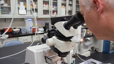
In vivo Imaging of the Mouse Spinal Cord Using Two-photon Microscopy

Stereotaxic Injection of a Viral Vector for Conditional Gene Manipulation in the Mouse Spinal Cord

Diffusion Imaging in the Rat Cervical Spinal Cord

Two-photon Imaging of Cellular Dynamics in the Mouse Spinal Cord

Imaging Serotonergic Fibers in the Mouse Spinal Cord Using the CLARITY/CUBIC Technique

Imaging Neural Activity in the Primary Somatosensory Cortex Using Thy1-GCaMP6s Transgenic Mice

An In Vivo Duo-color Method for Imaging Vascular Dynamics Following Contusive Spinal Cord Injury

Laminectomy and Spinal Cord Window Implantation in the Mouse
Preparation of Rhythmically-active In Vitro Neonatal Rodent Brainstem-spinal Cord and Thin Slice