
Method Article
Ensayos pronta entrada virales para la identificación y evaluación de compuestos antivirales
En este artículo
Resumen
Here, we present a protocol that examines specific steps of the viral entry to identify and evaluate novel antiviral agents.
Resumen
Cell-based systems are useful for discovering antiviral agents. Dissecting the viral life cycle, particularly the early entry stages, allows a mechanistic approach to identify and evaluate antiviral agents that target specific steps of the viral entry. In this report, the methods of examining viral inactivation, viral attachment, and viral entry/fusion as antiviral assays for such purposes are described, using hepatitis C virus as a model. These assays should be useful for discovering novel antagonists/inhibitors to early viral entry and help expand the scope of candidate antiviral agents for further drug development.
Introducción
Viral infections are a constant threat to the public health and a significant cause of epidemic diseases, morbidity, and deaths worldwide. Specific modes of control against viral infections include vaccine development and antiviral therapies. While vaccine efforts have proven successful in immunizing against several viruses, many viral pathogens remain without a protective vaccine including dengue virus (DENV), human cytomegalovirus (HCMV), hepatitis C virus (HCV), human immunodeficiency virus (HIV), and respiratory syncytial virus (RSV)1-5. Antivirals, on the other hand, play an important role for the management of these viral infections when prophylactic vaccines are unavailable. However, to date, only few licensed and cost-effective antiviral drugs are available compared to the number of viral pathogens that threatens the public health. More importantly, due to an increase in global travel and rapid urbanization, the situation is aggravated by risks of epidemic outbreaks from emerging/re-emerging viral infections that are being introduced into non-indigenous areas6. Recent outbreaks caused by severe acute respiratory syndrome (SARS) virus, influenza viruses (H1N1, H5N1, H3N2, and H7N9), DENV, West Nile virus (WNV), measles virus (MV), Middle East Respiratory Syndrome (MERS) virus, and Ebola virus6-12 are among the examples reflecting the need for antivirals development when immunization and/or therapeutics are unavailable. In addition, there is always a potential risk of generating drug-resistant infections with currently used antivirals. Thus, the continuous development and expansion of the scope of antivirals to these emerging/re-emerging viral infections are necessary to provide better management strategies and safeguard the public health.
Most antiviral therapies consist of direct acting antivirals (DAAs) which target a specific viral protein or cofactor that mediates important steps in the viral life cycle. For example, the nucleoside analogue Acyclovir inhibits herpesvirus DNA polymerase, protease inhibitors Boceprevir and Telaprevir antagonize the HCV NS3, and Oseltamivir and Zanamivir are neuraminidase inhibitors that block the release of influenza virus particles from infected cells13-15. There are however very few licensed viral entry inhibitors including Enfuvirtide, which targets HIV gp41 to prevent fusion, and Maraviroc, which blocks the HIV co-receptor CCR5, thereby preventing viral entry16. Exploring novel antagonists/inhibitors to viral entry could help provide additional therapeutics for prophylactic or therapeutic use, such as in combination with other antivirals with a different mechanism of action to better manage viral infections17-19.
Identification of antivirals can involve structure-guided drug design and candidate drug screening-based strategy. Methods for assessing antiviral activity of test agents include biochemical assays of enzymatic activity and evaluation by cell-based systems20-23. In cell-based systems, the viral life cycle can be dissected into distinct stages of infection, such as entry events (attachment, fusion, uncoating), the replication phase (viral genome replication and protein translation), and virion egress (assembly, maturation, and release). Since the assays can be adapted to investigate each specific stage using various tools and methods, this approach allows identification/examination of potential candidate antivirals with a specific mechanism of action targeting the distinct stage analyzed. For instance, to analyze drug effect specifically on the free virus particles prior to binding to the host cell, a ‘viral inactivation assay’ can be performed. In this assay, the virus is allowed to incubate with the test drug and then diluted to titrate out the drug before infecting a cell monolayer. Additional steps such as viral attachment and entry/fusion stages can also be analyzed individually, by shifting the temperature during the infection. For many enveloped viruses, viral entry/fusion at the host cell membrane is greatly facilitated at 37 °C, but is typically precluded at 4 °C which does not affect virus binding24-29. Finally, the use of reporter viruses or cell systems could facilitate these studies and permit a high-throughput analysis.
We previously employed the cell-based approach and dissected the early entry of various enveloped viruses for the examination of antiviral compounds that potentially antagonize viral entry30,31. Herein, the various methods used, including viral inactivation, viral attachment, and viral entry/fusion assays, are described.
Protocolo
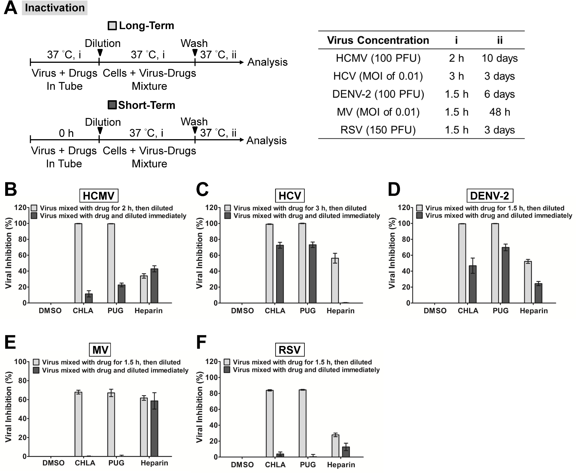
Nota: Asegúrese de que todos los procedimientos relacionados con el cultivo de células y de infección por el virus se llevan a cabo en las campanas de bioseguridad certificados que sean apropiados para el nivel de bioseguridad de las muestras que se manejan. Para el propósito de describir los protocolos, la luciferasa Gaussia HCV reportero de etiquetado se usa como un modelo de virus 32. En el contexto de los resultados representativos, el ácido compuestos chebulágico (CHLA) y punicalagina (PUG) se utilizan como antivirales candidatos que se dirigen las interacciones glicoproteína viral con glicosaminoglicanos superficie celular durante la entrada viral temprana pasos 31. La heparina, que se sabe que interfiere con la entrada de muchos virus 30,31,33,34, se utiliza como un tratamiento control positivo en tal contexto. Para base de fondo en las técnicas de virología, la propagación de virus, determinación del título de virus, y la expresión de la dosis infecciosa en unidades formadoras de placa (PFU), se centran unidades formadoras (FFU), o multiplicidad de infección (MOI), el lector es repreferido para hacer referencia 35. Para ejemplos anteriores y condiciones optimizadas utilizados para los virus que se muestran en los resultados representativos, se remite al lector a las referencias 30-32,36-39, así como detalles enumerados en la Tabla 1, la Figura 1A, y la Figura 2A.
1. Cultivo Celular, Preparación Compuesto y citotoxicidad Compuesto
- Crece la línea celular respectiva para el sistema de infección por el virus para ser analizados (Tabla 1). Para VHC, crecer las células Huh-7.5 en medio de Eagle modificado por Dulbecco (DMEM) suplementado con 10% de suero bovino fetal (FBS), 200 U / ml de penicilina G, 200 mg / ml de estreptomicina, y 0,5 mg / ml de anfotericina B.
- Preparar los compuestos de ensayo y controles utilizando sus respectivos disolventes: por ejemplo, disolver CHLA y PUG en sulfóxido de dimetilo (DMSO); preparar heparina en agua bidestilada estéril. Para todas las diluciones posteriores, utilizar medios de cultivo.
Nota: La última concentración de DMSO en los tratamientos compuesto de ensayo es menos de 1% en los experimentos; 1% de DMSO se incluye como un tratamiento de control negativo en los ensayos para la comparación. - Determinar la citotoxicidad de los compuestos de ensayo (por ejemplo, chla y PUG) sobre las células para la infección viral mediante el uso de una viabilidad celular determinar reactivo tal como XTT (2,3-bis [2-metoxi-4-nitro-5-sulfofenil] -5-fenilamino) carbonil] -2H-tetrazolio hidróxido):
- Para VHC, sembrar células Huh-7.5 en una placa de 96 pocillos (1 × 10 4 células por pocillo) y se incuba a 37 ° C en una incubadora de CO2 al 5% O / N para obtener una monocapa.
- Aplicar el control de DMSO (1%) o concentraciones crecientes del compuestos de ensayo y CHLA PUG (Ex. 0, 10, 50, 100, y 500 mM) a los pocillos de cultivo por triplicado.
- Incubar a 37 ° C durante 72 hr, luego descarta el medio en la placa y lavar las células con 200 l de solución salina tamponada con fosfato (PBS) dos veces.
- Añadir 100 l de assaying solución del kit de ensayo in vitro de la toxicología en basado en XTT a cada pocillo e incubar las placas a 37 ° C durante otras 3 horas para permitir la producción de formazan XTT.
- Determinar la absorbancia con un lector de microplacas a una longitud de onda de ensayo de 492 nm y una longitud de onda de referencia de 690 nm.
- Calcular el porcentaje de células supervivientes usando la siguiente fórmula: Viabilidad celular (%) = A / Como × 100%, donde "A" y "Como 'se refieren a la absorbancia de los compuestos de ensayo y el control de disolvente (ex 1% de DMSO. ) tratamientos, respectivamente. Determinar la concentración de 50% de citotoxicidad celular (CC 50) de los compuestos de ensayo a partir de un software de análisis tales como GraphPad Prism de acuerdo con el protocolo del fabricante.
2. Lectura de la infección viral
Nota: La lectura de la infección viral depende del sistema utilizado y virus puede implicar métodos tales como ensayos de placas o measuring señales indicadoras de virus reportero-etiquetado. El método para detectar la infección por VHC reportero basado en la actividad de informador de luciferasa se describe a continuación.
- Reunir los sobrenadantes de los pozos infectados y aclarar a 17.000 xg en una microcentrífuga durante 5 min a 4 ° C.
- Mezclar 20 l de sobrenadante de ensayo a 50 l de sustrato de luciferasa del kit de ensayo de luciferasa Gaussia y medir directamente con un luminómetro de acuerdo con las instrucciones del fabricante.
- Expresar la infectividad del VHC como log 10 de unidades relativas de luz (RLU) para la determinación de la inhibición viral (%) y calcular la concentración 50% eficaz (CE 50) de los compuestos de ensayo contra la infección por el VHC usando algoritmos de software GraphPad Prism de acuerdo con el protocolo del fabricante.
3. Inactivación Viral Ensayo
Nota: Los ejemplos de período de incubación y la dosis viral de diversos virus unare enumerados en la Figura 1A. Las concentraciones más altas del virus también pueden ser probados por el aumento de la MOI / PFU.
- Semilla de células en una placa de 96 pocillos de 7,5 Huh-(1 × 10 4 células por pocillo) y se incuba a 37 ° C en un incubador de CO2 5% de O / N para obtener una monocapa.
- Incubar los compuestos de ensayo o controles (concentraciones finales son: CHLA = 50 M; PUG = 50 M; heparina = 1,000 g / ml; DMSO = 1%) con las partículas de VHC a 37 ° C (Figura 1A, 'a largo plazo' ) en una proporción de 1: 1. Por ejemplo, para un inóculo de virus de 100 l que contiene 1 x 10 4 FFU, añadir 100 l de una dilución de trabajo 100 mM CHLA; esto produce el tratamiento CHLA a una concentración final de 50 mM.
- Diluir la mezcla de virus-fármaco para (ineficaz) de concentración "sub-terapéutica" de los compuestos de ensayo. Por ejemplo, la concentración ineficaz de CHLA y PUG contra el VHC está en 1 M 31; Por Consiguienteesto requiere una dilución de 50 veces de la mezcla de virus-fármaco que se puede lograr con 9,8 ml de medio basal (medio de cultivo celular con 2% de FBS).
Nota: La dilución a la concentración sub-terapéutica impide la interacción significativa entre los compuestos de ensayo y la superficie de la célula huésped y permite el examen del efecto del tratamiento en los viriones libres de células. Tenga en cuenta que esta dilución depende de la respuesta a la dosis antiviral de los compuestos de ensayo frente a la infección viral en particular, y se determina antes de realizar este ensayo en particular 31. - Para la comparación, mezclar el virus con los compuestos de ensayo y de inmediato diluir (sin período de incubación) a la concentración sub-terapéutica antes de la infección (Figura 1A, 'corto plazo').
- Añadir 100 l de la mezcla de HCV-fármaco diluida sobre el 7,5-Eh monocapa celular (la cantidad de virus se encuentra ahora en 1 x 10 2 FFU; MOI final = 0,01) e incubar durante 3 horas a 37 ° C para permitir viraladsorción.
- Tras la infección, eliminar el inóculo diluido y lavar suavemente los pocillos con 200 l de PBS dos veces.
Nota: Realizar los lavados con cuidado para evitar el levantamiento de las células. - Aplicar 100 l de medio basal a cada pocillo e incubar a 37 ° C durante 72 hr.
- Analizar la infección resultante sometiendo a ensayo el sobrenadante para la actividad de luciferasa como se describe en '2. Lectura de la infección viral '.
4. Ensayo Adjunto Viral
Nota: Los ejemplos de período de incubación y la dosis viral para varios virus se enumeran en la Figura 2A, el "Anexo". Las concentraciones más altas del virus también pueden ser probados por el aumento de la MOI / PFU.
- Semilla de células en una placa de 96 pocillos de 7,5 Huh-(1 × 10 4 células por pocillo) y se incuba a 37 ° C en un incubador de CO2 5% de O / N para obtener una monocapa.
- Pre-enfriar las monocapas de células en placas a 4 ° C for 1 hr.
- Co-tratamiento de las células con inóculo HCV (MOI = 0,01) y compuestos o controles de ensayo (concentraciones finales son: CHLA = 50 M; PUG = 50 M; heparina = 1,000 g / ml; DMSO = 1%) a 4 ° C durante 3 hr. Por ejemplo, para un inóculo de virus de 90 l que contiene 1 x 10 2 FFU, añadir 10 l de una dilución de trabajo 500 mM CHLA; esto produce el tratamiento CHLA a una concentración final de 50 mM y una infección por el VHC a MOI = 0,01 en la monocapa de células.
Nota: Es importante llevar a cabo el experimento a 4 ° C, ya que permite la unión del virus, pero se opone a la entrada que se produce de manera más eficiente a 37 ° C. Realizar la adición de compuestos de virus y de prueba en el hielo y la incubación subsiguiente en un refrigerador 4 ° C para asegurar que la temperatura se mantiene a 4 ° C. - Eliminar el sobrenadante y lavar suavemente la monocapa de células con 200 l de helado de PBS dos veces.
Nota: Realizar los lavados con cuidado para evitar el levantamiento de las células <./ li> - Aplicar 100 l de medio basal a cada pocillo e incubar a 37 ° C durante 72 hr.
- Analizar la infección resultante sometiendo a ensayo el sobrenadante para la actividad de luciferasa como se describe en '2. Lectura de la infección viral '.
5. Viral Entry / ensayo de fusión
Nota: Ejemplo de períodos de incubación y dosis viral para varios virus se enumeran en "Entrada / Fusión 'Figura 2A. Las concentraciones más altas del virus también pueden ser probados por el aumento de la MOI / PFU.
- Semilla de células en una placa de 96 pocillos de 7,5 Huh-(1 × 10 4 células por pocillo) y se incuba a 37 ° C en un incubador de CO2 5% de O / N para obtener una monocapa.
- Pre-enfriar las monocapas de células en placas a 4 ° C durante 1 hora.
- Infectar las células con HCV (MOI = 0,01) a 4 ° C durante 3 hr. Por ejemplo, utilice un inóculo de virus 100 l que contiene 1 x 10 2 FFU.
Nota: Realice el adición del inóculo viral en hielo y la incubación subsiguiente en un refrigerador 4 ° C para mantener la temperatura a 4 ° C, la cual permite la entrada de unión pero no viral. - Eliminar el sobrenadante y lavar suavemente las monocapas de células con 200 l de helado de PBS dos veces.
Nota: Realizar los lavados con cuidado para evitar el levantamiento de las células. - Tratar los pocillos con los compuestos de ensayo o controles (concentraciones finales son: CHLA = 50 M; PUG = 50 M; heparina = 1,000 g / ml; DMSO = 1%) y se incuba a 37 ° C durante 3 hr. Por ejemplo, añadir 10 l de una dilución de trabajo 500 M CHLA a 90 l de los medios de comunicación, mezclar y tratar los pozos; esto produce el tratamiento CHLA a una concentración final de 50 mM.
Nota: El cambio de 4 ° C a 37 ° C ahora facilita el evento de entrada / fusión viral y por lo tanto permite la evaluación del efecto de los compuestos de ensayo 'en este paso particular. - Aspirar el sobrenadante que contiene el fármaco y quitar no internalizadovirus extracelulares ya sea por lavado con 200 l de tampón citrato (citrato de sodio 50 mM, cloruro de potasio mM 4, pH 3,0) o PBS. Aplicar 100 l de medio basal antes de la incubación a 37 ° C durante 72 hr.
- Analizar la infección resultante sometiendo a ensayo el sobrenadante para la actividad de luciferasa como se describe en '2. Lectura de la infección viral '.
Resultados
En la Figura 1, se realizó el "ensayo de inactivación viral 'para examinar si dos compuestos naturales CHLA específica y PUG podrían inactivar los virus con envoltura diferentes en el estado libre de células y prevenir la infección subsiguiente. La respuesta a la dosis antiviral y la citotoxicidad de estos compuestos se han determinado antes de realizar el estudio mecanicista 31. Los virus fueron pre-tratados con los compuestos de ensayo y después las mezclas de virus a los fármacos se diluyeron a concentraciones sub-terapéuticas antes de la inoculación sobre la respectiva monocapa de células para cada sistema de virus. Como se muestra en la Figura 1, tanto CHLA y PUG aparecieron para interactuar con los viriones libres de células, dando lugar a efectos irreversibles que protegían la monocapa de células de la infección subsiguiente. Los dos compuestos de ensayo alcanzan alrededor de 100% de inhibición contra HCMV, VHC y DENV-2, mientras que un 60 - 80% de bloque se observó contra MV y RSV. Estos resultados Suggest que CHLA y PUG tienen impacto directo sobre estas partículas virales libres al inactivar ellos y neutralizando su infectividad.
En la Figura 2, el apego y ensayos de entrada / fusión se llevaron a cabo para explorar el efecto de CHLA y PUG en contra de estos principios de los acontecimientos relacionados con la entrada-virales de HCMV, VHC, DENV-2, MV, y RSV. Tanto CHLA y PUG impidieron efectivamente la unión de los virus investigados en la célula huésped respectiva, como se muestra por la inhibición de la infección viral resultante (Figura 2, el "Anexo ': barras de color gris claro). El efecto inhibidor sobre la fijación del virus por ambos compuestos era similar contra HCMV (Figura 2B), HCV (Figura 2C), DENV-2 (Figura 2D), y RSV (Figura 2F), que van desde 90 hasta 100%. Por otro lado, PUG parecía ser más eficaz que CHLA contra MV de unión (Figura 2E), con la tasa de inhibición de la two compuestos que varían entre 50 - 80%. La heparina tratamiento de control, que es conocido para bloquear la entrada de muchos virus, también inhibió la unión de HCMV, DENV-2, RSV, ad MV, pero fue menos eficaz contra el VHC. La subsiguiente 'ensayo de entrada / fusión viral' examinó si CHLA y PUG mantuvieron su actividad durante la fase de entrada de virus / fusión (Figura 2, 'Entrada / Fusión': barras de color gris oscuro). Una vez más, tanto CHLA y PUG se observaron perjudicar efectivamente el paso de entrada / fusión viral de los virus examinados (Figura 2 B - F), produciendo un 50 - efecto protector del 90% en la respectiva monocapa celular. La heparina también inhibe potentemente la entrada / fusión de DENV-2 y las infecciones por VRS, pero fue menos eficaz contra HCMV, VHC y MV (<40% de inhibición en promedio).
| Virus | Tipo de la célula |
| HCMV | HEL |
| VHC | Huh-7.5 |
| DENV-2 | Vero |
| MV | CHO-SLAM |
| RSV | HEp-2 |
Tabla 1:. Tipo de célula huésped para la infección viral El tipo celular utilizado para cada sistema de la infección viral se describe en los resultados representativos se indica. Los detalles adicionales con respecto a las células se pueden encontrar en la referencia 31.

Figura 1. Inactivación de infecciones virales por el CHLA compuestos de ensayo y PUG Diferentes virus se trataron con los compuestos de ensayo durante un período largo. (Se incubaron durante 1.5 - 3 horas antes de la titulación; barras de color gris claro) o corto período de tiempo (inmediatamente diluido; gris oscuro bares) a 37 ° C antes de una dilución a concentraciones sub-terapéuticación y posterior análisis de la infección en las respectivas células de acogida. (A) Esquema del experimento (que se muestra a la izquierda) con la concentración de virus final (PFU / pocillo o MOI), período de incubación del virus con la droga a largo plazo (i), y el tiempo de incubación posterior (ii) indicado para cada virus en el cuadro a la derecha. Los análisis para (B) HCMV, (C) del VHC, (D) DENV-2, (E) MV, y (F) de RSV se indican en cada panel adicional. Los resultados se representan frente al tratamiento de control negativo DMSO para la infección por virus y los datos mostrados son las medias ± error estándar de la media (SEM) de tres experimentos independientes. Esta cifra se ha modificado de la referencia 31. Haga clic aquí para ver una versión más grande de esta figura.

Figura 2. Evaluación de las actividades antivirales de la CHLA compuestos de ensayo y PUG contra la fijación del virus y la entrada / fusión. (A) El procedimiento experimental, la concentración de virus (PFU / pocillo o MOI), y el tiempo de adición y el tratamiento con los compuestos de prueba (i, ii, iii) se presentan para cada virus en los esquemas y las tablas asociadas. En el análisis de la fijación del virus (barras de color gris claro), las monocapas de diferentes tipos de células fueron pre-refrigerados a 4 ° C durante 1 hr, a continuación, co-tratado con los respectivos virus y compuestos de ensayo a 4 ° C (1,5 - 3 h; i) antes de lavar los inóculos y los compuestos de ensayo para su posterior incubación (37 ° C; ii) y el examen de la infección por virus. En la entrada del virus / análisis de fusión (barras gris oscuro), monocapas de células sembradas se pre-refrigerada a 4 ° C durante 1 hora y después se estimularon con los virus respectivos a 4 ° C durante 1,5 - 3 h (i). Las células fueron entonceslavada y tratada con los compuestos de ensayo durante un periodo de incubación adicional (ii) durante el cual la temperatura se cambió a 37 ° C para facilitar el evento de entrada / fusión viral. Al final de la incubación, los virus extracelulares fueron retirados por cualquiera de tampón citrato (pH 3,0) o lavados con PBS y las células se incubaron adicionalmente (iii) para el análisis de la infección por virus. Resultados para (B) HCMV, (C) del VHC, (D) DENV-2, (E) MV, y (F) de RSV se indican en cada panel adicional. Los datos se representan frente al tratamiento de control negativo de la infección por el virus de DMSO y se presentan como medias ± SEM de tres experimentos independientes. Esta cifra se ha modificado de la referencia 31. Haga clic aquí para ver una versión más grande de esta figura.
Discusión
In this report the methods to identify and evaluate antiviral compounds based on a mechanistic approach of dissecting the early viral entry events were described. Specifically, the assays allowed us to examine the effect of test compounds on free virus particles, viral attachment, and viral entry/fusion. Critical steps were implemented to distinctly evaluate the drug effect on the specific stage of early viral entry. For instance, in the ‘viral inactivation assay’, the dilution of the virus-drug mixture to sub-therapeutic concentration prevents significant interaction between the test compound and the host cell surface by ‘titrating out’ the drug. This ensures that the inhibitory effect observed on the subsequent infection of the host cell is due to a direct interaction between the test compound and the cell-free virions, rather than an effect from the test compound on host cell membrane or membrane-associated molecules, including viral receptors30. Similarly, the shift in temperature between 4 °C (which allows for virus binding but not entry) and 37 °C (which facilitates virus entry/fusion) in the ‘viral attachment assay’ and ‘viral entry/fusion assay’ are crucial to determine the test compound’s effect on each of these specific events. This is feasible due to the temperature sensitivity of enveloped viruses during these steps in the infection24-29. It is therefore important that the assays are performed at the indicated temperature to ensure the accuracy of the results; for example, by carrying out the experiment on ice to maintain at 4 °C and by placing the sample directly in a 37 °C incubator for the temperature shift. In addition, the use of negative (ex. DMSO solvent for drug preparation) and positive (ex. heparin treatment) controls also help further establish the assays’ accuracy. The utility and applicability of such methods have been demonstrated in many antiviral studies26,30,31,40,41. Note that while heparin is included as a control for all three assays in the context of the representative results, it typically blocks the initial virus binding rather than the ensuing fusion/entry step (as reflected by the data in Figure 2). Additional controls could also be used, such as neutralizing antibodies directed against the virus (for viral inactivation assay), antibodies that mask the cell surface receptors for the virus (for viral attachment assay), and membrane fusion inhibitors (for viral fusion/entry assay).
The assays described in this report, which are specific to the early stages of the viral infection, are useful in terms of application as secondary tests to characterize the mechanism of action of candidate drugs from primary screens which typically target the viral infection more broadly. Alternatively, they could also be incorporated in primary screens if one is specifically looking for inhibitors of early viral entry, including virus inactivating agents, viral attachment antagonists, and inhibitors to viral entry/fusion. In this case, their use allows a more focused and precise screen analysis for the identification of mechanism-specific antiviral candidates, which, in turn, would expedite downstream drug development.
The use of cell-based assays in identifying antiviral agents provides several important advantages compared to biochemical assays, including revealing potential off-target effects (such as cytotoxicity) and adding physiological relevance to the bioactivity of the test agents42. These issues are important considerations for deciding whether a candidate agent is of value for continuation in subsequent phases of drug development. Similarly, the early viral entry-specific assays described in this report allow examination of the drug effect on the distinct viral entry stage at the cellular level, and more specifically in the context of an authentic viral infection in vitro. The results obtained from such assays would therefore help better predict the antiviral efficacy of the test compounds and also identify potentially unwanted off-target effects against the host cell. One potential limitation though, is that an in vitro cell-based assay may not completely reflect the actual in vivo entry step in the context of a natural viral infection. Nonetheless, the assays presented in this protocol do serve as an analytical platform for mechanism-based identification and evaluation of novel antiviral agents.
The development of reporter viruses or reporter cell systems to quantitate the amount of viral infection has greatly facilitated cell-based screening and evaluation of antiviral compounds. Examples include the use of recombinant viruses carrying a reporter gene or by means of recombinant human cell lines containing a reporter gene driven by the specific virus promoter31,43. In this report, the infection from luciferase-tagged HCV can be easily monitored by quantitating the reporter signal, thus facilitating data analysis. By incorporating these useful reporter-based tools, the early viral entry assays described here can essentially be adapted into high-throughput format for mechanism-based screening of small molecule libraries.
In conclusion, a protocol was described for assays dissecting the early viral entry as a means of identifying and evaluating mechanism-specific antiviral compounds. Such assays would be useful for discovering novel antagonists/inhibitors to viral entry and help expand the scope of antiviral agents for development as prophylactic and/or therapeutic treatments.
Divulgaciones
The authors declare that no competing interests exist.
Agradecimientos
This study is supported by funding from Taipei Medical University Hospital (102TMU-TMUH-19) and the Ministry of Science and Technology of Taiwan (MOST103-2320-B-038-031-MY3).
Materiales
| Name | Company | Catalog Number | Comments |
| DMEM | GIBCO | 11995-040 | |
| FBS | GIBCO | 26140-079 | |
| Penicillin-Streptomycin | GIBCO | 15070-063 | |
| Amphotericin B | GIBCO | 15290-018 | |
| DMSO | Sigma | D5879 | |
| In vitro toxicology assay kit, XTT-based | Sigma | TOX2 | |
| PBS pH 7.4 | GIBCO | 10010023 | |
| Microplate reader | Thermo Scientific | 89087-320 | |
| Microcentrifuge | Thermo Scientific | 75002420 | |
| BioLux Gaussia luciferase assay kit | New England Biolabs | E3300L | |
| Luminometer | Promega | GloMax-20/20 | |
| Sodium citrate, dihydrate | Sigma | 71402 | |
| Potassium chloride | Sigma | P5405 |
Referencias
- Munier, C. M., Andersen, C. R., Kelleher, A. D. HIV vaccines: progress to date. Drugs. 71, 387-414 (2011).
- Rothman, A. L. Immunity to dengue virus: a tale of original antigenic sin and tropical cytokine storms. Nat Rev Immunol. 11, 532-543 (2011).
- Sung, H., Schleiss, M. R. Update on the current status of cytomegalovirus vaccines. Expert Rev Vaccines. 9, 1303-1314 (2010).
- Torresi, J., Johnson, D., Wedemeyer, H. Progress in the development of preventive and therapeutic vaccines for hepatitis C virus. J Hepatol. 54, 1273-1285 (2011).
- Wright, M., Piedimonte, G. Respiratory syncytial virus prevention and therapy: past, present, and future. Pediatr Pulmonol. 46, 324-347 (2011).
- Christou, L. The global burden of bacterial and viral zoonotic infections. Clin Microbiol Infect. 17, 326-330 (2011).
- Cascio, A., Bosilkovski, M., Rodriguez-Morales, A. J., Pappas, G. The socio-ecology of zoonotic infections. Clin Microbiol Infect. 17, 336-342 (2011).
- Grais, R. F. Measles vaccination in humanitarian emergencies: a review of recent practice. Confl Health. 5, 21 (2011).
- Gautret, P. Emerging viral respiratory tract infections-environmental risk factors and transmission. Lancet Infect Dis. 14, 1113-1122 (2014).
- Sampathkumar, P. Middle East respiratory syndrome: what clinicians need to know. Mayo Clin Proc. 89, 1153-1158 (2014).
- Burd, E. M. Ebola Virus: a Clear and Present Danger. J Clin Microbiol. 53, 4-8 (2015).
- Bishop, B. M. Potential and Emerging Treatment Options for Ebola Virus Disease. Ann Pharmacother. , (2014).
- Arduino, P. G., Porter, S. R. Oral and perioral herpes simplex virus type 1 (HSV-1) infection: review of its management. Oral Dis. 12, 254-270 (2006).
- Mitrasinovic, P. M. Advances in the structure-based design of the influenza A neuraminidase inhibitors. Curr Drug Targets. 11, 315-326 (2010).
- Soriano, V. Directly acting antivirals against hepatitis C virus. J Antimicrob Chemother. 66, 1673-1686 (2011).
- Haqqani, A. A., Tilton, J. C. Entry inhibitors and their use in the treatment of HIV-1 infection. Antiviral Res. 98, 158-170 (2013).
- Melby, T., Westby, M. Inhibitors of viral entry. Handb Exp Pharmacol. , 177-202 (2009).
- Vanderlinden, E., Naesens, L. Emerging antiviral strategies to interfere with influenza virus entry. Med Res Rev. 34, 301-339 (2014).
- Antoine, T. E., Park, P. J., Shukla, D. Glycoprotein targeted therapeutics: a new era of anti-herpes simplex virus-1 therapeutics. Rev Med Virol. 23, 194-208 (2013).
- Pawlotsky, J. M., Chevaliez, S., McHutchison, J. G. The hepatitis C virus life cycle as a target for new antiviral therapies. Gastroenterology. 132, 1979-1998 (2007).
- Beyleveld, G., White, K. M., Ayllon, J., Shaw, M. L. New-generation screening assays for the detection of anti-influenza compounds targeting viral and host functions. Antiviral Res. 100, 120-132 (2013).
- Kilianski, A., Baker, S. C. Cell-based antiviral screening against coronaviruses: developing virus-specific and broad-spectrum inhibitors. Antiviral Res. 101, 105-112 (2014).
- Caillet-Saguy, C., Lim, S. P., Shi, P. Y., Lescar, J., Bressanelli, S. Polymerases of hepatitis C viruses and flaviviruses: structural and mechanistic insights and drug development. Antiviral Res. 105, 8-16 (2014).
- Frey, S. Temperature dependence of cell-cell fusion induced by the envelope glycoprotein of human immunodeficiency virus type 1. J Virol. 69, 1462-1472 (1995).
- Tscherne, D. M. Time- and temperature-dependent activation of hepatitis C virus for low-pH-triggered entry. J Virol. 80, 1734-1741 (2006).
- Madan, R. P. Molecular umbrellas: a novel class of candidate topical microbicides to prevent human immunodeficiency virus and herpes simplex virus infections. J Virol. 81, 7636-7646 (2007).
- Haywood, A. M., Boyer, B. P. Time and temperature dependence of influenza virus membrane fusion at neutral pH. J Gen Virol. 67 (Pt 12), 2813-2817 (1986).
- Haywood, A. M., Boyer, B. P. Sendai virus membrane fusion: time course and effect of temperature, pH, calcium, and receptor concentration). Biochemistry. 21, 6041-6046 (1982).
- Wang, G., Hernandez, R., Weninger, K., Brown, D. T. Infection of cells by Sindbis virus at low temperature. Virology. 362, 461-467 (2007).
- Lin, L. T. Hydrolyzable tannins (chebulagic acid and punicalagin) target viral glycoprotein-glycosaminoglycan interactions to inhibit herpes simplex virus 1 entry and cell-to-cell spread. J Virol. 85, 4386-4398 (2011).
- Lin, L. T. Broad-spectrum antiviral activity of chebulagic acid and punicalagin against viruses that use glycosaminoglycans for entry. BMC Microbiol. 13, 187 (2013).
- Marukian, S. Cell culture-produced hepatitis C virus does not infect peripheral blood mononuclear cells. Hepatology. 48, 1843-1850 (2008).
- Baba, M., Snoeck, R., Pauwels, R., de Clercq, E. Sulfated polysaccharides are potent and selective inhibitors of various enveloped viruses, including herpes simplex virus, cytomegalovirus, vesicular stomatitis virus, and human immunodeficiency virus. Antimicrob Agents ChemotheR. 32, 1742-1745 (1988).
- Barth, H. Cellular binding of hepatitis C virus envelope glycoprotein E2 requires cell surface heparan sulfate. J Biol CheM. 278, 41003-41012 (2003).
- Flint, S. J., Enquist, L. W., Racaniello, V. R., Skalka, A. M. . Principles of Virology. , (2008).
- Brown, M. G. Dramatic caspase-dependent apoptosis in antibody-enhanced dengue virus infection of human mast cells. J Leukoc Biol. 85, 71-80 (2009).
- Huang, Y., Cyr, S. L., Burt, D. S., Anderson, R. Murine host responses to respiratory syncytial virus (RSV) following intranasal administration of a Protollin-adjuvanted, epitope-enhanced recombinant G protein vaccine. J Clin Virol. 44, 287-291 (2009).
- Isaacson, M. K., Compton, T. Human cytomegalovirus glycoprotein B is required for virus entry and cell-to-cell spread but not for virion attachment, assembly, or egress. J Virol. 83, 3891-3903 (2009).
- Leonard, V. H., et al. Measles virus blind to its epithelial cell receptor remains virulent in rhesus monkeys but cannot cross the airway epithelium and is not shed. J Clin Invest. 118, 2448-2458 (2009).
- Ciesek, S. The green tea polyphenol, epigallocatechin-3-gallate, inhibits hepatitis C virus entry. Hepatology. 54, 1947-1955 (2011).
- Lin, L. T. Saikosaponin b2 is a naturally occurring terpenoid that efficiently inhibits hepatitis C virus entry. J Hepatol. 62, 541-548 (2015).
- Atkins, C., Evans, C. W., White, E. L., Noah, J. W. Screening methods for influenza antiviral drug discovery. Expert Opin Drug Discov. 7, 429-438 (2012).
- Zhang, J. Identification of novel virus inhibitors by influenza A virus specific reporter cell based screening. Antiviral Res. 93, 48-54 (2012).
Reimpresiones y Permisos
Solicitar permiso para reutilizar el texto o las figuras de este JoVE artículos
Solicitar permisoThis article has been published
Video Coming Soon
ACERCA DE JoVE
Copyright © 2025 MyJoVE Corporation. Todos los derechos reservados