Recording Forelimb Muscle Activity in Head-Fixed Mice with Chronically Implanted EMG Electrodes
In This Article
Summary
This protocol describes the hand fabrication and surgical implantation of electromyographic (EMG) electrodes in the forelimb muscles of mice to record muscle activity during head-fixed behavior experiments.
Abstract
Powerful genetic and molecular tools available in mouse systems neuroscience research have enabled researchers to interrogate motor system function with unprecedented precision in head-fixed mice performing a variety of tasks. The small size of the mouse makes the measurement of motor output difficult, as the traditional method of electromyographic (EMG) recording of muscle activity was designed for larger animals like cats and primates. Pending commercially available EMG electrodes for mice, the current gold-standard method for recording muscle activity in mice is to make electrode sets in-house. This article describes a refinement of established procedures for hand fabrication of an electrode set, implantation of electrodes in the same surgery as headplate implantation, fixation of a connector on the headplate, and post-operative recovery care. Following recovery, millisecond-resolution EMG recordings can be obtained during head-fixed behavior for several weeks without noticeable changes in signal quality. These recordings enable precise measurement of forelimb muscle activity alongside in vivo neural recording and/or perturbation to probe mechanisms of motor control in mice.
Introduction
In recent decades, mice have become an attractive model organism for studying the mammalian motor system. Common experimental approaches involve head-fixed mice performing motor tasks alongside the monitoring and/or perturbation of neural activity1,2,3,4,5. Motor system studies in larger species (such as cats and primates) have traditionally relied upon electromyography (EMG) to measure motor output directly during such experiments6,7,8. However, recording muscle activity in mice is challenging because their musculature is too small for commercially available EMG electrodes used in large mammal experiments9. Many researchers opt to track limb kinematics through video4,10,11 and/or behavioral performance2,4,12 to probe motor output indirectly, but these methods lack the resolution to detect the millisecond-timescale influence of neural activity and perturbation thereof on muscles. Thus, recording EMG is desirable for researchers interested in direct neural control of muscles.
EMG involves measuring the voltage between two points, typically separated by a short distance roughly parallel to the fibers of the muscle being recorded. EMG electrodes come in surface (or "patch") and intramuscular (or "needle") varieties. Surface electrodes are placed atop the skin or overlaid on the muscle tissue and secured with adhesive or suturing. As such, surface electrodes are less invasive than intramuscular electrodes and are most popular with humans, cats, and primates due to their relative ease of use. Surface electrodes have also been used successfully with rats and mice13,14; however, they must be hand-fabricated and surgically implanted under the skin due to the tendency of rodents to try to remove foreign objects while grooming. Intramuscular EMG electrodes, on the other hand, are surgically implanted within the muscle tissue. Because they are engulfed in muscle tissue, they provide high spatial resolution and remain fixed in position indefinitely. Thus, implanted intramuscular EMG electrodes are ideal over surface electrodes for long-term experiments using rodents. To record intramuscular EMG reliably in mice, researchers have developed a method for hand fabricating and implanting EMG electrodes in muscles as small as those in an adult mouse's forearm. These electrodes enable chronic muscle recording during motor behavior in rodents over several weeks.
The protocol described here is the result of a decade-long refinement of established methods15,16,17,18, which has yielded a procedure for hand fabricating, implanting, and recording from wire EMG electrodes chronically implanted in flexor/extensor muscle pairs of the elbow and wrist in behaving mice. The first section describes the hand fabrication of an electrode set with four electrode pairs and an 8-pin connector for the head stage interface. The next section details the surgical implantation of the electrodes intramuscularly in upper and lower arm muscles in the same surgery as headplate implantation. Finally, representative recordings from mice performing a variety of behaviors are discussed. Overall, this method is a cost-effective and customizable way to include muscle activity measurements in head-fixed behavior experiments that is ideal for labs with some electrode fabrication experience.
Protocol
All experiments and procedures were performed according to NIH guidelines and approved by the Institutional Animal Care and Use Committee of Northwestern University. Other countries and/or institutions may have different regulations that require modifications to this procedure. Animals included in the present study were C57BL6/J adult males (see Table of Materials) aged 12-20 weeks with a minimum body weight of 20 g.
1. Electrode set fabrication
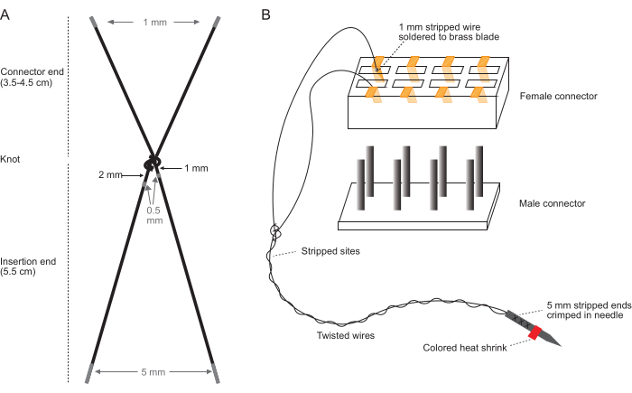
NOTE: Perform these steps on a clean benchtop using a stereomicroscope with a magnification range of 10x-40x and clean, bare hands. See Figure 1 for diagrams detailing electrode wire stripping (Figure 1A) and connector assembly (Figure 1B).
- Cut wires: For each electrode pair, cut two pieces of PFA-coated braided stainless-steel wire (7-strand, 0.0055" diameter) (see Table of Materials). For upper arm muscles, cut each wire 9.5 cm long. For lower arm muscles, cut each wire 10.5 cm long.
- Tie the two wires together with a single knot -this will become the knot sitting just outside the insertion site (proximal knot) when implanted. The tied wires comprise one electrode pair.
- Using an 18 G needle inserted into a piece of corrugated cardboard, position the knot 6 cm from the insertion end and tighten around the needle by pulling the electrode strands against the cardboard. The insertion end will shrink to about 5.5 cm, with the remaining 0.5 cm tied up in the knot.
- Carefully remove the needle and pull the knot tight with bare hands twice to tighten further.
NOTE: The knot should not be as tight as possible; tightening this way will result in a proximal knot that is the correct size to anchor the implanted electrodes in tissue. - For upper arm muscles, ensure the connector ends are 3.5 cm long. For lower arm muscles, ensure the connector ends are 4.5 cm long.
- Strip 0.5 mm of insulation off each wire: 1-1.5 mm from the knot on one wire and 2-2.5 mm from the knot on the other wire. Figure 1 illustrates where to strip each wire.
- Tape the electrode taut against a piece of flat cardboard with the two connector end wires together and the insertion end wires spread apart.
- Using a scalpel, make nicks in the insulation, marking the ends where the insulation will be removed.
- At each nick, make a series of ~6 cuts with a scalpel blade at each nick: ~2 on top, ~2 on the side, and ~2 underneath the wire.
NOTE: It is critical to avoid cutting the wire itself too much. Otherwise, the strands may break. Practice is required to achieve the appropriate amount of pressure to cut through the insulation completely with limited damage to the wire. - Rotate the electrode pair 180 degrees and repeat the 6 cuts in each nick.
- Place additional cuts as needed to separate the 0.5 mm of insulation from the underlying wire. The number of cuts needed here will depend on the pressure applied and the sharpness of the scalpel blade.
- Angle the scalpel blade to cut along the loosened insulation lengthwise and remove it from the wire using forceps.
- Inspect the exposed wire for breakage and/or frayed insulation that could cause damage during insertion into tissue.
- Strip 1 mm off the end of each wire at the connector end.
- Strip 5 mm off the end of each wire at the insertion end.
- Twist the insertion end wire segments together and crimp the 5 mm exposed ends together in the shaft of a 0.5 in. 27 G needle. Typical hypodermic needles can be used after removing the luer lock ending.
- Repeat steps 1.1-1.6 for each electrode pair.
- Assemble the connector.
- Cut the female end of the 12-pin connector (see Table of Materials) down to size: # electrode pairs x 2 pin slots.
- Remove the brass fittings from each connector port (these come pre-attached to the 12-pin connector) gently with pliers. Save these fittings for the next step.
- Solder each 1 mm exposed end of each wire to the outside surface of one of the blades of a brass fitting.
CAUTION: Soldering releases fumes that can cause skin, eye, and respiratory irritation. Wear gloves (or wash your hands afterward), use eye protection, and use a local fume extraction device to limit exposure.- Secure the brass fitting in a toothless alligator clip attached to a helping hand tool with the exterior surface of one of its blades facing up.
- Position the fitting under the microscope to allow high visual control during soldering. Using an unfolded paper clip or a piece of scrap wire, dab a small amount of stainless steel compatible flux on the surface of the blade.
- Apply enough solder to the blade to cover the lower ~1.5 mm of the brass blade using a narrow conical solder tip.
NOTE: Too much solder here will interfere with connector assembly, whereas too little solder could leave an inadequate connection. Have extra brass fittings available to start over if necessary. - Liberally coat the surface of the solder on the blade with stainless steel compatible flux, but avoid having flux drip into the space between the two blades.
- Hold the 1 mm of exposed electrode wire flush against the solder on the blade and heat the solder with the iron to fuse the connection.
- Inspect the connection: The majority of the exposed wire should be submerged in the solder, and the wire should be firmly attached to the brass fitting. Ensure that no solder has ended up between the two blades of the brass fitting - this could make it hard to insert a male connector later on. Lastly, ensure that the connection lies flush with the brass blade to allow the fitting to be reinserted into the connector in the next step.
- Using straight forceps, reinsert each soldered brass fitting back into the connector, ensuring the wires of each electrode pair are adjacent to each other and are not tangled with other electrode pairs. See Figure 1B to visualize the orientation of a single electrode pair in the connector.
NOTE: The ideal orientation from left to right is: biceps (3.5 mm at the connector end), triceps (3.5 mm at the connector end), extensor carpi radialis (ECR; 4.5 mm at the connector end), and palmaris longus (PL; 4.5 mm at the connector end). Mark one side with a marker or white-out to keep track of the connector's orientation during implantation. - Cut down a male 12-pin connector to the same size (# electrode pairs x 2) as the female end and connect it to the female connector. If fittings are displaced, they can be reinserted with straight forceps after the male connector is seated.
- Remove tabs emanating from brass fittings with pliers.
- Coat the pin slots with epoxy, ensuring all metal or wire near the connector will be insulated from tissue.
CAUTION: Epoxy can cause skin, eye, and respiratory irritation with prolonged exposure. Wear gloves, eye protection, and only use epoxy in a well-ventilated area or under a local fume extraction device. - Allow the connector to air-dry for at least 30 min.
- Test the resistance of each electrode pair and label needles with small colored segments of heat shrink for easy identification during surgery.
NOTE: Resistance should be between 18-50 ohms. Lower resistance may indicate a short. Higher resistance may indicate too much damage to the wire strands. However, high resistance often stems from imperfect connection between the needle shaft and the wire (made in step 1.6), which can be solved by additional crimping at this junction. - Ensure the electrode set is free of fibers and other debris before implantation. Spray duster can be used for this. Inspecting under the microscope can be helpful to verify.
2. Electrode implantation surgery
NOTE: This section describes a single surgical procedure to implant a headplate and electrodes fabricated in the previous section into the triceps, biceps, extensor carpi radialis (ECR), and palmaris longus (PL). For the latter two muscles, it is very difficult to implant the electrode exclusively in these individual muscles without going through nearby synergist muscles. See the discussion below regarding the caveats of attempting to isolate recordings from individual muscles. Headplates are typically custom-designed and fabricated for specific experiments. The present study used 3D-printed plastic RIVETS headplates19. Many open-source headplate designs are available online through Janelia, the Allen Institute, and independent research groups. The headplate procedure described here has been used successfully with titanium and plastic headplates. The surgical procedure must be performed on a stereotaxic instrument (see Table of Materials) with a stereomicroscope ranging from 10-40x magnification.
- Cold sterilize the electrodes and headplate using 1.5% glutaraldehyde overnight or for 8 h. Rinse briefly in sterile water and let air-dry completely before implanting.
CAUTION: Glutaraldehyde is harmful if swallowed and can cause irritation to the eyes, skin, and respiratory tract. Handle with gloves in a ventilated fume hood. - Induce anesthesia with 2-4% isoflurane in medical grade oxygen in an induction chamber until loss of righting reflex (approximately 3 min) following institutionally approved protocols.
- Transfer the animal to the movable nose cone to continue receiving anesthesia. Under anesthesia with 2% isoflurane in medical grade oxygen, shave the animal's head, neck, and limb prior to surgery. Perform all remaining steps in this section under anesthesia. Adjust the dosage of isoflurane as needed to maintain a breathing rate of 1 Hz and the absence of a toe-pinch reflex.
- Apply eye lubricant and reapply every 1 h throughout the surgery.
- Administer injectable analgesic (such as carprofen, 5 mg/kg) and antibiotic medication subcutaneously (such as enrofloxacin, 10 mg/kg) (see Table of Materials) at the beginning of surgery.
- Implant the headplate using the following steps or an alternative institution-approved method.
- Secure the animal's head in ear bars on the stereotaxic instrument and provide anesthesia (2% isoflurane) through the nose cone attached to the stereotaxic instrument. Add a sterile drape to maintain asepsis.
- Clean the head and neck with povidone-iodine prep pads and sterile alcohol prep pads.
Reapply eye lubricant if necessary. - Inject lidocaine (4 mg/kg) (see Table of Materials) subcutaneously at the site of incision.
- Ensure adequate anesthetic induction by performing a toe pinch. If the righting reflex is absent, make an incision down the midline of the animal's head from the caudal edge of the eyes to the caudal edge of the ears.
- Clamp out the skin with tissue clips: two at the rostral edge on each side just behind the eyes and two at the caudal edge just behind the ears.
- Clean the skull surface by gently scraping with a scalpel blade to remove dried fascia.
- Apply a thin layer of dental cement (see Table of Materials) to the surface of the skull and allow it to dry for 5 min.
- Position the headplate on the animal's skull. How and where the headplate is implanted will vary by headplate and research question.
- Use dental cement to adhere the headplate to the skull. Ensure that the skull is fully covered. Allow dental cement to dry for 10 min.
- Transfer the animal to a movable plastic nose cone to provide anesthesia for the remainder of the surgery; this will permit the repositioning of the animal regularly during surgery to access different muscles while maintaining anesthesia.
- Extend the incision on the back of the neck (made during headplate implantation) so that it reaches 1 cm caudal to the ears.
- Using a blunt bone scraper, separate the skin under the neck incision from the underlying tissue to clear a path from the neck incision to the forelimb, where electrodes will be implanted.
- Secure the EMG connector to the headplate temporarily using a small piece of tape to keep it in place during electrode implantation.
- Clean the animal's forelimb with povidone-iodine prep pads and sterile alcohol prep pads.
- Make the triceps incision.
- Position the animal on its side with the triceps facing up.
- Inject lidocaine (4 mg/kg) at the planned site of incision. This will relieve local pain and keep muscles moist during the implantation.
NOTE: Muscles should remain moist but not dripping wet throughout surgery. Apply sterile saline topically as needed if muscles or skin seem dry. - Cut 7mm over the triceps parallel to the bone.
- Separate the skin surrounding the incision from the underlying tissue with the blunt bone scraper. Work the scraper under the skin and back to the neck incision to clear a path for the electrodes.
- Trim away any fascia obscuring the muscle.
- Ensure the path from the neck to the triceps is large enough by inserting closed scissors through the triceps incision and pushing out the neck hole, opening slightly after exiting the hole.
- Bring the triceps electrode to the triceps incision: Insert the tip of the large needle driver through the triceps incision and out of the neck incision. Clamp the needle driver around the electrode needle lengthwise and pull through to the triceps incision.
- Triceps insertion: Follow step 3 "inserting electrodes into muscles".
- Make the distal arm incision.
- Position the animal on its back.
- Tape the arm along the animal's side with its palm facing down.
- Inject lidocaine (4 mg/kg) at the planned site of incision.
- Make a 1 cm incision above the biceps and ECR from the bottom of the deltoid to midway down the lower arm, parallel to the bone. The distal end of the incision should be ~2 mm above the end of the lower arm muscles.
- Clear away the fascia to expose the biceps muscle.
- Clear a path from the distal arm incision back to the triceps incision above (proximal to) the large blood vessel running along under the skin of the upper arm.
- Thread the biceps electrode under the skin through the neck incision to the triceps incision, then from the triceps incision to the distal arm incision.
- Biceps insertion: Follow step 3 "inserting electrodes into muscles".
- Re-tape the animal's arm in the same position but with the palm facing down.
- Insert as close as possible to the proximal end of the exposed biceps.
- To submerge at least 3 mm of the electrode in the muscle tissue, ensure to insert slightly diagonally to the muscle fibers depending on the mouse's size.
- Clear a path from the triceps incision to the distal arm insertion site below (distal to) the large blood vessel running under the skin of the upper arm, creating a distinct path from that of the biceps electrode.
- ECR insertion: Follow step 3 "inserting electrodes into muscles".
- Insert into the most proximal part of ECR.
- Exit in the crease between ECR and its antagonist and tie the knot in this crease.
- Clear a path from the triceps insertion to the PL insertion site under the elbow.
- PL insertion: Follow step 3 "inserting electrodes into muscles".
- Position the animal on its back and tape its arm over its head with the palm facing up.
- Insert just distal to the elbow.
- Exit sufficiently proximal to wrist tendons so that the knot at the exit site will lie on the muscle and not the wrist tendons.
- Reposition the animal on its side and suture the triceps incision using 6-0 silk sutures.
- Reposition the animal on its back and suture the distal arm incision using 6-0 silk sutures.
- Affix the connector to the back of the headplate using dental cement.
- Suture the neck incision using 6-0 silk sutures.
- Apply topical antibiotic cream to incision sites to reduce inflammation.
- Affix an Elizabethan collar (see Table of Materials) on the animal to prevent it from disturbing sutures during recovery.
3. Inserting electrodes into muscles
- Slightly curve the needle (27 G, step 1) by bending.
- Hold the needle with the needle driver (see Table of Materials) and press it against the handle of a pair of forceps to add a 5-10-degree bend.
- Add three total bends at different positions along the length of the needle.
- Visualize where to enter and exit the muscle.
- Remove any fat and fascia obscuring the entry and exit site by cutting or pulling with fine forceps. Try to avoid vascular damage to limit bleeding.
- Aim for 3-5 mm of submerged wire running parallel to the muscle fibers. This will ensure that the exposed stretches of electrode wire are submerged within the muscle.
- Using the needle driver, insert the needle in the proximal end of the muscle while applying counter pressure with blunt curved forceps in your other hand. Push the needle through the muscle to the exit site.
- Once the needle exits the muscle, grab the tip with blunt forceps and pull the needle through. Keep pulling until the proximal knot sits atop the insertion site.
- Make the distal knot.
- Using forceps, tie a loose knot distal to the exit site. Tighten the knot down to a 1 cm loop.
- Push the loop with forceps and position it over the exit site.
- Visualize where the distal knot should be closed down to before it is fully tightened, about 0.5 mm distal to the exit site. Gently grasp the loop with the fine bent forceps at this position and pull the loop tight over your forceps.
NOTE: Do not tighten the distal knot immediately over the exit site in this step, or the muscle will be squeezed when the knot is fully tightened in the next step. - Remove the fine forceps from the knot and finish tightening the knot by pushing the knot toward the exit site with the fine bent forceps and pulling the needle end with the fingers. Ensure both the proximal and distal knots are properly positioned outside of the insertion and exit sites, respectively, to anchor the inserted electrode in place.
- Grasp the exit knot with straight, fine forceps and tightly curl the distal wire around the forceps to bend the wire around the knot and toward the muscle/away from the skin.
- Cut off wire 0.5 mm distal to the distal knot, leaving a small nub curled around the knot.
The previous step ensures that the cut end of the nub will not poke into the animal's skin, which can cause irritation.
4. Post-operative care
- Immediately after surgery, perform the following steps.
- Singly-house the animal so its cage mates do not disturb its sutures.
- Place the animal in a clean cage with low bedding. Remove any nestlet and enrichment material that might interfere with the animal's mobility while wearing the Elizabethan collar.
- Give the animal water and wet food that it can access while wearing the collar.
- Perform the following steps 24 h and 48 h post-surgery.
- Remove the Elizabethan collar to allow the animal to groom itself for 20 min.
- Induce anesthesia with isoflurane, as mentioned in step 2.2 and step 2.3.
- Administer injectable analgesic and antibiotic medication.
- Inspect incision sites for missing sutures, open wounds, and signs of infection or irritation. Replace sutures and apply more topical antibiotics if needed.
- Replace wet food every 48 h until the Elizabethan collar is removed.
- Perform the following steps 6 days post-surgery.
- Check wounds for complete healing.
- If wounds are closed, remove the sutures. If wounds are open, wait two more days to remove the sutures. Remove the Elizabethan collar when sutures are removed.
- Return the mouse to a new, clean cage with full bedding and enrichment.
NOTE: Animals can proceed to experimentation or water deprivation 7 days after surgery.
Representative Results
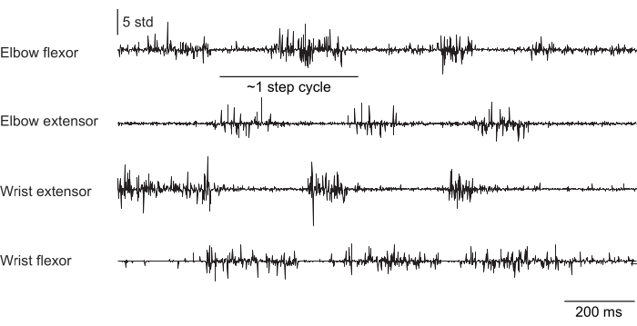
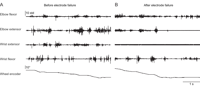
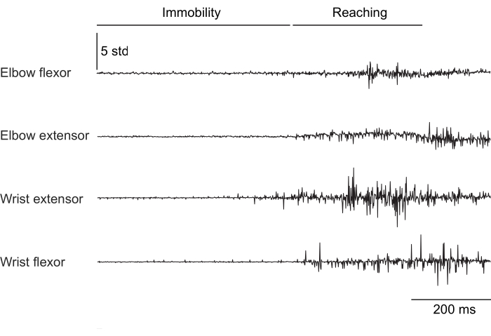
Figure 2, Figure 3, and Figure 4 show normalized muscle activity recorded from the forelimb muscles of mice performing different behaviors: treadmill walking without head-fixation (Figure 2), climbing a rotating wheel under head-fixation (Figure 3), and reaching for water droplets under head-fixation (Figure 4). Figure 2 shows 1.5 s of treadmill locomotion with an approximate step cycle estimated from the time between two elbow flexor activations. Figure 3 shows 5 s of EMG data from an animal that had the wrist extensor electrode fail 6 weeks after implantation. In Figure 3A, all four electrodes produce a clean EMG signal that aligns with the turning of the wheel (which indicates climbing). Figure 3B shows the signal from the same electrodes after failure: the wrist extensor electrode produces a noisy signal that does not change with the animal's movement. Figure 4 shows 1 s of EMG from the four forelimb muscle groups during a task in which the mouse transitioned from immobility to reaching for a water droplet.
In Figure 2, Figure 3, and Figure 4, the voltage signals were amplified and bandpass filtered (250-20,000 Hz) using a differential amplifier. Raw voltage was then subsampled to 1 kHz and z-scored for comparison across datasets. Note again that while electrodes were implanted in the four muscles specified in the protocol (biceps, triceps, ECR, and PL), it is not guaranteed that adjacent synergistic muscles did not influence EMG signal; therefore, each recording is assigned to its synergy group (elbow flexor, etc.) for accuracy. Verifying isolated recordings from single muscles would require simultaneous recordings in multiple synergists to assay for crosstalk between muscle recordings, which may be prohibitively difficult, especially in the lower arm of mice.

Figure 1: Schematics of the electrode set fabrication. (A) Diagram of a single electrode pair. Gray areas indicate where to strip. (B) Diagram of the connector assembly with a single completed electrode pair inserted into the connector. The diagram in (B) is not to scale. Please click here to view a larger version of this figure.

Figure 2: Representative EMG recording from four muscles of a freely moving (not head-fixed) mouse walking on a treadmill. The total duration is 1.5 s. The step cycle was estimated from the time between sequential elbow extensor activations. Please click here to view a larger version of this figure.

Figure 3: Representative EMG recording from four muscles of a head-fixed mouse performing a naturalistic climbing behavior. The 5th row shows the position of the climbing wheel read out by a rotary encoder; changes in this value indicate that the wheel is turning and the animal is actively climbing. The total duration is 5 s. (A) Recording 36 days after implantation during climbing. (B) Recording 72 days after implantation in the same mouse after the wrist extensor electrode failed. Please click here to view a larger version of this figure.

Figure 4: Representative EMG recording from four muscles of a head-fixed mouse transitioning from immobility to performing a reaching movement. The total duration is 1 s. Please click here to view a larger version of this figure.
Discussion
This protocol enables stable muscle activity recordings from head-fixed mice performing a variety of behaviors for several weeks. Recently, this method has been employed to examine neural control of limb musculature during behaviors such as treadmill locomotion18,20, a joystick pulling task18, and a co-contraction task21. While the protocol described here is specific for mouse elbow and wrist muscles, it is easily modified to record from different muscles or a different number of muscles by changing the length and/or total number of electrode pairs. The method described here was adapted from those used previously to record forelimb and hindlimb muscle activity in mice without head restraint15,16,17.
Electrode fabrication requires significant practice to master. Daily practice for 1-2 h is recommended while learning. Stripping the electrodes is the most challenging step due to the precise level of force required to cut the insulation without damaging the underlying wire. This level of force is dependent on the sharpness of the blade, so frequently replacing the scalpel blade can help ensure reproducibility during learning. Soldering the wires to the brass blades of the connector can also be difficult because stainless steel does not readily solder. Applying a liberal amount of stainless steel-compatible flux helps promote the connection.
The main challenge during the implantation surgery is tying the distal knot without disturbing the implanted wire or proximal knot. The proximal knot must be large enough to resist slipping into muscle at the insertion site - thus, avoid tying the knot too tight in step 2 of electrode set fabrication. If the proximal knot migrates after implantation, use carbon fiber-tipped forceps to reposition it carefully. Tighten the distal knot slowly while maintaining a firm grip on the wire with forceps to avoid pulling the entire electrode through. This step is critical to ensure the longevity of implanted electrodes: too much tension placed on the electrode can cause it to break when the animal moves, while a loose electrode can shift during recovery and lose contact with its associated muscle as the tissue heals.
Animals recover remarkably well from the surgery, though there are potential complications to note. First, mice will chew on their sutures and electrodes if given the chance. While the Elizabethan collar prevents this, it also prevents the animal from grooming itself. Some mice develop a mucus-like build-up around their eyes. Occasional male mice, particularly older ones, experience urethra blockages that can be distressing to the animal. Allowing the animal to groom itself for 20 min each day before inspecting sutures should give the animal enough time to prevent these issues.
There are important limitations of this method to note. First, these custom electrodes generally cannot resolve single motor unit activity. Furthermore, the electrical signal is not guaranteed to emanate exclusively from a specific muscle (i.e., biceps), as it is difficult to rule out crosstalk from activity in nearby synergist muscles. Therefore, in publications, researchers commonly refer to the recorded muscles by their synergy group (i.e., elbow flexor). It is recommended to perform post-mortem dissections after each experiment to verify the position of each electrode, as they could shift in the tissue during recovery.
Researchers interested in single motor unit activity should consider trying newly developed EMG electrodes by the Center for Advanced Motor Bioengineering Research (CAMBER) at Emory University. These electrodes are still being developed, but CAMBER will provide the latest electrode design. The main drawback of these electrodes is longevity: the hand-fabricated electrodes described in this protocol generally allow recordings for several weeks, whereas CAMBER electrodes work best for short-term experiments. Researchers selecting an EMG recording method can contact CAMBER directly to determine if their electrodes will be suitable for a given experiment.
Disclosures
None.
Acknowledgements
The authors would like to acknowledge Dr. Claire Warriner for contributing to the development of this method. Mark Agrios and Sajishnu Savya assisted with preparing figures. This research was supported by a Searle Scholar Award, a Sloan Research Fellowship, a Simons Collaboration on the Global Brain Pilot Award, a Whitehall Research Grant Award, The Chicago Biomedical Consortium with support from the Searle Funds at The Chicago Community Trust, NIH grant DP2 NS120847 (A.M.), and NIH grant 2T32MH067564 (A.K.).
Materials
| Name | Company | Catalog Number | Comments |
| #11 Scalpel Blades | World Precision Instruments | 504170 | For EMG electrode fabrication |
| #3 Scalpel Handle | Fine Science Tools | 10003-12 | For EMG electrode fabrication |
| 1 mL Sub-Q Syringe (100 pack) | Becton Dickinson | 309597 | For administering injectable drugs |
| 12-pin connector | Newark | 33AC2371 | 12-pin connector with brass fittings; for EMG electrode fabrication |
| 18 G Needles | Exel International | 26419 | For EMG electrode fabrication |
| 27 G Needles | Exel International | 26426 | For EMG electrode fabrication |
| 3 M Transpore Surgical Tape | 3M | 1527-0 | For taping animal's limbs out during surgery |
| 6-0 silk sutures | Henry Schein | 101-2636 | These sutures work well with delicate skin around the wrists |
| C&B Metabond Complete Kit | Pearson Dental | P16-0126 | Dental cement to affix connector to headplate |
| C57BL6/J Mice | Jackson Laboratories | #000664 | Wild type mice |
| Carbofib 5-CF Tweezers (2) | Aven tools | 18762 | Carbon fiber tipped forceps, used to manipulate delicate parts of electrode (stripped or inserted sections) |
| Carprodyl (Carprofen) 50 mg/mL Injection | Ceva Animal Health, LLC | G43010B | Injectable analgesic for pain management during and after surgery |
| Castroviejo Micro Needle Holder | Fine science tools | 12060-01 | For suturing |
| Castroviejo Needle Holder (large) | Fine science tools | 12565-14 | For inserting needle into muscle |
| Delicate Bone Scraper | Fine science tools | 10075-16 | To separate skin from underlying tissue |
| Dietgel 76A Dietary Supplement | Clear H2O | 72-07-5022 | For post-operative care |
| Dumont #5/45 Forceps | Fine science tools | 11251-35 | To remove fascia overlying muscle |
| Elizabethan collar for mouse | Kent Scientific Corporation | EC201V-10 | For post-operative care |
| Enrofloxacin 2.27% | Covetrus | #074743 | Injectable antibiotic for use during and after surgery |
| Epoxy gel | Devcon | 14265 | For EMG electrode fabrication |
| Hopkins Bulldog Clamp (4) | Stoelting | 10-000-481 | Tissue clamps for headplate implantation |
| Isoflurane Solution | Covetrus | 11695067771 | Inhalable anesthesia |
| Lidocaine Hydrochloride Injectable - 2% | Covetrus | #002468 | Topical analgesic for pain management during surgery |
| Medical Grade Oxygen | Airgas | OX USP200 | For administering isoflurane during surgery |
| MetriCide 1 Gallon | Metrex | 10-1400 | Glutaraldehyde solution for cold-sterilization of headplate and electrodes |
| MetriTest Strips 1.5% | Metrex | 10-303 | Test strips for monitoring glutaraldehyde solution (recommended) |
| Model 900LS Small Animal Stereotaxic Instrument | Kopf Instruments | 900LS | Stereotax with lazy susan feature that allows platform rotation during surgery |
| PFA-coated 0.0055" braided stainless steel wire | A-M systems | 793200 | For EMG electrode fabrication |
| Povidone-iodine prep pads | Dynarex | 1108 | For cleaning skin |
| Puralube Vet Ointment | Dechra | 37327 | Eye ointment for surgery |
| Sterile alcohol prep pads | Dynarex | 1113 | For cleaning skin |
| Straight fine #5 forceps | Fine science tools | 11295-10 | For curling wire after insertion |
| Straight fine scissors | Fine science tools | 14060-11 | For cutting wire |
| Student Vannas Spring Scissors | Fine science tools | 91500-09 | For making incisions, trimming fat and fascia, and suturing |
| Technik Tweezers 7B-SA (2) | Aven tools | 18074USA | Curved blunt forceps, for general use during surgery |
| Triple Antibiotic Ointment | Walgreens | 975863 | Topical antibiotic for surgery |
| V-1 Tabletop Laboratory Animal Anesthesia System | VetEquip | 901806 | Contains all necessary equipment for anesthesia induction and scavenging including vaporizer, induction chamber, moveable plastic nose cone, and tubing |
References
- Dombeck, D. A., Khabbaz, A. N., Collman, F., Adelman, T. L., Tank, D. W. Imaging large scale neural activity with cellular resolution in awake mobile mice. Neuron. 56 (1), 43-57 (2007).
- Guo, Z. V., et al. Flow of cortical activity underlying a tactile decision in mice. Neuron. 81 (1), 179-194 (2014).
- Guo, J. Z., et al. Cortex commands the performance of skilled movement. eLife. 4, e10774 (2015).
- Morandell, K., Huber, D. The role of forelimb motor cortex areas in goal directed action in mice. Sci Rep. 7 (1), 15759 (2017).
- Galiñanes, G. L., Bonardi, C., Huber, D. Directional reaching for water as a cortex-dependent behavioral framework for mice. Cell Rep. 22 (10), 2767-2783 (2018).
- Evarts, E. V., Tanji, J. Reflex and intended responses in motor cortex pyramidal tract neurons of monkey. J Neurophysiol. 39 (5), 1069-1080 (1976).
- Hounsgaard, J., Hultborn, H., Jespersen, B., Kiehn, O. Bistability of alpha-motoneurones in the decerebrate cat and in the acute spinal cat after intravenous 5-hydroxytryptophan. J Physiol. 405, 345-367 (1988).
- Murphy, P. R., Hammond, G. R. The role of cutaneous afferents in the control of gamma-motoneurones during locomotion in the decerebrate cat. J Physiol. 434, 529-547 (1991).
- Manuel, M., Chardon, M., Tysseling, V., Heckman, C. J. Scaling of motor output, from mouse to humans. Physiol Bethesda Md. 34 (1), 5-13 (2019).
- Sauerbrei, B. A., et al. Cortical pattern generation during dexterous movement is input-driven. Nature. 577 (7790), 386-391 (2020).
- Barrett, J. M., Raineri Tapies, M. G., Shepherd, G. M. G. Manual dexterity of mice during food-handling involves the thumb and a set of fast basic movements. PLoS One. 15 (1), e0226774 (2020).
- Serradj, N., et al. Task-specific modulation of corticospinal neuron activity during motor learning in mice. Nat Commun. 14, 2708 (2023).
- Scholle, H. C., et al. Spatiotemporal surface EMG characteristics from rat triceps brachii muscle during treadmill locomotion indicate selective recruitment of functionally distinct muscle regions. Exp Brain Res. 138 (1), 26-36 (2001).
- Scholle, H. C., et al. Kinematic and electromyographic tools for characterizing movement disorders in mice. Mov Disord off J Mov Disord Soc. 25 (3), 265-274 (2010).
- Pearson, K. G., Acharya, H., Fouad, K. A new electrode configuration for recording electromyographic activity in behaving mice. J Neurosci Methods. 148 (1), 36-42 (2005).
- Akay, T., Acharya, H. J., Fouad, K., Pearson, K. G. Behavioral and electromyographic characterization of mice lacking EphA4 receptors. J Neurophysiol. 96 (2), 642-651 (2006).
- Akay, T., Tourtellotte, W. G., Arber, S., Jessell, T. M. Degradation of mouse locomotor pattern in the absence of proprioceptive sensory feedback. Proc Natl Acad Sci USA. 111 (47), 16877-16882 (2014).
- Miri, A., et al. Behaviorally selective engagement of short-latency effector pathways by motor cortex. Neuron. 95 (3), 683-696 (2017).
- Osborne, J., Dudman, J. RIVETS: A mechanical system for in vivo and in vitro electrophysiology and imaging. PloS One. 9 (2), e89007 (2014).
- Santuz, A., Laflamme, O. D., Akay, T. The brain integrates proprioceptive information to ensure robust locomotion. J Physiol. 600 (24), 5267-5294 (2022).
- Warriner, C. L., Fageiry, S., Saxena, S., Costa, R. M., Miri, A. Motor cortical influence relies on task-specific activity covariation. Cell Rep. 40, 111427 (2022).
This article has been published
Video Coming Soon
ACERCA DE JoVE
Copyright © 2025 MyJoVE Corporation. Todos los derechos reservados