
A subscription to JoVE is required to view this content. Sign in or start your free trial.
Method Article
Ex vivo הדמיה חיה של ריאות גרורה microenvironment שלהם
In This Article
Summary
אנו מתארים שיטה פשוטה יחסית עבור הדמיה vivo לשעבר החי של אינטראקציות תא-stroma הגידול בתוך גרורות ריאה, ניצול כתבים ניאון בעכברים. באמצעות מיקרוסקופ confocal ספינינג-דיסק, טכניקה זו מאפשרת הדמיה של תאים חיים לפחות 4 שעות ויכול להיות מותאם ללמוד תנאים ריאות דלקתיות אחרות.
Abstract
גרורה היא אחד הגורמים העיקריים לתחלואה ותמותה מסרטן. גרורה הוא תהליך רב שלב ובשל מורכבותו, התהליכים התאיים והמולקולריים המדויקים שולטי הפצה וצמיחה גרורתי עדיין חמקמקים. הדמיה חיה מאפשרת הדמיה של האינטראקציות הדינמיות המרחבי של תאי microenvironment שלהם. גידולים מוצקים גרורות בדרך כלל אל הריאות. עם זאת, המיקום האנטומי של הריאות מציבה אתגר בפני הדמיה intravital. פרוטוקול זה מספק שיטה פשוטה ומהירה יחסית הדמיה vivo לשעבר החי של אינטראקציות דינמי בין תאים סרטניים stroma הסובבת אותם בתוך גרורות ריאה. באמצעות שיטה זו, תנועתיות של תאים סרטניים כמו גם יחסי הגומלין בין תאים סרטניים ותאים סטרומה ב microenvironment שלהם ניתן דמיינו בזמן אמת במשך כמה שעות. באמצעות עכברי כתב ניאון מהונדסים, שורת תאי פלורסנט, בזריקות שכותרתו fluorescentlyמולקולות ו / או נוגדנים, מספר רכיבים של microenvironment הריאות יכולות להיות חזותי, כגון כלי דם ותאי חיסון. כדי לדמות את סוגי התאים השונים, דיסק מסתובב מיקרוסקופ confocal המאפשר הדמיה רציפה ארוכת טווח עם מהירה, רכישת תמונה בארבעה צבעים נוצל. הזמן לשגות סרטים מלוקטים מתמונות שנאספו במשך מספר רב של עמדות ומטוסים מוקדי להראות אינטראקציות בין גרורה חיים ותאי חיסון במשך שעה לפחות 4. טכניקה זו יכולה לשמש כדי להוסיף ולבדוק כימותרפיה או טיפול ממוקד. יתר על כן, שיטה זו יכולה להיות מותאמת לחקר פתולוגיות הקשורות ריאה אחרת שעלולים להשפיע על המיקרו-סביבת הריאות.
Introduction
The deadliest aspect of cancer is metastasis, which accounts for more than 90% of cancer-related morbidity and mortality1. Metastasis is a multistep process and due to its complexity, the exact cellular and molecular mechanisms that govern metastatic dissemination and growth are still elusive. To metastasize, tumor cells in the primary tumor must detach from their neighboring cells and basement membrane, cross through the extracellular matrix, intravasate, travel via blood or lymphatic vessels, extravasate at the secondary site, and finally, survive and establish secondary tumors. In addition to the properties of the tumor cells, the contribution from the microenvironment, which includes the adjacent stroma along with the normal counterparts of the cancer cells, is crucial for the seeding and establishment of metastatic lesions2.
Traditional methods to study metastatic seeding and growth examine static states, as tissues are excised and sectioned for histology. These data only generate a snapshot of this highly dynamic process. Although some useful information can be gained from these studies, the complicated process by which tumor and stromal cells interact during metastatic formation cannot be adequately assessed by these methods. Furthermore, it is not possible to gain insights into tumor or stromal cell migration patterns, which are important in establishing a colony at the distant site. In order to effectively study the metastatic process, it is essential to visualize various interactions between cancer cells and their microenvironment in a continuous manner and at real time.
The lung is a common site for metastases from solid tumors as breast, colorectal, pancreatic cancer, melanoma and sarcoma3. Intravital imaging was previously used to study cell-cell interaction in various primary tumor and metastatic models4,5. Methods of lung imaging in mice, including intravital imaging, lung section imaging, and an ex vivo pulmonary metastasis assay have been published6–9. Intravital imaging of mouse lungs utilizes a thoracic suction window to stabilize the lungs6. This method is used for time-lapse imaging of the lung microcirculation and alveolar spaces. The anatomical location of the lungs poses a challenge to intravital imaging. In order to access the lungs, the chest cavity must be opened which leads to loss of negative pressure and collapsed lungs. This method only allows the visualization of a small part of the lungs and is technically demanding; an unnecessary complication in studies that examine processes that are independent of blood flow. Moreover, this method also requires gating out movement caused by breathing. This is done either by collecting images between breaths or during post image acquisition analyses10. The alternative ex vivo lung section imaging provides stability and depth, and also prepares lung parenchyma for immunostaining7. However, the lengthy sectioning process leads to an extensive delay between the time of animal sacrifice and the start of the imaging session. Moreover, the process of sectioning a mouse lung causes considerable amount of cell death8, thus interfering with the quality and quantity of imaging samples and perhaps needlessly altering tumor-stroma interactions. In order to technically bridge between the methods of intravital imaging and lung section imaging, while exploiting the advantages of the two techniques, a relatively fast and easy method for ex vivo lung imaging was developed. This method was achieved by imaging of non-sectioned whole lung lobes. Using this method, the motility of cancer cells as well as interactions between cancer cells and stromal cells in their microenvironment can be visualized in real time for several hours.
Protocol
כל ההליכים המתוארים חייב להתבצע בהתאם להנחיות והתקנות לשימוש בעלי חיים בעלי חוליות, כולל אישור מראש על ידי טיפול מוסדי בעלי חיים הוועדה המקומית שימוש (IACUC).
דור 1. ריאות גרורות עבור vivo לשעבר הדמית חיה (מהונדס או זנב וריד הזרקה)
הערה: גרורות ריאות יכול להיווצר על ידי ניצול מודלים עכבר מהונדס גנטית או על ידי תוך ורידי (IV) הזרקה של תאים סרטניים.
- צור גרורות סרטניות בריאות הדמיה ידי חצייה במודל של עכברי גידול מהונדס גנטית לעכבר כתב מהונדס, למשל, לחצות את במודל העכברי של סרטן שד, המסוף ארוך וירוס גידול חלב העכבר החוזר-polyoma אנטיגן באמצע T (MMTV-PyMT) 11 לתוך ACTB-ECFP עכבר מודל 12.
הערה: מודל ACTB-ECFP מבטא משופרת חלבון פלואורסצנטי ציאן (ECFP) תחת β-המעשהב אמרגן כך שכל התאים לזרוח בערוץ כחול, CFP. עם זאת, תאים סרטניים הם ללא ספק הבולטים ונראים כמו הארי של תאים ECFP חיובי תחת מיקרוסקופ. מודל עכבר MMTV-PyMT מפתחת מחלה מתקדמת, שבה צמיחת גידול חלב קשורה להפצת תאים סרטניים לפריפריה, במיוחד אל ריאות. בעכברים MMTV-PyMT על FVB / n רקע, micrometastases ניתן לצפות סביב 10-11 שבועות של גיל. באופן כללי, התקדמות אלה macrometastases בסביבות 14 שבועות של גיל 13.
אוֹ - צור גרורות ניסיון באמצעות תאים ראשוניים או שורות תאי syngeneic. השתמש בתאי גידול ראשוני מניפולציות במבחנה או שורות תאים (למשל., תמרה) ואחריו הזרקת iv 14.
- בקצרה, בפרוטוקול זה, להזריק חלבון פלואורסצנטי ירוק (GFP) -expressing (+) שורת תאי MMTV-PyMT לעכברי כתב ניאון (ACTB-ECFP) או עכברי wildtype. אז,לדמיין תאים אלה מכונים 15 תאים VO-PyMT באמצעות המסלול הירוק, GFP.
הערה: שורת תאי VO-PyMT המקורי נגזרו על אורתופדיה ונדרבילט בנשוויל, טנסי. VO מייצג אורתופדיה ונדרבילט. - בעקבות הזרקה של 10 6 תאים (200 μl), להתבונן extravasation תא סרטני מיד ועד כמה שעות לאחר ההזרקה; להתבונן micrometastases בין 1-3 שבועות לאחר ההזרקה וכדי לזהות macrometastases 3 שבועות לאחר ההזרקה 15.
הערה: פחות תאים ניתן להזריק להאריך את הזמן בין הזרקה לצמיחה גרורתי.
- בקצרה, בפרוטוקול זה, להזריק חלבון פלואורסצנטי ירוק (GFP) -expressing (+) שורת תאי MMTV-PyMT לעכברי כתב ניאון (ACTB-ECFP) או עכברי wildtype. אז,לדמיין תאים אלה מכונים 15 תאים VO-PyMT באמצעות המסלול הירוק, GFP.
2. תיוג של רכיבי ענייני microenvironment גרורתי (טרנסגניים ו / או הזרקה)
הערה: תיוג יכול להיות מושגת על ידי עכברים מהונדסים ו / או על ידי הזרקה שונה. הקפד להשתמש בצבעי ניאון שונים עבור תיוג של סוגי תאים שונים.
- רכיבי תווית של microenvironment גרורתי באמצעות עכברים מהונדסים. נחצה את מודל הגידול העכבר שהוזכרו לעיל (למשל., MMTV-PyMT x ACTB-ECFP) למודל עכבר מהונדס שבו תאים סטרומה עניין מסומנים על ידי חלבון פלואורסצנטי שהוא לא ECFP, למשל., ג-FMS-EGFP 4,16.
הערה: בנוסף להדמיה של תאים סרטניים בערוץ CFP, זו מאפשרת הדמיה של תאים מיאלואידית ב GFP ערוץ 4.
ו / או - לייבל רכיבים שונים של microenvironment גרורתי באמצעות הזרקה לעכברי כתב ניאון מהונדסים או (לא ניאון) עכברי wildtype.
הערה: תרכובות כמה ניתן להזריק לתייג רכיבים שונים של microenvironment גרורתי, למשל, נוגדן AF647 מצומדות Gr-1 משמש כאן לתייג נויטרופילים וכמה מונוציטים 13 ו dextrans המשקל המולקולרי שונים משמשלתייג נימי ריאות. להכנת ההזרקה אלה ראו שלב 4.
3. הכנת חומרים לפני החיתוך
- 2% Agarose
- לשקול 0.2 גרם של agarose ולהוסיף 10 מ"ל 1 x PBS. מחממים את הפתרון לפזר את agarose. Agarose יהיה לגבש ב RT, אז לשמור אותו באמבט מים 37 מעלות צלזיוס עד בשימוש לאינפלציה.
- CO 2 ו בקר טמפרטורה
- בדוק DDH 2 O בתא humidification. מילוי בעת הצורך. הכנס צלחת תצורה לתוך בעל צלחת במת טמפרטורה (תא האקלים). הפעל את בקר ה- CO 2 ולהגדיר 2 CO ב -5%. ודא שקצב זרימת האוויר מוגדר 0.4 NL / min.
- פתח את האוויר ושסתום 2 CO. הפעל את בקר הטמפרטורה. צריך להקפיד שהטמפרטורה של החדר האקלים והמכסה נקבעים על 37 מעלות צלזיוס.
- שחרר לחץ אוויר על מטר 2 CO. בדוק CO 2 הגדלה, דוארquilibration עשוי להימשך עד 30 דקות.
- דיסק confocal ספינינג מיקרוסקופ
הערה: פרטים על הגדרת מיקרוסקופ תוארו בעבר 4,17.- הפעל לייזרים (הלייזר ארגון עבור עירור 488 ננומטר ננומטר של מצב מוצק 405, 561 ננומטר ו 640 nm לייזרים). הפעל את המיקרוסקופ, מצלמה, יחידת בקרת דיסק ספינינג, AOTF, יחידת בקרת הליזר לבין בקר המצלמה.
- פתח את תריס מיקרוסקופ, להדליק את המחשב פועל המיקרוסקופ ולפתוח את התוכנה.
- הכנת הכלים והפלטפורמה לנתיחה.
- הפעל מעקר את החרוז החם ולתת לו להגיע 250 מעלות צלזיוס. 2 זוגות נקיים של מספרי מלקחיים כירורגית עם מים וסבון. לעקר את הכלים שניים 30 לפחות. תנו את הכלים להתקרר. השתמש מכסה פוליסטירן כפלטפורמה לנתיחה. כסה אותה בפיסת שתיין במעבדה.
4. הכנת זריקות
הערה: בהתאם את זמן מחצית החיים ואת התגובה המועדפת, להזריק fluorescently שכותרתו נוגדנים ו / או מולקולות ניאון או מיד לפני להקריב בעלי חיים או כמה שעות עד ימים לפני.
- חזרה לתמונה Gr1 חיובי נויטרופילים ומונוציטים, להכין מזרק עם 7 μl של AF647 מצומדות המניות Gr-1 נוגדנים (1 מ"ג / מ"ל) לתוך 100 μl של PBS סטרילי מתחת למכסה המנוע. מניחים מחט G ½ 27 על המזרק.
- כדי נימים ריאות תמונה, להכין מזרק השני והשלישי עם 100 μl של 70 או dextran rhodamine מצומדות KD (4 מ"ג / מ"ל) או 10 KD dextran AF647 מצומדות (4 מ"ג / מ"ל). מניחים מחט G ½ 27 על המזרקים.
- להזריק את hr iv 5 פתרון נוגדן AF647 מצומדות לפני כריתה של הריאות.
- להזריק אחד או שני פתרונות dextran iv הערב 'כריתת הריאות.
5. הכנת הריאות עבור vivo לשעבר הדמיה חיה
הערה: נסה לעבוד סטרילי וזהירה ככל האפשר כדי להימנע אתגרים מיותרים של תאים חיסוניים בתוך הריאות.
- להזריק את intraperitoneal העכבר (IP) באמצעות מנת יתר קטלנית של הרדמה המותר על פי פרוטוקול חיה שאושר על ידי IACUC, למשל., 1 מ"ל של 2.5% Avertin. חכה בעכבר כדי להפסיק לנשום ולהיות שאינם מגיבים לחלוטין לגירויים מזיקים (קמצוץ כפה אחורי).
הערה: נקע בצוואר הרחם המתת חסד פחמן דו חמצני יש להימנע כפי שהוא יכול להשפיע לרעה על כדאיות התא ריאות. - לשתק את העכבר על קרש חיתוך לעקר את העכבר עם אתנול 70%.
- השתמש במספריים כירורגית שיחולל תחילה חתך רוחבי ברום הבטן דרך העור, ואחריו חתך דומה דרך הצפק. החזק את קרש החיתוך במצב אנכי וחתך את אבי העורקים יורד, כך מצטבר דם למטה בבטן ולא בתוך חלל החזה.
- Sלגדוע פתח קטן בסרעפת לשחרר ואקום. גזור לאורך הצלע -10 וה -12 שיחתוך את הסרעפת ולקבל גישה ויזואלית אל הריאות.
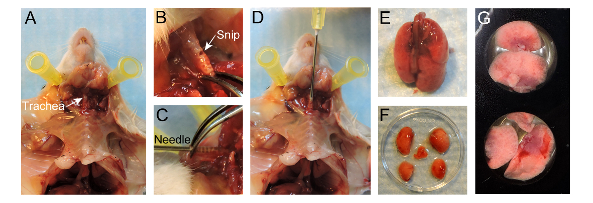
- השתמש במספריים כירורגית לחתוך את העור עד קנה נשימה מעל צלעותיו אבל לעזוב את בית החזה ללא פגע. הפרד את העור מפני בית החזה. לחשוף את קנה הנשימה על ידי הסרת רקמת החיבור שמסביב, נזהר שלא לפגוע קנה הנשימה עצמו (איור 1 א).
- לגזור פתח קטן כ 1 מ"מ הקוטר במקביל קנה נשימה החשופה טבעות הסחוסים, קרוב הגרון ככל האפשר (איור 1 ב). היזהר שלא לחתוך לחלוטין דרך קנה הנשימה.
- קח מחט 20 G ו בעדינות להכניס את המחט מ"מ 4-5 לתוך קנה הנשימה ללא כוח נגדי (1D איור). סופו של מחט צריך להיות גלוי דרך קנה הנשימה (איור 1 ג). שימוש במלקחיים כדי לייצב את המחט בתוך קנה הנשימה. לחלופין, תפר עשוי להיות קשור Around קנה הנשימה להחזיק את המחט במקום.
הערה: על ידי הוספה עמוקה מדי, קארינה ניתן טראומה או רק צד אחד של ריאות עלולים להיות מנופח. - ממלאים מזרק עם 400 μl של 37 ° C 2% agarose התכה נמוכה בטמפרטורה (נלקח ישירות מאמבט טמפרטורה קבועה). ודא קרש החיתוך בעמידה ולאט לאט להחדיר את agarose חם דרך המחט אל תוך הריאות, השתמש ~ 400 μl כדי לנפח את הריאות.
הערה: צופה ריאות הניפוח בתוך בית החזה. אל מעל לנפח את הריאות כפי שהוא יהיה קרע. - לאחר הריאות מנופחות, מילוי ~ ⅔ של כלוב צלעות, לנתק את המזרק ולשמור את המחט בתוך קנה הנשימה כדי למנוע כל agarose מ דולפת.
- יוצק כ 50 מיליליטר של 20 ° C PBS מעל הריאות המנופחות כדי לאפשר agarose בתוך הריאות להגדיר לחזק. לאט להוציא את המחט ולסגור את קנה הנשימה עם מלקחיים כדי למנוע כל אי-הקרושה agarose מ דולף.
- לחשוף את הריאות על ידי ביצוע sternotomy ובהמשך שיחתוך את הריאות. עבור כריתה של הריאות, על מנת להחזיק את קנה הנשימה תוך חיתוך דרך קנה הנשימה לחלוטין. משוך בעדינות את קנה הנשימה, לחתוך את רקמת החיבור והוושט תוך משייכת הריאות מתוך חלל החזה עד הריאות מופרדות העכבר (איור 1E).
- לטבול את הריאות ב RPMI-1640 חם לשטוף דם מוגזם בעדינות להפריד את האונות באמצעות מספריים מלקחיים לחתוך את הסמפונות הגבעול המרכזי 'האונות ב hilum (איור 1F).
- מניח את האונות, עם המשטח השטוח למטה על מנת למקסם את שטח הדמיה, בעוד גם צלחת הדמיה 24 גם (1G איור). הוספת 100 μl של 37 ° C RPMI-1640 על גבי האונות. מניחים כמה 15 מ"מ שקופיות כיסוי מיקרוסקופ עגול על גבי האונות כדי למנוע ממנה צף.
- יוצקים PBS חם לתוך הבארות שמסביב למנוע התקשורת RPMI-1640 מ EVAPנואם. הכנס את צלחת 24 גם לתוך תא אקלים equilibrated ולתחזק את אונה של הריאות ב 37 מעלות צלזיוס עם% אוויר ו -5 CO 2. הכנס את קאמרית האקלים על הבמה של המיקרוסקופ confocal.
הערה: תערובות גז אחרות (למשל, 5% O 2, 5% CO 2 ב N 2 לבחון התנהגות תא בתנאים של היפוקסיה / חמצן נמוך) יכולה להיות גם נחשבת.

איור 1. פרוטוקול להכנת ריאות הדמיה לחיות. (א) חשיפה של קנה נשימה לאחר הכנה של עכבר. (ב) חיתוך קטן שנעשה במקביל קנה נשימה החשופה טבעות הסחוסים. (ג) 20 מחט G מוכנס 4-5 מ"מ לתוך קנה הנשימה. (ד) החדרה של 400 μl 2% נמוכים היתוך בטמפרטורה agarose לריאות. (E) Inflריאות ated מופרדות העכבר. (F) אונות נפרדו לאחר האינפלציה. (G) לאונות להציב גם צלחת הדמיה 24 באר. אנא לחץ כאן כדי לצפות בגרסה גדולה יותר של דמות זו.
6. רכישה וניתוח של תמונות
הערה: תמונות ניתן לרכוש עם מגוון רחב של ספינינג מיקרוסקופים confocal דיסק נתמך על ידי תוכנות שונות. בפרוטוקול זה, או μManager עם מיקרוסקופ confocal דיסק מסתובב מחוייט או זן עם מיקרוסקופ confocal דיסק מסתובב, זמין מסחרית משמש לרכישת התמונה, בעוד Imaris משמש לעריכת סרטים וניתוח.
- לרכוש תמונות באמצעות μManager. צעד מפורט על-ידי פרוטוקול צעד לרכישת תמונות באמצעות תוכנת μManager מתואר בעבר 18.
אוֹ - לרכוש תמונותתוכנת ניתוח תמונה באמצעות כגון זן (ראה איור S1).
- לחץ על הכרטיסייה 'אתר', ולבחור אובייקטיבי (10x או 20x) בכלי 'נתיב האור' (איור S1A, תיבה אדומה). בהמשך לכך, לחץ על 'עיניים - DAPI' להסתכל על ערוץ CFP דרך עיני (האיור S1A, קופסא כחולה). למקם את המדגם באופן ידני באמצעות מיקרוסקופ. לחץ על 'כל Off'after הרקמה במרכז שדה הראייה.
- לחץ על הכרטיסייה 'רכישה' כדי להגדיר את כל הפרמטרים לרכישת התמונה.
- בכלי 'ערוצים', לחץ על '+' כפתור (איור S1B, תיבה אדומה). תפריט מוקפץ מופיע ולחפש את צבען (ים) הנוכחי במדגם ב 'מסד דיי' (איור S1B). בחר את הצבע ולחץ על 'הוסף'.
הערה: התוכנית תקבע את כל המסננים להיות מותאם. ניתן למחוק לצבועעל ידי בחירה בו ואחריו לחיצה על כפתור פח האשפה (האיור S1B, תיבה צהובה). - בתפריט 'מצב רכישה', להגדיר 'binning' כדי 5x5. לחץ לחיצה כפולה על ECFP בתפריט הערוצים כדי לבחור בו. מנמיכים את כוח לייזר עד 20% כך המדגם לא יהיה מולבן בעת הגדרת הפרמטרים לרכישת התמונה.
- סמנו את התיבה 'האריחים' בקטע 'מנהל ניסוי' וכלי האריחים מופיע בקבוצת הכלי 'הרכישה רבה הממדית "(האיור S1C). לחץ על כפתור 'הגדרות מתקדמות' כדי להציג את התמונה בזמן אמת מהמצלמה. לחץ על הכפתור 'הוסף' בקטע 'ועמדות להוסיף 4 עד 6 עמדות הניסוי. להסיר עמדה, בחר מיקום כי ולחץ על כפתור פח האשפה.
- בקבוצת הכלי 'פרמטר הרכישה', פתח את הכלי "אסטרטגית הפוקוס ', ובחר' אבסולוטדואר קבוע Z-עמד מהרשימה נפתחת.
- סמן את התיבה Z-Stack במקטע 'מנהל ניסוי' והכלי-סטאק Z מופיע בקבוצת כלי 'רב מימדי רכישת' (איור S1D). לחץ לחיצה כפולה על אחת מעמדות במקטע 'הפוזיציות' ולחץ על 'בשידור חי'. הגדר באופן ידני הראשונה ולהגדיר בתפקידו האחרון של בתחום ההדמיה. הגדר את המרווח ב 4 מיקרומטר.
הערה: התכנית תקבע את מספר הפרוסות עבור מגוון מרווח הנבחר. באופן אידיאלי, 5-7 פרוסות נוחות כדי לאפשר ויזואליזציה מספיק ורכישת תמונה מהירה. - סמנו את התיבה 'סדרות עתיות' בקטע 'מנהל ניסוי'. בקבוצה הרצויה 'משך' ושעות 'מרווח' בכלי 'סדרות עתיות "שהופיעו בקבוצת כלי' רכישת רב ממדית" (איור S1E).
- בשנות ה Acquisition מצב 'התפריטים, להגדיר' binning 'כדי 2x2. לחץ לחיצה כפולה על fluorophore בתפריט ערוצים כדי לבחור בו ולהגביר את כוח לייזר עד 100%. לחץ 'בשידור חי' ולהתאים את 'זמן חשיפה'. חזור על פעולה זו עבור כל fluorophore.
- סמן את התיבה 'אפשר שמירה אוטומטית'. בחר תיקייה ולהקליד את שם הקובץ. כל התמונות רכשה יישמרו באופן אוטומטי בתיקייה זו.
- לחץ על 'התחלת ניסוי' בקטע 'מנהל ניסוי' כדי להתחיל רכישת התמונה.
- לאחר רכישת תמונה, לקמפל את הנתונים הגולמיים בתוכנת Imaris. המרת תמונות .ims קבצים יכולות להתבצע התאמות. צעד מפורט על-ידי פרוטוקול צעד לגיור של קבצים, ביצוע התאמות וסרטי חיסכון באמצעות Imaris מתואר בעבר 18.
- בעת שמירת הסרט, להגדיר את 'מסגרת השיעור' עד 5 פריימים לשנייה (fps).
תוצאות
באמצעות מיקרוסקופ confocal ספינינג-דיסק, מערכות במודל של עכברים שונים הזרקה, במיקרו-סביבה של גרורתי ניתן דמיינו מסומנים לאורך זמן. שימוש MMTV-PyMT; ACTB-ECFP; ג-FMS-EGFP במודל עכבר משולשת מהונדס, מרכיבים תאיים שונים מסומנים fluorescently (איור 2 א, סרט 1). המבנה הטיפוסי ...
Discussion
כתב יד זה מתאר שיטה מפורטת עבור הדמית vivo לשעבר חיים של גרורות ריאה בעכברי מודל של גרורות. פרוטוקול הדמיה זו מספקת להדמיה ישירה של אינטראקציות stroma תאים סרטניים דינמית מרחבית במיקרו-סביבה של הריאות. זוהי השיטה קלה ומהיר יחסית המאפשר הדמיה אמינה של גרורות ריאה במשך...
Disclosures
The authors have no conflicts of interest to disclose. All animal experiments were conducted in accordance with IACUC approved protocols, UCSF.
Acknowledgements
We thank Nguyen H. Nguyen for her technical help and Audrey O’Neill for support with the Zeiss Cell Observer spinning-disk confocal microscope. This work was supported by a Department of Defense postdoctoral fellowship (W81XWH-11-01-0139) and the Weizmann Institute of Science-National Postdoctoral Award Program for Advancing Women in Science (to V.P.).
Materials
| Name | Company | Catalog Number | Comments |
| MMTV-PyMT/FVB mice | Jackson Laboratory | 2374 | Female mice |
| ACTB-ECFP/FVB mice | UCSF Werb lab | Female mice | |
| c-fms-EGFP/FVB mice | UCSF Werb lab | Female mice | |
| FVB mice | Jackson Laboratory | 1800 | Female mice |
| GFP+ VO-PyMT cells | UCSF Werb lab | ||
| 70,000 kDa Dextran, rhodamine-conjugated | Invitrogen | D1818 | Dilute to 4mg/ml in 1 x PBS and store at -20 °C. Use 0.4 mg per animal. |
| 10,000 kDa Dextran, Alexa Fluor 647 conjugated | Invitrogen | D22914 | Dilute to 4mg/ml in 1 x PBS and store at -20 °C. Use 0.4 mg per animal. |
| Anti-mouse Gr-1 antibody Alexa Fluor 647 | UCSF Monoclonal antibody core | Stock 1mg/ml. Use 7 ug per animal. | |
| Anesthetic | Anesthesia approved by IACUC, used for anesthesia and/or euthanesia | ||
| 1X PBS | UCSF cell culture facility | ||
| PBS, USP sterile | Amresco INC | K813-500ML | Ultra pure grade for i.v. injection |
| Styrofoam platform | Will be used as dissection board | ||
| Fine scissors sharp | Fine Science Tools | 14060-11 | |
| Forceps | Roboz Surgical Store | RS-5135 | |
| Hot bead sterilizer | Fine Science Tools | 18000-45 | Turn ON 30min before use |
| Air | UCSF | ||
| Oxygen | UCSF | ||
| Carbon dioxide | UCSF | ||
| 1 mL syringe without needle | BD | 309659 | |
| 27 G x 1/2 needle | BD | 305109 | for i.v. injection |
| 20 G x 1 needle, short bevel | BD | 305178 | |
| Low-melting-temperature agarose | Lonza | 50111 | To make 10 ml of solution, weigh 0.2 g of agarose, add to 10 ml 1 x PBS, and heat to dissolve. Agarose will solidify at room temperature, so maintain in a 37 °C water bath until used for inflation. |
| RPMI-1640 medium without phenol red | Life Technologies | 11835-030 | |
| 24 well Imaging plate | E&K scientific | EK-42892 | |
| Glass cover slides, 15 mm | Fisher Scientific | 22-031-144 | |
| Digital CO2 and temperature controller | Okolab | DGTCO2BX | http://www.oko-lab.com |
| Climate chamber | Okolab | http://www.oko-lab.com | |
| Cell Observer spinning disk confocal microscope | Zeiss | ||
| Zen software | Zeiss | ||
| Inverted microscope | Carl Zeiss Inc | Zeiss Axiovert 200M | |
| ICCD camera | Stanford Photonics | XR-Mega-10EX S-30 | |
| Spinning disk confocal scan-head | Yokogawa Corporation | CSU-10b | |
| Imaris | Bitplane | ||
| mManager | Vale lab, UCSF | Open-source software |
References
- Chaffer, C. L., Weinberg, R. A. A perspective on cancer cell metastasis. Science. 331 (6024), 1559-1564 (2011).
- Plaks, V., Kong, N., Werb, Z. The cancer stem cell niche: how essential is the niche in regulating stemness of tumor cells. Cell stem cell. 16 (3), 225-238 (2015).
- Nguyen, D. X., Bos, P. D., Massague, J. Metastasis: from dissemination to organ-specific colonization. Nat Rev Cancer. 9 (4), 274-284 (2009).
- Egeblad, M., Ewald, A. J., et al. Visualizing stromal cell dynamics in different tumor microenvironments by spinning disk confocal microscopy. Dis Model Mech. 1 (2-3), 155-167 (2008).
- Ellenbroek, S. I. J., van Rheenen, J. Imaging hallmarks of cancer in living mice. Nat Rev Cancer. 14 (6), 406-418 (2014).
- Looney, M. R., Thornton, E. E., Sen, D., Lamm, W. J., Glenny, R. W., Krummel, M. F. Stabilized imaging of immune surveillance in the mouse lung. Nat Methods. 8 (1), 91-96 (2011).
- Thornton, E. E., Krummel, M. F., Looney, M. R. Live imaging of the lung. Cur Protoc Cytom. , (2012).
- Thornton, E. E., Looney, M. R., et al. Spatiotemporally separated antigen uptake by alveolar dendritic cells and airway presentation to T cells in the lung. J Exp Med. 209 (6), 1183-1199 (2012).
- Mendoza, A., Hong, S. -. H., et al. Modeling metastasis biology and therapy in real time in the mouse lung. J Clin Invest. 120 (8), 2979-2988 (2010).
- Lelkes, E., Headley, M. B., Thornton, E. E., Looney, M. R., Krummel, M. F. The spatiotemporal cellular dynamics of lung immunity. Trends Immunol. 35 (8), 379-386 (2014).
- Guy, C. T., Cardiff, R. D., Muller, W. J. Induction of mammary tumors by expression of polyomavirus middle T oncogene: a transgenic mouse model for metastatic disease. Mol Cell Biol. 12 (3), 954-961 (1992).
- Hadjantonakis, A. -. K., Macmaster, S., Nagy, A. Embryonic stem cells and mice expressing different GFP variants for multiple non-invasive reporter usage within a single animal. BMC Biotechnol. 2, (2002).
- Casbon, A. -. J., Reynaud, D., et al. Invasive breast cancer reprograms early myeloid differentiation in the bone marrow to generate immunosuppressive neutrophils. Proc Natl Acad Sci USA. 112 (6), 566-575 (2015).
- Donovan, J., Brown, P. Parenteral injections. Curr Protoc Immunol. , (2006).
- Halpern, J., Lynch, C. C., et al. The application of a murine bone bioreactor as a model of tumor: bone interaction. Clin Exp Metastas. 23 (7-8), 345-356 (2006).
- Sasmono, R. T., Oceandy, D., et al. A macrophage colony-stimulating factor receptor-green fluorescent protein transgene is expressed throughout the mononuclear phagocyte system of the mouse. Blood. 101 (3), 1155-1163 (2003).
- Ewald, A. J., Werb, Z., Egeblad, M. Dynamic, long-term in vivo imaging of tumor-stroma interactions in mouse models of breast cancer using spinning-disk confocal microscopy. Cold Spring Harb Protoc. (2), (2011).
- Bonnans, C., Lohela, M., Werb, Z. Real-time imaging of myeloid cells dynamics in ApcMin/+ intestinal tumors by spinning disk confocal microscopy. J Vis Exp. (92), (2014).
- Nakasone, E. S., Askautrud, H. A., et al. Imaging tumor-stroma interactions during chemotherapy reveals contributions of the microenvironment to resistance. Cancer cell. 21 (4), (2012).
- Cheng, N., Lambert, D. L. Mammary transplantation of stromal cells and carcinoma cells in C57BL/6J mice. J Vis Exp. (54), (2011).
- Al-Mehdi, A. B., Tozawa, K., Fisher, A. B., Shientag, L., Lee, A., Muschel, R. J. Intravascular origin of metastasis from the proliferation of endothelium-attached tumor cells: a new model for metastasis. Nat Med. 6 (1), 100-102 (2000).
- Wong, C. W., Song, C., et al. Intravascular location of breast cancer cells after spontaneous metastasis to the lung. Am J Pathol. 161 (3), 749-753 (2002).
- Liang, C. -. C., Park, A. Y., Guan, J. -. L. In vitro scratch assay: a convenient and inexpensive method for analysis of cell migration in vitro. Nat protoc. 2 (2), 329-333 (2007).
- Nelson, K., Bobba, C., Ghadiali, S., Hayes, D. J., Black, S. M., Whitson, B. A. Animal models of ex vivo lung perfusion as a platform for transplantation research. World J Exp Med. 4 (2), (2014).
- Magness, S. T., Bataller, R., Yang, L., Brenner, D. A. A dual reporter gene transgenic mouse demonstrates heterogeneity in hepatic fibrogenic cell populations. Hepatology. 40 (5), 1151-1159 (2004).
- Motoike, T., Loughna, S., et al. Universal GFP reporter for the study of vascular development. Genesis. 28 (2), (2000).
- Srivastava, M. K., Andersson, A., et al. Myeloid suppressor cells and immune modulation in lung cancer. Immunotherapy. 4 (3), (2012).
- Craig, A., Mai, J., Cai, S., Jeyaseelan, S. Neutrophil recruitment to the lungs during bacterial pneumonia. Infect Immun. 77 (2), 568-575 (2009).
- Kreisel, D., Nava, R. G., et al. In vivo two-photon imaging reveals monocyte-dependent neutrophil extravasation during pulmonary inflammation. Proc Natl Acad Sci USA. 107 (42), 18073-18078 (2010).
Reprints and Permissions
Request permission to reuse the text or figures of this JoVE article
Request PermissionThis article has been published
Video Coming Soon
Copyright © 2025 MyJoVE Corporation. All rights reserved