
A subscription to JoVE is required to view this content. Sign in or start your free trial.
Method Article
לחקר endoplasmic reticulum ואת המיטוכונדריה אינטראקציות על ידי
In This Article
Summary
כאן, אנו מתארים הליך לחזות ולכמת עם רגישות גבוהה האינטראקציות אנדוגני בין הרטיקולום ואת המיטוכונדריה endoplasmic בתאים קבוע. הפרוטוקול כולל אופטימיזציה assay קשירת הקרבה באתרו מיקוד אינוסיטול 1,4,5-אדנוזין קולטן / גלוקוז מוסדר חלבון 75 / ערוץ אניון תלוי מתח / cyclophilin D מורכבים על ממשק הקרום קשור המיטוכונדריה.
Abstract
Structural interactions between the endoplasmic reticular (ER) and mitochondrial membranes, in domains known as mitochondria-associated membranes (MAM), are crucial hubs for cellular signaling and cell fate. Particularly, these inter-organelle contact sites allow the transfer of calcium from the ER to mitochondria through the voltage-dependent anion channel (VDAC)/glucose-regulated protein 75 (GRP75)/inositol 1,4,5-triphosphate receptor (IP3R) calcium channeling complex. While this subcellular compartment is under intense investigation in both physiological and pathological conditions, no simple and sensitive method exists to quantify the endogenous amount of ER-mitochondria contact in cells. Similarly, MAMs are highly dynamic structures, and there is no suitable approach to follow modifications of ER-mitochondria interactions without protein overexpression. Here, we report an optimized protocol based on the use of an in situ proximity ligation assay to visualize and quantify endogenous ER-mitochondria interactions in fixed cells by using the close proximity between proteins of the outer mitochondrial membrane (VDAC1) and of the ER membrane (IP3R1) at the MAM interface. Similar in situ proximity ligation experiments can also be performed with the GRP75/IP3R1 and cyclophilin D/IP3R1 pairs of antibodies. This assay provides several advantages over other imaging procedures, as it is highly specific, sensitive, and suitable to multiple-condition testing. Therefore, the use of this in situ proximity ligation assay should be helpful to better understand the physiological regulations of ER-mitochondria interactions, as well as their role in pathological contexts.
Introduction
מיטוכונדריה reticulum endoplasmic (ER) אינה אברונים עצמאי בתא, אבל הם פועלים מבניים והן מבחינה תפקודית באתרי קשר המוגדרים ממברנות reticulum endoplasmic הקשורים המיטוכונדריה (MAM). למעשה, MAMs מתאימות באזורים שבהם קרום של ER ו המיטוכונדריה apposed מקרוב, המאפשר אינטראקציות בין חלבונים משני הצדדים. עם זאת, את הקרומים של אברונים אלה אינם מתאחים בתוך אזורים אלה, כך שהם שומרים ישויות נפרדות שלהם. MAMs לשחק תפקיד מכריע סידן (Ca 2 +) והעביר פוספוליפידים מ ER מיטוכונדריה, המשפיע חילוף אנרגיה ותא הישרדות 1-3.
העמותה בין ER ואת המיטוכונדריה היה דמיינו לראשונה בשנות ה -1970 עם מיקרוסקופ אלקטרונים. מאז, מיקרוסקופי אלקטרוני הילוכים 4,5, טומוגרפיה אלקטרון 6,7 או חיסוני לוקליזציה של fluorophore ER ואת המיטוכונדריה ספציפיs / חלבוני ניאון 8 שימשו קלאסי ללמוד אינטראקציות ER-המיטוכונדריה. כלי שימושי נוסף לניתוח MAM מבוסס על השימוש חלוק subcellular. זה מאפשר בידוד של שברים MAM ידי ultracentrifugation ההפרש מצמידים את שיפוע Percoll 9. עם זאת, המוצר הסופי מכיל שברי MAM מועשרים, ולא שברים טהורים. בסך הכל, האסטרטגיות אלה אינן רגישות במיוחד ו / או כמותית, והם לא מקובל בקלות הקרנה גדולה. לחלופין, גישות גנטיות באמצעות linkers הבין-אברון ניאון סמים מושרה צמחו, אבל הם אינם מאפשרים ניתוח של אינטראקציות אברון ברמות ביטוי אנדוגני של חלבונים 10.
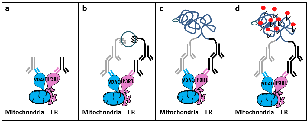
בהתבסס על תגלית של Szabadkai של המתחם IP3R / GRP75 / VDAC בבית MAM 11, פיתחנו שיטה כמותית לנתח אינטראקציות ER-המיטוכונדריה. השתמשנו ב ligati הקרבה באתרועל assay כדי לזהות ולכמת אינטראקציות בין VDAC1 ו IP3R1, שני חלבונים אברון-משטח מעורב במתחם -channeling Ca 2+ על הממשק MAM בתאים קבוע 12. בקצרה, העמקנו VDAC1 על קרום המיטוכונדריה החיצוני (עכבר אנטי VDAC1 נוגדן ראשוני) ו IP3R1 על הממברנה ER (ארנב נוגדן ראשוני נגד IP3R1) (איור 1, לוח א '). לאחר מכן, על פי assay, הוספנו שני אנטי עכבר IgG נגד ארנב (עכבר בדיקות assay קשירת ארנב קרבה), אשר מצומדות כדי רחבות oligonucleotide משלימות. אם שני החלבונים הממוקדים הם במרחק מתחת ל -40 ננומטר, oligonucleotides יכול להכליא עם oligos מחבר הנוסף לאחר מכן לאפשר את היווצרות של תבנית ה- DNA מעגלי (איור 1, לוח ב '). מולקולת הדנ"א חוזר זה ligated מוגבר, יצירת מוצר דנ"א חד-גדילי המצורפת קוולנטית לאחד חלליות הקרבה (איור 1, ג פאנל) . היות והמרחק בין ER ואת המיטוכונדריה בממשק MAM נע בין 10 ננומטר ל -25 6 ננומטר, קשירת הקרבה והגברה ניתן לעשות, מה שמוביל לגילוי הבאים עקב הכלאה של בדיקות oligonucleotides אדום שכותרתו טקסס (איור 1, פאנל ד ). נקודה פלורסנט מייצגת אינטראקציות בין VDAC1 / IP3R1, ובכך מאפשר כימות של באתרו ER-המיטוכונדריה אינטראקציות בתאים בודדים.

איור 1: איור סכמטי של איתור של endoplasmic אינטראקציות reticulum-המיטוכונדריה ידי ב assay קשירת Situ קרבה. א) נוגדן עכבר עיקרי המכוון נגד VDAC1 ו נוגדן ראשוני ארנב המכוון נגד IP3R1 יכול להיקשר אפיטופים שלהם בסמיכות בממשק MAM, ב) התוספת של זוג חלליות קשירת קרבהמכוון נגד עכבר IgG ארנב. בדיקות אלה יש מצורפות גדילי דנ"א שיכול ליצור תבניות עבור קשירת oligos מחבר. ג) גדיל DNA המעגלי שיקום לאחר הקשירה יכול להיות מוגבר ו ד) מדמיין ידי מיקרוסקופ כנקודת פלורסנט באמצעות oligonucleotides האדום שכותרתו טקסס. אנא לחץ כאן כדי לצפות בגרסה גדולה יותר של דמות זו.
דומה בניסויים assay קשירת הקרבה באתרו ניתן לבצע עם זוג GRP75 / IP3R1 של נוגדנים, כמו גם cyclophilin D (CypD) / נוגדנים IP3R1, בהתחשב בכך CypD הוצגה אינטראקציה עם מורכבות IP3R / GRP75 / VDAC בממשק MAM 12-14.
Protocol
1. הכנת פתרונות
- הכן 10 פורמלדהיד% ב PBS (מלח נמוכה) על ידי דילול 5.5 מ"ל של פורמלדהיד 37% ב 14.5 מ"ל של PBS. הכן 1 M גליצין, pH 8.0, על ידי המסת 3.8 גרם של גליצין ב 50 מ"ל של PBS; לדלל את הפתרון הזה כדי לקבל גליצין 100 מ"מ 1x PBS.
- כן 0.1% Triton-X100 ב 1x PBS. כן 20x המלוח סודיום ציטראט (SSC) באמצעות 3.0 M נתרן כלורי 0.30 M ציטראט Trisodium ערוך חיץ מים ללא יונים עם pH 7.0 במהירות של 25 ° C. לדלל חיץ זה ל X 1 ו 0.01x באמצעות מים ללא יונים.
קיבוע 2. של תאים
הערה: השתמשנו הקו הסלולרי HuH7 hepatocarcinoma במחקר זה, אך שיטה זו ישימה בתרביות תאים חסידות אחרות.
- פלייט תאי HuH7 (בתרבית גלוקוז DMEM 1 גר '/ ל, בתוספת 10% עוברי עגל בסרום 0.01% מניות פניצילין, סטרפטומיצין) ב 150,000 תאים לכל צלחת זכוכית תחתונה בגודל 35 מ"מ ללא ציפוי. כשעובד על תא ראשוניתרבויות, משתמשים מנות מצופות קולגן.
- למחרת, להסיר את המדיום לתרבות. שוטפים את התאים עם 1 מ"ל של PBS ואת לשאוב. תקן את התאים על ידי הוספת 1 מ"ל של פורמלדהיד 10% ו דגירה במשך 10 דקות בטמפרטורת החדר (RT) תחת תסיסה.
- עצור את התגובה עם 1 מ"ל של 1 M גליצין ומערבבים ידי סיבוב. הסר את הפתרון התגובה להפסיק לשטוף את התאים על ידי הוספת 1 מ"ל של 1x PBS; להתסיס ידי סיבוב לשאוב. הוסף 1 מ"ל של 100 מ"מ גליצין לתאים דגירה אותם במשך 15 דקות ב RT תחת תסיסה, ולאחר מכן לשאוב.
הערה: הפרוטוקול ניתן לעצור כאן ואת הצעדים הבאים יכולים להידחות ליום אחר. במקרה זה, להוסיף 1 מ"ל של 100 גליצין מ"מ ולשמור על 4 מעלות צלזיוס, אם יהיה צורך בכך.
3. Permeabilization של תאים
- הוסף 1 מ"ל של 0.1% Triton-X100 ב PBS 1x, דגירה במשך 15 דקות ב RT תחת תסיסה, ולאחר מכן לשאוב. זמן דגירה זה יכול להיות מוגבר עד 15 - 20 דקות, כאשר עובדים על cultur תא הראשוניes (למשל, hepatocytes עכברים העיקרי). שוטפים את התאים על ידי הוספת 1 מ"ל של 1x PBS; לשאוב.
4. חסימה
- הוסף 40 μl של פתרון לחסימת מדגם זה (המסופק על ידי בערכה); נפח זה יכול להיות מוגבר על מנת לכסות את המדגם. דגירת המנות למשך 30 דקות ב 37 מעלות צלזיוס בתא לחות.
- הקש את פתרון חסימת הנחה של המנות. נסה להשיג כרכים שיורית שווים עבור כל שקופית, מאחר שהדבר ישפיע על שחזור. אל תתנו דגימות להתייבש!
5. נוגדנים ראשוניים
- לדלל את הנוגדנים הראשוניים ב 1x PBS ולהוסיף פתרון המנות (נוגדן עכבר VDAC1: 1/100, נוגדן ארנבת IP3R1: 1/500). לחלופין, נוגדני GRP75 או CypD (נוגדני העכבר הן השתמשו ב 1/500) יכולים לשמש במקום VDAC1.
- דגירת הלילה בתא לחות ב 4 ° C.. שטפו את השקופיות פעמיים באמצעות טריס בופר עם 0.01% Tween (TBS-T).
- בדיקות assay קשירת סמיכות ניתנות על ידי ההערכה. בחר בדיקות על פי המין של נוגדנים ראשוניים.
- הכן את בדיקות assay קשירת שתי הקרבה 1: 5 ב diluent נוגדן. אפשר לתערובת לשבת במשך 20 דקות ב RT. מוסיף את פתרון בדיקת assay בדילול קרבת קשירה. דגירה מנות בתא לחות מחומם מראש עבור שעה 1 ב 37 מעלות צלזיוס. לשטוף את הכלים פעמיים עם-T TBS.
7. קשירה
- השתמש ב מגיב איתור באתרו ערכת אדום טקסס.
- לדלל את המניות קשירת 5x (המסופקים על ידי בערכה) 1: 5 במים טוהר גבוהה ומערבבים היטב. לדלל את האנזים בפתרון קשירת 1x (המסופק על ידי בערכה) 01:40 ו מערבולת. חכה להוסיף את האנזים עד שהערב בנוסף הדגימות.
- הוסף פתרון זה מדגם זה (40 μl עבור מנות זכוכית תחתונה בגודל 35 מ"מ) דגירה את השקופיות צ'אם לחות מחוממת מראשבער במשך 30 דקות ב 37 מעלות צלזיוס. שטפו את השקופיות פעמיים עם-T TBS.
הגברה 8.
הערה: היזהר, ריאגנטים רגיש לאור.
- לדלל את מניות הגברת 5x (המסופקות על ידי בערכה) 1: 5 במים טוהרים גבוהה. הסר את פולימראז מהמקפיא באמצעות לחסום הקפאה (-20 ° C). לדלל את פולימראז (המסופק על ידי בערכה) 1:80 בתמיסת מערבולת הגברת 1x. מוסיפים את פולימראז מיד לפני השימוש בתערובת.
- הוסף פתרון זה מדגם זה (40 μl עבור מנות זכוכית תחתונה בגודל 35 מ"מ). דגירה השקופיות בתא לחות מחומם מראש ל -100 דקות ב 37 מעלות צלזיוס. קש פתרון הגברה-פולימראז ההנחה של השקופיות.
כביסה 9. סופי
- שטפו את השקופיות כביסה חיץ 1x SSC למשך 2 דקות. שטפו את השקופיות חיץ כביסה SSC 0.01x למשך 2 דקות. תנו את הכלים לייבוש ב RT בחושך.
הכנת 10. הדמיה
- הר השקופיות באמצעות בהיקף מינימאלי של הרכבה בינונית המכילים DAPI (מימי לא לחזק), להבטיח כי אין בועות אוויר להיתפס תחת להחליק את המכסה. ניתן להשתמש לק כדי לאטום את הקצוות. חכה כ -15 דקות לפני הניתוח באמצעות קרינה או מיקרוסקופ confocal (עירור: 594 ננומטר, פליטה: 624 ננומטר, גדלה: 63X).
- לאחר הדמיה, אחסן את השקופיות ב -20 ° C בחושך.
תוצאות
בהתבסס על הניסיון שלנו באמצעות פרוטוקול זה, אנחנו יכולים בבטחה ממליצים על שיטה זו עבור להדמיה וכימות של אינטראקציות ER-המיטוכונדריות בתאים קבועים. נציג תמונות של באינטראקציות ER-המיטוכונדריה assay-דמיינו קשירת הקרבה באתרו הקו הסלולרי HuH7 hepatoca...
Discussion
Collectively, our studies indicate that the in situ proximity ligation assay is truly a relevant strategy to follow and quantify endogenous ER-mitochondria interactions in fixed cells, without the need for using organelle-specific fluorophores or fluorescent proteins. The specific use of VDAC1/IP3R1 antibodies has been adapted to study ER-mitochondria interactions in HuH7 cells. However, alternative isoforms of VDAC and IP3R may be used, depending on the cell type. In this case, antibodies need to be validated b...
Disclosures
The authors declare that they have no competing financial interests.
Acknowledgements
אנו מודים לכל האנשים במעבדה שלנו שתרמו לייעל ולאמת את הפרוטוקול. עבודה זו נתמכה על ידי INSERM ואת סוכנות המחקר הלאומי (ANR-09-JCJC-0116 ו ANR-11-BSV1-033-02). ET נתמכה במהלך הדוקטורט שלה על ידי מענק מחקר מהמשרד הצרפתי של השכלה גבוהות ומחקר.
Materials
| Name | Company | Catalog Number | Comments |
| Formaldehyde | Sigma | F-8775 | |
| Glycine | Sigma | G-8898 | |
| Triton | Sigma | T8532 | |
| 35 mm Glass bottom culture dishes | MatTeK corporation | P35G-0-14-C | |
| Blocking solution | Sigma | DUO-92004 or DUO-92002 | provided in the Duolink PLA probes, Sigma |
| VDAC1 antibody | Abcam | ab14734 | |
| IP3R1-H80 antibody | Santa Cruz | sc28614 | |
| CypD antibody | Abcam | ab110324 | |
| Grp75 antibody | Santa Cruz | sc13967 | |
| TBS 10x | euromedex | ET220 | Dilute to obtain 1x |
| Tween 100x | euromedex | 2001-B | dilute in TBS to obtain 0,01% |
| PLA Probes Mouse MINUS | Sigma | DUO-92004 | Duolink, Sigma |
| PLA Probes Rabbit PLUS | Sigma | DUO-92002 | Duolink, Sigma |
| Duolink detection reagents red | Sigma | DUO-92008 | Duolink, Sigma |
| Ligation solution | Sigma | DUO-92008 | Part of the Duolink detection reagents red, Sigma |
| Ligase | Sigma | DUO-92008 | Part of the Duolink detection reagents red, Sigma |
| Amplification solution | Sigma | DUO-92008 | Part of the Duolink detection reagents red, Sigma |
| Polymerase | Sigma | DUO-92008 | Part of the Duolink detection reagents red, Sigma |
| Duolink Mounting Medium | Sigma | DUO80102 | Duolink, Sigma |
| Softwares | |||
| Blob-finder software | BlobFinder is a freely distributed software that can perform calculations on cells from fluorescence microscopy images. This software can be downloaded for free from The Centre for Image Analysis at Uppsala University who have developed the software and the work was supported by the EU FP6 Project ENLIGHT and Olink Bioscience. http://www.cb.uu.se/~amin/BlobFinder/index_files/Page430.htm | ||
| ImageJ software | Can be downloaded for free from: http://rsb.info.nih.gov/ij/download.html | ||
References
- Bravo-Sagua, R., et al. Organelle communication: signaling crossroads between homeostasis and disease. The international journal of biochemistry & cell biology. 50, 55-59 (2014).
- Giorgi, C., et al. Mitochondria-associated membranes: composition, molecular mechanisms, and physiopathological implications. Antioxidants & redox signaling. 22, 995-1019 (2015).
- Phillips, M. J., Voeltz, G. K. Structure and function of ER membrane contact sites with other organelles. Nature reviews. Molecular cell biology. 17, 69-82 (2016).
- Cosson, P., et al. The RTM resistance to potyviruses in Arabidopsis thaliana: natural variation of the RTM genes and evidence for the implication of additional genes. PLoS One. 7, 39169 (2012).
- Mannella, C. A. Structure and dynamics of the mitochondrial inner membrane cristae. Biochim Biophys Acta. 1763, 542-548 (2006).
- Csordas, G., et al. Structural and functional features and significance of the physical linkage between ER and mitochondria. The Journal of cell biology. 174, 915-921 (2006).
- Mannella, C. A., Buttle, K., Rath, B. K., Marko, M. Electron microscopic tomography of rat-liver mitochondria and their interaction with the endoplasmic reticulum. Biofactors. 8, 225-228 (1998).
- Rizzuto, R., et al. Close contacts with the endoplasmic reticulum as determinants of mitochondrial Ca2+ responses. Science. 280, 1763-1766 (1998).
- Wieckowski, M. R., Giorgi, C., Lebiedzinska, M., Duszynski, J., Pinton, P. Isolation of mitochondria-associated membranes and mitochondria from animal tissues and cells. Nat Protoc. 4, 1582-1590 (2009).
- Csordas, G., et al. Imaging interorganelle contacts and local calcium dynamics at the ER-mitochondrial interface. Mol Cell. 39, 121-132 (2010).
- Szabadkai, G., et al. Chaperone-mediated coupling of endoplasmic reticulum and mitochondrial Ca2+ channels. J Cell Biol. 175, 901-911 (2006).
- Tubbs, E., et al. Mitochondria-associated endoplasmic reticulum membrane (MAM) integrity is required for insulin signaling and is implicated in hepatic insulin resistance. Diabetes. 63, 3279-3294 (2014).
- Paillard, M., et al. Depressing Mitochondria-Reticulum Interactions Protects Cardiomyocytes From Lethal Hypoxia-Reoxygenation Injury. Circulation. 128, 1555-1565 (2013).
- Rieusset, J., et al. Disruption of calcium transfer from ER to mitochondria links alterations of mitochondria-associated ER membrane integrity to hepatic insulin resistance. Diabetologia. 59, 614-623 (2016).
- Allalou, A., Wahlby, C. BlobFinder, a tool for fluorescence microscopy image cytometry. Computer methods and programs in biomedicine. 94, 58-65 (2009).
- Theurey, P., et al. Mitochondria-associated endoplasmic reticulum membranes allow adaptation of mitochondrial metabolism to glucose availability in the liver. Journal of molecular cell biology. , (2016).
- de Brito, O. M., Scorrano, L. Mitofusin 2 tethers endoplasmic reticulum to mitochondria. Nature. 456, 605-610 (2008).
- Soderberg, O., et al. Direct observation of individual endogenous protein complexes in situ by proximity ligation. Nature methods. 3, 995-1000 (2006).
- De Pinto, V., Messina, A., Lane, D. J., Lawen, A. Voltage-dependent anion-selective channel (VDAC) in the plasma membrane. FEBS letters. 584, 1793-1799 (2010).
- Kaul, S. C., Taira, K., Pereira-Smith, O. M., Wadhwa, R. Mortalin: present and prospective. Experimental gerontology. 37, 1157-1164 (2002).
Reprints and Permissions
Request permission to reuse the text or figures of this JoVE article
Request PermissionExplore More Articles
This article has been published
Video Coming Soon
Copyright © 2025 MyJoVE Corporation. All rights reserved