このコンテンツを視聴するには、JoVE 購読が必要です。 サインイン又は無料トライアルを申し込む。
Method Article
ゼブラフィッシュ胚眼の組織の顕微解剖
要約
この記事では、1〜3日postfertilizationの胚から、接続されている網膜色素上皮がある場合とない場合のゼブラフィッシュ網膜を顕微解剖するアプローチを説明します。
要約
ゼブラフィッシュは、その急速の眼の発生に関する研究のための人気の動物モデルである
プロトコル
パート1:マイクロダイセクション前の準備
- ソリューション
- E3培地4(5 mMのNaCl、0.17 mMの塩化カリウム、0.33 mMのCaCl 2および0.33 mMのMgSO 4を )。
- リンゲル溶液1、5は (116 mMのNaCl、2.9 mMの塩化カリウム、1.8 mMのCaCl 2を 、5mMのHEPES、pH7.2)、フィルタは滅菌。
- 5N NaOHで。
- タングステン針の化学エッチング
- 粘土でペトリ皿に5N NaOHを含む小さなビーカーを固定します。
- それは、NaOH溶液と接触するように、ビーカーの側面にペーパークリップを取り付けます。
- ペーパークリップやタングステン線の短い断片(〜2センチメートル)の一端に正極へのDC電源から負極を接続します。
- 小さな粘土のボールを持つタングステン線のもう一方の端をラップし、NaOH溶液にこの端を浸し。
- 約3Vまで電圧を増加し、良好なエッチング速度が得られるまでNaOH溶液で上下線を浸漬。良い品質の針は通常10分でエッチングすることができる。
- ソリューションにさらされるタングステン線は徐々に薄くなるでしょう。粘土のボールがオフに低下すると、まだ正極に接続されているワイヤの反対側には、鋭い針の形状を持つことになります。
- 塩の付着物を除去するためにリンゲル液またはE3に鋭い針を洗浄します。
- テープや取り扱いを容易にするためニードルホルダーが木製のアプリケーターに針を取り付けます。
- 培養プレート
- ファルコン60 × 15mmのポリスチレン培養プレートは、網膜のマイクロダイセクションのために使用する準備が整いました。
- RPE -添付の網膜を切開するため、30分解剖する前に表面の接着性を取り除くためにリンゲル液を含有する培養プレートで完全にtwo胚をつぶす。まもなく解剖する前に、新鮮なリンゲル液で広範囲にプレートを洗浄。
- 胚のコレクションとステージング
- 大人のゼブラフィッシュは、4、5を説明するように維持されます。
- 分周器によって繁殖の前に夜をペアワイズ静的な繁殖タンク内の親魚を区切ります。
- 部屋のライトが次の朝にオンになって後に分周器を取り外します。両親は10分間隔で30分おきに交差することができます。各交差点の後に魚を区切ります。
- 28で別々にE3培地に各交差点から収集された胚を維持℃に
- 確立された基準(1)(6)によれば、特定の時点で10〜12時間後に受精(HPF)でと再演するすぐに解剖する前段階の胚、。
パート2:解剖-脳の除去と眼のばく露1
- このセクションで説明する手順は、網膜(パート3A)及びRPE -添付の網膜を(パート3B)解剖のための共通です。
- すべての微細な解剖はデュモン鉗子の先端でおこなわれており、組織を位置決めするためのデュモン鉗子の別のペアによって援助されています。必要に応じて化学的にエッチングされたタングステンの針は、細かい操作に使用することができます。
- すべての微細な解剖は1X目的または同等の装備オリンパスSZX16顕微鏡下で〜5倍速の倍率で実行されます。
- 28でベンチトップインキュベータにE3培地で胚を維持℃の解剖時に簡単に胚のアクセスのための顕微鏡の隣にある。
- 顕微鏡のステージは、遺伝子発現に及ぼす温度変動の影響を最小限に抑えるためにサーモプレートで28℃に加温している。
- 第1部#3で説明したように調製されたファルコン60 × 15mmの培養プレートでリンゲル液に特定の発達段階にある胚を移す。
- 体内から速やかに前方トランクの部分で頭を切り落とします。
- 支援鉗子で培養プレートにこの組織の後端を固定。
- 前額から始まる背側に頭を開きます。
- 目の内側が露出され、さらなる操作のために上向きになるように脳を取り外します。
パート3a:網膜切開1
- として第2部で説明されている胚を準備する。
- 網膜に小さな開口部が見られるまで慎重に鉗子の先端が上向きに直面している眼球の内側に露出したRPEを磨きます。
- ブラッシングと網膜の内側はほぼすべてが露出するまでアクションを剥離を継続する。露出網膜を傷つけないし、パンチは避けてください。
- 今下向き直面している網膜、アプローチに横方向のRPEを削除することと培養プレートの表面から約45 °の角度でブラシ。
- cにRPEのデタッチ部分を固定することによって、鋸状縁に比較的しっかりと接続されてRPEを削除します。ulture郭清のためのプレート、そして次にゆっくりと網膜を持ち上げます。
- 残留RPEをクリーンアップするために培養皿の底に網膜をロールバックします。
- 我々は、胚が粉砕されると、この特定のファルコンのポリスチレン培養プレートは、約20分間RPEの優先的な密着性を持っていることに気づいた。このプロパティは、RPEの残党の除去のために利用されている。しかし、網膜細胞は、高倍率下で密接に検査することができるより少ない程度に表面に棒を行います。一つは、RPEの除去と網膜の整合性の完全性とのバランスを持っています。
- レンズは、しばしば培養プレートに付着し、圧延工程中に網膜から切り離します。時折、それはレンズの表面に渦巻くアクションでエッチングされたタングステン針でレンズを取り外すときに必要です。
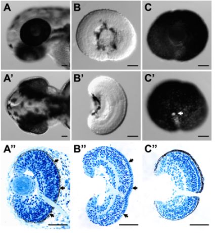
- これらの手順は、正常組織の整合性を損なうことなく、網膜からRPEを削除することができます。これは、良好な全体的な形態(図1B及びB')と組織学(図1B")で示される、矢印特に、光受容体層とRPEとの間の細胞外マトリックスの保全が(図1B非常に良いです。";全胚(図1A"))から、組織型で比較する。
一部3B:RPE接続網膜解剖3
- として第2部で説明されている胚を準備する。
- 助手の鉗子によって培養プレートに頭部を固定。後部側面からそっと目を持ち上げて、前方側にロールバックします。
- RPE層の外側に推定脈絡膜と強膜組織は、皮膚に、比較的しっかりと接続されており、主に前方側に注意深く目を圧延して剥離することができる。
- 目の側面が露出していると眼がまだ肌に接続されているときに後のタングステン針でレンズを外します。
- これらのプロシージャは、正常網膜で全体RPE層を保持することができます。推定脈絡膜と強膜組織は、主に(図1C、C'とC")は削除することができます。
パート4:RNAの作業のための組織サンプルの収集
- 解剖のサンプルは1日より前に説明したように下流のRNAの特性評価のためのRNaseフリーのマイクロチューブにTRIZOLに集めることができます。
代表的な結果

図1()解剖前の52 HPFにおけるゼブラフィッシュ幼生の頭部の横方向と(')背ビュー。幼虫の頭の中で()&(')の(")に対応する組織切片(B)側面と(B')54 HPFにおける解剖網膜の背ビュー。網膜の表面には、両方の側面から無傷だったと背側ビュー。(B)&(B')の解剖網膜の(B")に対応する組織切片。網膜と網膜積層構造は無傷であった。特に、光受容体層とRPE(間細胞外マトリックスは、"(、矢印、矢印)も解剖網膜B)に保存されていました"。 (C)側方および52 HPFにおける解剖RPEに接続された網膜の(C')内側のビュー。 RPE層はまた、切除した組織の組織切片(C")で示された、無傷と連続だった。C'の白い領域は、視神経(矢印)である。組織は、組織試料を4%で収集し、修正された3を説明するようにパラホルムアルデヒド。埋め込 み、これらの試料のセクショニングのプラスチックを行った。スケールバー=50μmのは。
ディスカッション
ゼブラフィッシュの眼の組織の顕微解剖は、効果的に無傷の網膜とRPEに接続された網膜を取得することができます。これは実質的に特定の眼組織(すなわち、網膜や網膜色素上皮)に関連する発現研究を支援します。実際に、我々が正常に全体の網膜1とRPE 3のRNAの発現プロファイルを取得するには、次の手順を利用してきた。これらのプロファイルのユーティリティが強く網...
開示事項
謝辞
この作品は、パーデュー大学で生物科学科からのスタートアップ資金によってサポートされています。
資料
| Name | Company | Catalog Number | Comments |
| Cordless pestle motor | VWR international | 47747-370 | |
| DC power supply | Lascar | PSU130 | Any DC supply would work. The specific voltage of a different machine will need further optimization. |
| Disposable pestle & microtube, 1.5 mL (DNase, RNase and pyrogen-free) | VWR international | 47747-366 | These are used for tissue collection in TRIzol for expression analysis. |
| Dumont #5 forceps, Tips: 0.05 x 0.01mm, Inox | World Precision Instruments, Inc. | 500341 | Fine tip dimension is desirable but is not inflexible, as one may need to sharpen the tip from time to time. |
| Dumont #5SF forceps, Tips: 0.025 x 0.005mm, Inox | Fine Science Tools | 11252-00 | Fine tip dimension is desirable but is not inflexible, as one may need to sharpen the tip from time to time. |
| Falcon polystyrene culture plates, 60 X 15 mm | BD Biosciences | 351007 | These plates are used as dissection plates. |
| Olympus SZX16 Stereomicroscope | Olympus Corporation | SZX16 | Any stereomicroscope would work. We used Leica stereomicroscope in previous studies1-3 without any issues. We also use the 1X objective exclusively for the dissection even though we have a 2X objective installed. |
| Sharpening stone | Fine Science Tools | 29008-01 | Use this to sharpen the tip of the forceps if necessary |
| Thermo plate | Tokai Hit | MATS-U55SZX2B | This is used to maintain the temperature of the tissue throughout dissection and minimize the influence of temperature fluctuation on gene expression. We also put the whole microscope in an environmentally controlled room at 28°C during dissection in previous studies1-3 with good success. |
| Trizol, 100 mL | Invitrogen | 15596-026 | |
| tungsten wire, 0.015 inch diameter | World Precision Instruments, Inc. | TGW1510 | |
| Wooden Applicator | Puritan | 807 | This is used for holding the chemically-etched tungsten needle. |
参考文献
- Leung, Y. F., Dowling, J. E. Gene expression profiling of zebrafish embryonic retina. Zebrafish. 2, 269-283 (2005).
- Leung, Y. F., Ma, P., Link, B. A., Dowling, J. E. Factorial microarray analysis of zebrafish retinal development. Proc Natl Acad Sci U S A. 105, 12909-12914 (2008).
- Leung, Y. F., Ma, P., Dowling, J. E. Gene expression profiling of zebrafish embryonic retinal pigment epithelium in vivo. Invest Ophthalmol Vis Sci. 48, 881-890 (2007).
- Nusslein-Volhard, C., Dahm, R. . Zebrafish : a practical approach. , (2002).
- Westerfield, M. The zebrafish book : a guide for the laboratory use of zebrafish (Danio rerio). , (2000).
- Kimmel, C. B., Ballard, W. W., Kimmel, S. R., Ullmann, B., Schilling, T. F. Stages of embryonic development of the zebrafish. Dev Dyn. 203, 253-310 (1995).
- Fadool, J. M., Dowling, J. E. Zebrafish: a model system for the study of eye genetics. Prog Retin Eye Res. 27, 89-110 (2008).
転載および許可
このJoVE論文のテキスト又は図を再利用するための許可を申請します
許可を申請さらに記事を探す
This article has been published
Video Coming Soon
Copyright © 2023 MyJoVE Corporation. All rights reserved