
Method Article
上の機能的研究のための3Dヒト肺組織モデル
要約
Human tuberculosis infection is a complex process, which is difficult to model in vitro. Here we describe a novel 3D human lung tissue model that recapitulates the dynamics that occur during infection, including the migration of immune cells and early granuloma formation in a physiological environment.
要約
結核(TB)は依然として世界の人々の健康に大きな脅威を保持し、私たちは病気のメカニズムを理解し、新たな治療選択肢の発見を進める手助けするためのコスト効率に優れたが、信頼性の高いモデルの必要性がある。単層のインビトロ細胞培養または共培養は、三次元(3D)環境および組織応答を欠いています。ここで、我々は結核菌 ( 結核菌 )の感染の間に起こる複雑な事象を研究するための有効なツールであることが期待されているヒト肺組織のインビトロモデルで革新的なについて説明します。 3次元組織モデルは、多孔質膜の上に、コラーゲンマトリックス中で培養された組織特異的上皮細胞および線維芽細胞からなります。空気暴露されると、上皮細胞層別化し、頂端側の粘液を分泌します。 M.に感染したヒト初代マクロファージを導入することにより組織モードに結核lは、我々は、免疫細胞が感染組織中に移動し、TBの肉芽腫の初期段階を形成することが示されています。これらの構造は根本的に異なるか、一般的に広く使用されている実験動物モデルにおいて観察されていない人間の結核、肉芽腫の明確な特徴を再現。この器官培養法は、宿主細胞病原体相互作用の空間的および時間的機能に関する重要な情報を提供する3次元可視化と堅牢な定量分析を可能にします。まとめると、肺組織モデルは、結核の研究のための生理学的に関連する組織の微小環境を提供します。従って、肺組織モデルは、基本的な機構的および応用研究の両方のための潜在的な意味を持ちます。重要なことには、モデルは、それによって、肺に影響を与える感染症の様々なモデル化するためのその使用が広がり、個々の細胞型の添加または操作を可能にします。
概要
ヒトでは、感染症、組織炎症、細胞の動員、組織リモデリングおよび組織の恒常性の調節に応答が異なる細胞型が関与する複雑な事象です。したがって、これらの方法は、最高の局所組織環境において研究されています。以前は、これは、主に実験動物モデルを使用して可能でした。彼らはしばしば、人間とは異なる方法で病原体に対応し、また、疾患1の別のコースを表示しかし、広く使用されている実験動物は、多くの制限を保持します。 インビトロの肺組織モデル中のヒトはヒト肺で特異的免疫応答を研究するための可能性を保持しています。
ヒト結核感染(TB)は、主に肺に影響を与える病気である。 結核菌 ( 結核菌 )、結核の原因物質は、細菌が肺dendriに飲み込まれている肺胞腔に輸送されるエアロゾル液滴を経由して肺に達します感染2,3に対する自然免疫応答の一部として、チック細胞や肺胞マクロファージ。病原体の食作用はファゴソーム内のバグの区画化につながる、理想的には食細胞による中和および病原体を死滅させることになります。 Mに曝露された個体の50%まで結核は、先天性免疫応答4を介して感染をクリアすることができると考えられています。感染の他の結果は後の段階で適応免疫系によるクリアランス、潜伏感染またはアクティブな疾患5慢性最悪のケースです。
以前は人間の結核の研究のために何のインビトロ組織モデルは存在していません。ヒトマクロファージまたは他の末梢血細胞の単一細胞培養物は、多くの場合、6,7を使用されています。このアプローチの欠点は、Mに露出し、肺組織内で一緒に動作して、異なる細胞型の動態を反映することができないことです結核 。したがって、TB上の機能とメカニズムの研究を行うことができるようにするインビトロモデルが必要とされています。セル・ベースのここで説明する体外ヒト肺組織モデルでは、もともとの樹状細胞の機能8の研究のために私たちのグループによって設立されました。私たちは、結核の研究のために、この方法を適応しています。
ここに提示されたヒト肺組織モデルは、組織特異的な上皮細胞および線維芽細胞8から成ります。これらの細胞は、トランスウェルインサート中の多孔質膜の上に、コラーゲンマトリックス中で培養され、フォーム構造は、正常なヒトの肺組織( 図1)に似ています。空気にさらされると細胞は頂端側8に粘液を分泌し始めます。 M.に感染したヒト初代マクロファージを注入することにより、モデルへの結核 、我々は、免疫細胞が組織に移行し、結核性肉芽腫9の初期段階を形成する方法を観察しています。これは最初のヒト組織モデルDESCRです結核ibed、それが結核と肺の他の疾患に対する自然免疫応答を研究するための有望なツールをもたらします。これまでに、我々は、モデルにおいて免疫細胞としてのみ単球およびマクロファージを使用しているが、複雑さのレベルは、追加の関連する細胞型を含めることによって増加させることができます。

肺組織モデルの概略1.概略図。 (A)モデルM.、ヒト肺特異的上皮細胞で構成されています結核は、初代マクロファージをに感染し、赤色の色素ラベルされた単球をトランスウェルフィルター上に作製したコラーゲン埋め込 まれた線維芽細胞上に播種。空気への組織モデルの露出は、上皮によって細胞外マトリックスタンパク質の産生、粘液分泌と層化を開始します。こうして開発された3次元組織モデルは 、M を研究するための便利なツールですCLO環境における結核感染SELYヒトの肺。(B)組織モデルの製造における様々なステップの代表的な顕微鏡像。(C)肺モデル組織切片の完全な構造に似ています。スケール- 。100ミクロンこの図の拡大版をご覧になるにはこちらをクリックしてください。
プロトコル
注:リンショーピン大学病院の血液銀行で購入健康匿名血液ドナーからのヒト末梢血、スウェーデンは、この研究のための免疫細胞の供給源として使用しました。このプロトコルは、24 mmの6ウェルプレートの挿入のために設計されています。組織モデルは、開発中に垂直方向と水平方向の両方に収縮するので、他のウェルフォーマットへの直接適応が推奨されていません。
細菌/細胞系の材料、メディアと文化の作製
- 細菌の培養:
- マイコバクテリア菌株Mを成長0.05%を含むミドルブルック7H9培地中で構成的に緑色蛍光タンパク質(GFP)を発現するpFPV2プラスミドを保有する結核菌H37Rv、トゥイーン80、0.5%グリセロール、カナマイシン(20μgの/ ml)を、そして(ミドルアルブミン、ブドウ糖及びカタラーゼ濃縮を補っミドルADC濃縮)、7〜10日間、5%CO 2で37℃で。
注:全ての実験手順ライブ病原性M.を含む S 結核菌株は、BSL-3施設で実施すべきです。
- マイコバクテリア菌株Mを成長0.05%を含むミドルブルック7H9培地中で構成的に緑色蛍光タンパク質(GFP)を発現するpFPV2プラスミドを保有する結核菌H37Rv、トゥイーン80、0.5%グリセロール、カナマイシン(20μgの/ ml)を、そして(ミドルアルブミン、ブドウ糖及びカタラーゼ濃縮を補っミドルADC濃縮)、7〜10日間、5%CO 2で37℃で。
- 1×ダルベッコ改変イーグル培地(DMEM)完全培地を調製し(1mMのピルビン酸ナトリウム、2mM L-グルタミン、100 U / mlのペニシリン、100μg/ mlのストレプトマイシン、10mMのHEPES、0.1mMの非必須アミノ酸及び10を補っ%のウシ胎児血清(FBS))を熱で不活性化。また、抗生物質を含まないDMEM完全培地を準備します。
- 1×最小必須培地(MEM)、完全培地を調製し(1mMのピルビン酸ナトリウム、2mM L-グルタミン、100 U / mlペニシリン、100μg/ mlのストレプトマイシン、10mMのHEPES、0.1mMの非必須アミノ酸及び10%熱不活性化ウシ胎児血清(FBS))。
- フィブロネクチン/コラーゲンコートフラスコ(合計10ml)中の調製
- 清浄なチューブにピペットで8.8 mlの滅菌1×リン酸緩衝生理食塩水(PBS)。 1 mlのウシ血清アルブミン(1ミリグラム/ ml)を、100μlのI型コラーゲン(3 mg / mlの)ウシおよび100μlの組換えHUを追加人間のフィブロネクチン(1mg / mlの)。
- 逆さま5回チューブを回して溶液を混ぜます。フラスコ(T-25、T-75フラスコのために2 mlのために1 ml)を、フィブロネクチン/コラーゲン溶液でコーティングされます。 37℃でO / Nそれを残します。インキュベーション後、溶液を除去し、室温でコーティングしたフラスコを格納します。
注:回収した溶液を4℃で保存し、3回再利用することができます。 2週間以上のストレージは、液体がそれを廃棄しなければならない時に、茶色(結晶の形成)を、有効にする原因となることがあります。
- 線維芽細胞の培養:
- 37℃、5%CO 2で、完全DMEM中で、成長し、MRC-5、(14週齢の雄の胎児の正常肺組織に由来するヒト肺線維芽細胞株)を維持します。通路24-26で線維芽細胞を使用し、70〜80%コンフルエントになるまで成長します。
注:通路> 30でMRC-5細胞株は、形態を失う傾向があると組織モデルでの使用は推奨されません。
- 37℃、5%CO 2で、完全DMEM中で、成長し、MRC-5、(14週齢の雄の胎児の正常肺組織に由来するヒト肺線維芽細胞株)を維持します。通路24-26で線維芽細胞を使用し、70〜80%コンフルエントになるまで成長します。
- 上皮細胞の培養:
- 16HBE14o-(16HBE)、正常なヒト気道上皮の分化した形態と機能を保持する不死化ヒト気管支上皮細胞株を取得し、(これは博士ディーターGruenert、山シオンがんセンター、カリフォルニア大学、サンフランシスコからの贈り物でした、米国10。)。フィブロネクチン/コラーゲンコートフラスコで培養16HBE細胞と37℃で5%CO 2に完全MEMで細胞を維持します。
- 5倍のDMEMの調製
- DMEM粉末13.4 gであり、滅菌蒸留水150ml中の炭酸水素ナトリウム3.7グラムを溶解することによって5倍DMEMを準備します。 、7.3に培地のpHを調整する200ミリリットルにボリュームを構成し、0.22μmのメンブレンフィルターを用いてろ過します。滅菌容器中で濾過培地を収集し、室温で使用するまで保存されました。
コラーゲン埋め込まれた線維芽細胞の調製
- 37℃の水浴中にFBSおよびL-グルタミンの融解凍結アリコート。解凍後にサンプルを氷上で保冷。 4℃で炭酸水素ナトリウム(71.2 mg / mlの)とゲンタマイシン(50 mg / mlの)を配置します。前クール50mlの遠心分離チューブ、10 mlの滅菌ピペット4℃です。
注:(5×DMEMを除く)で使用される全ての材料は、使用前に氷上で冷却され、すべてのステップは氷上で行っています。これはコラーゲンの凝固を防ぐように私ウシコラーゲン(1.1 mg / mlの)は、寒さに保持する必要があります入力してください。 - 線維芽細胞を準備します。
- 温かい37℃の水浴中でトリプシンと37℃で5%CO 2で10分間、肺線維芽細胞(MRC-5)細胞を十分な量をインキュベートします。 1×DMEMを完全に追加することにより、トリプシンを中和してください。 5分間300×gで細胞懸濁液と遠心分離機を吸引します。上清を吸引除去し、完全DMEM中2.3×10 5細胞/ mlで細胞を再懸濁します。使用するまで氷上に細胞を配置します。
- プレミックスを調製します:
- 「プレミックス」と表示されたチューブに以下を追加します。 3955倍のDMEMμlを、40μlのL-グルタミン、120μlの炭酸水素ナトリウム(71.2 mg / mlの)、440μlのFBS、5μlのゲンタマイシン(50 mg / mlの)、総容積千μL、その後、よく事前に混ぜて置く渦巻きます氷。
注:指定されたボリュームは、1 24ミリメートル6ウェルカルチャーインサート用です。インサートの総数に必要な具体的な金額を計算しますが、必ず、十分なプレミックスを用意することは、余分なものを追加します。
- 「プレミックス」と表示されたチューブに以下を追加します。 3955倍のDMEMμlを、40μlのL-グルタミン、120μlの炭酸水素ナトリウム(71.2 mg / mlの)、440μlのFBS、5μlのゲンタマイシン(50 mg / mlの)、総容積千μL、その後、よく事前に混ぜて置く渦巻きます氷。
- 無細胞のコラーゲンの混合物を準備します。
- 各培養物に無細胞のコラーゲンの混合物の1ミリリットルを追加します。与えられた順に氷上で50 mlのコニカルチューブに以下を追加します。 1.1 mg / mlのコラーゲンの686μlを、250μlのプレミックス1,000μLの全体積64μlの1×完全DMEM。よく空気の泡を確保していない溶液を混合。高速動作及び気泡を回避するために、チューブの壁にコラーゲンを加えます。
- 6ウェルプレートに入れインサートに無細胞層混合物の1ミリリットルを追加します。インサート外ウェルに任意のメディアを追加しないでください。における37℃のインキュベーター中で30分間cubate。無細胞混合物は、気泡なしで、インサート全体をカバーしていることを確認します。
- 携帯コラーゲン混合物を準備します。
- 次の順序で氷上に保持50mlコニカルチューブに細胞層の成分を混合。 2ミリリットルコラーゲン、615μlのプレミックス、1×完全DMEMの58μLと327μlの細胞懸濁液肺線維芽細胞(MRC-5)3000μlに全量をします。各培養物は、細胞のコラーゲン混合物の3ミリリットルを必要とします。
重要なステップ:細胞懸濁液を添加する前に、慎重にコラーゲンとプレミックスを混合することを確認してください。これは、線維芽細胞に毒性作用を回避するために、コラーゲンのpHを中和します。 - 無細胞のコラーゲン層の上に細胞層(3 ml)を加え、37℃のインキュベーターで2時間インキュベートします。高速動作及び気泡を回避するために、チューブの壁にコラーゲンを加えます。
- 重合後、トンへの完全DMEM 2ミリリットルを追加します彼(挿入中)6ウェルプレートの底部と24時間インキュベートします。
注:重合は発生しなかった場合は、線維芽細胞のコラーゲンマトリックスを含むプレートインサートを破棄し、起動オーバー再び。最も可能性の高い原因は、追加または上記試薬の1つの誤ったボリューム内のエラーです。
- 次の順序で氷上に保持50mlコニカルチューブに細胞層の成分を混合。 2ミリリットルコラーゲン、615μlのプレミックス、1×完全DMEMの58μLと327μlの細胞懸濁液肺線維芽細胞(MRC-5)3000μlに全量をします。各培養物は、細胞のコラーゲン混合物の3ミリリットルを必要とします。
線維芽細胞のコラーゲンマトリックスの3連続培養
- 慎重にきれいなピンセットを用いて、インサートを持ち上げて、ウェルの底から培地を吸引除去します。インサート内に2ミリリットル完全DMEMによりうまく続くの底に2ミリリットル完全DMEMを追加します。これは、外側と内側のチャンバ間の栄養素の拡散を防ぐことができますように、インサートの下に気泡を導入することは避けてください。マイクロピペットチップを用いて気泡を除去します。
- 約5〜7日間培養培地(内側とインサート以下)一日おきと文化を変更します。インサートから培地を除去する際には注意してください。コンタを回避するために、線維芽細胞のコラーゲンマトリックスとCT、少しきれいなピンセットを用いて挿入を傾け、インサート壁からメディアを吸引除去します。
重要なステップ:コラーゲンマトリックス内の線維芽細胞は細長い表現型を取得し、コラーゲン、その後契約を改造する必要があります。約5〜7日では、マトリックスは、インサートの中央にプラットホーム(直径10〜14 mm)を形成するために契約しました。契約した行列は、次のステップで使用するための準備ができています。マトリックスの均一な収縮を得るためには、線維芽細胞は前に(ステップ2.6.1)を播種するコラーゲンで十分に混合されていることが重要です。
免疫細胞(感染/非感染単球 - マクロファージ混合物)の4シード
注:以下の実験手順は、病原性マイコバクテリアを含んでいるため、BSL-3施設で実施されなければなりません。
- 主要な単球及びマクロファージの調製
- establiを使用して、ドナーの血液から末梢血単核球を分離プロトコルを流しました。組織モデルと文化の設定と同じ日に、単球を分離し、Mと感染する前に、約7日間マクロファージにそれらを区別する結核 。
注:これは、マクロファージと契約し、線維芽細胞のコラーゲンマトリックスの両方が7日後に使用可能であることを確認します。また、感染したマクロファージと一緒に追加されます新鮮な単球を分離します。
- establiを使用して、ドナーの血液から末梢血単核球を分離プロトコルを流しました。組織モデルと文化の設定と同じ日に、単球を分離し、Mと感染する前に、約7日間マクロファージにそれらを区別する結核 。
- Mの準備結核に感染したマクロファージ
- 培養菌体を収穫、0.05%のTween-80を含む1×PBS、抗生物質を含まない完全DMEM中に再懸濁して洗浄し、細菌の塊を分散させ、光学密度を測定するために切り詰められた無菌の27 G針を通過します。
注:事前決定M.結核のコロニーは、実験室での光学密度の単位同等物を形成します。これは、感染のために使用される細菌の数の推定値を与えます。 - 4時間マクロファージをインキュベートのM。感染多重度(MOI)10)で結核。感染後、細胞外細菌を除去するために、1×PBSで3回洗浄します。同じ方法ではなく、Mなしで培養された非感染マクロファージを使用してください結核 、コントロールとして。
- 37℃で10分間、2mMのEDTAで処理することにより培養プレートからマクロファージを取り外し、抗生物質を含まない完全DMEM中で細胞を再懸濁しました。
- 培養菌体を収穫、0.05%のTween-80を含む1×PBS、抗生物質を含まない完全DMEM中に再懸濁して洗浄し、細菌の塊を分散させ、光学密度を測定するために切り詰められた無菌の27 G針を通過します。
- 単球の標識
注:単球の単離及び標識はBSL-2ベンチで行われ、その後、さらなる処理のためにBSL-3施設に取り込むことができます。- 製造業者の指示に従って、5分間、2μMPKH26赤色色素の最終濃度を有する新たに調製した単球(2×10 7細胞)を染色します。洗浄3回、および1×10 7細胞/ mlの密度で、抗生物質を含まない完全DMEMで細胞を再懸濁します。
- 線維芽細胞のコラーゲンマトリックスへの免疫細胞の添加
- AFTER線維芽細胞のコラーゲンマトリックスの文化の5-7日は、外側と内側のチャンバーから培地を吸引し、外側チャンバへの完全な新鮮な抗生物質を含まないDMEMの1.5ミリリットルを追加します。
- MOの比で標識された単球 - マクロファージ(未感染/感染)の混合物を準備します。MQ(5:1)50μlのDMEM中で完了します。 50,000マクロファージのために25万の標識単球を取ります。
- 線維芽細胞のコラーゲンマトリックスへのMQ混合物を37℃で5%CO 2で1時間インキュベート:50μlのMOを追加します。インキュベーション後、穏やかに挿入培地2 mlを加え、37℃で5%CO 2でさらに24時間インキュベートします。
注意:追加された細胞が緩く付着しているように、メディアの追加は優しくインサートの壁に追加することで、遅くする必要があります。
肺上皮細胞の5播種(16HBE)
注意:次の手順は、BSL-3施設で実施されなければなりません。
- シード肺電子MQ-線維芽細胞のコラーゲン層:MOの上pithelial細胞(16HBE)。これを実行するには、まず(2.2.1のように)、トリプシンで処理することにより、フラスコから16HBE細胞を解離し、抗生物質を含まないDMEM中で4×10 6細胞/ mlで再懸濁します。
- 内側とインサートの外に培地を吸引除去します。 [挿入外に井戸の底で完全な1.5ミリリットルの抗生物質を含まないDMEMを追加します。
- 免疫細胞、線維芽細胞のコラーゲンマトリックスの上に16HBE50μlのを追加します。フード内で2分間のままにして、5%CO 2で37℃のインキュベーター中で1時間インキュベートします。インキュベーションの後、37℃で3日間インサートと文化の中で完全な抗生物質を含まないDMEMをゆっくりと2ミリリットルを追加します。培養工程は、組織モデルにおける上皮細胞の増殖を促進します。
注意:追加された細胞が緩く付着しているように、メディアの追加はインサートの壁をスライドさせることによって遅く、穏やかでなければなりません。
6. 3D肺の空気暴露モデル
注意:感染したマクロファージの5日後に添加した後、組織モデルは、大気に露出しており、以下の手順では、BSL-3施設で実施されなければなりません。
- 内側とインサートの外に培地を吸引除去します。
注:このステップでは上清を分泌因子の検出のために回収することができます。 -70℃での遠心分離、滅菌フィルターとストア上清。 - 外室に1.8ミリリットル抗生物質を含まないDMEM完全を追加し、2日間37℃インキュベーターで5%CO 2でインキュベートします。インサート内の培地を追加しないでください。
注:組織モデルのエアリフトは、ヒトの肺組織への組織と生理学的な類似する強度を提供重層上皮および粘液分泌の形成を促進します。
7.収穫と3D肺組織モデルのマウント
注意:次の手順は、BSL-3施設で実施されなければなりません。
- 感染したマクロファージの7日後の移植では、組織モデルは、収穫の準備ができています。組織モデルから完全に培地を除去します。室温で暗所で30分間、4%パラホルムアルデヒドで組織モデルを修正。このステップでは、細菌を殺し、さらなる処理のための組織/細胞形態を修正します。
- メスを用いて、ウェルインサートからの膜を分離します。よく含む1×PBSに組織を含む膜を転送します。
- きれいなメスを用いて組織モデルの側面をカットし、削除します。そして、4ほぼ等しい正方形のピースに組織モデルをスライス。スーパーフロストスライドガラス上に組織片を転送します。 4℃で1×PBS中の組織片を保管してください。
- 5分のための組織を乾燥させ、DAPIとカバーガラスで延長ゴールド褪色防止を使用してマウントします。乾燥するまで室温で暗所に妨げることなく、スライドのままにしておきます。
注:組織の厚さがわずかtiltiを起こし、中心部と周辺部との間で異なる場合がありますカバーガラスのNG。これを回避するために、(例えばパラフィルムのための)スペーサは、カバースリップの角に配置されてもよいです。 - カバーガラスの端にマニキュアを適用し、それを乾燥させます。 BSL-3施設の引き出すために、それらを安全にするために、70%エタノールでスライドを浸し。
8.可視化、買収および定量的3D解析
- レーザーはそれぞれ、GFP(緑色チャンネル)、PKH26標識単球のためのDAPI(青)のための420 nmおよび555 nmの(赤)の励起のための488nmで発光すると共焦点顕微鏡システムを用いた組織スライドを可視化。
- スタック間の1.5ミクロンの分離 - Z-スタックが20ミクロンの厚さの最小値でカバーし、1を有する512×512の解像度で3D画像を取得します。組織の作品全体をカバー5-10異なるフィールドを取得します。
注:光学解像度(波長、レーザパワー/露出、ピクセルサイズとズーム)の最適な設定のためにこのようなNyqvistような構成を使用してください。国連を避けますDERまたはピクセルの彩度を超えます。 - 三次元画像処理ソフトウェアを使用して共焦点画像を分析します。細胞クラスターの3次元定量のために、以下のステップは、最適な分析のために推奨されています。
- 3D画像処理ソフトウェアを開いて、イメージをロードします。核の大きさ、個々の単球及び単細菌、例えば、画像中に分析される対象物の寸法を測定します。これらの観察は、オブジェクトを定義するか、フィルタリングするために有用です。
- 表示調整ツールを使用して、画像のコントラスト、明るさ、不透明度をブレンドするための各チャネルを調整することにより、ボリュームレンダリングを最適化します。このステップは、ボリュームレンダリングのノイズの干渉を最小にすることです。
注:ガンマ補正は、画像の操作につながる可能性があり、したがって、避けるべきです。 - 赤チャンネル(単球)を選択してサーフェスを作成し、しきい値(自動または手動選択)を設定します。必要であれば、赤単球の選択を制限するフィルタを使用したり、Bを除外することackground。前述したように同様に、緑( 結核菌 )と青面(核)のチャネルを作成します。
- MS-Excelファイルにデータをエクスポートします。これは、特定のパラメータまたはすべてのデータをエクスポートすることが可能です。セルのクラスタリングの分析に関連するパラメータは、ボリューム、強度、オブジェクトの数、ボクセルと真球の数です。
- 適切な画像フォーマット、好ましくはTIFFで画像を保存してエクスポートします。
注:アニメーションはまた、アニメーションメニューを使用して行われ、メディアファイルとして保存することができます。 - 追加パラメータオプションを使用して、各チャネルの解析設定を保存し、再構築機能を使用して後で検索することができます。同時にバッチ処理ツールを使用して複数のファイルを分析します。
注:比較されるすべての画像が同じように、取得された処理され、分析されなければなりません。例えば8ビットの画像(256の整数)は12ビット画像(4096整数)と比較すべきではありません。
結果
ヒトの結核のための3D肺組織モデルは、効果的にMの宿主-病原体相互作用を研究するために使用することができ結核感染 。この方法の基本的な手順は、異なるステップ及び組織切片の全体的な微細構造の代表的な顕微鏡写真を図1に概説されている。このモデルは肺線維芽細胞、気管支上皮細胞および初代単球/マクロファージを含むヒト肺組織のいくつかの構成要素を有しています3D組織環境内に埋め込まれました。ヒト肺組織の構成要素を組み込むだけでなく、モデルは、生理的条件に上皮および粘液分泌のすなわち成層に似ています。
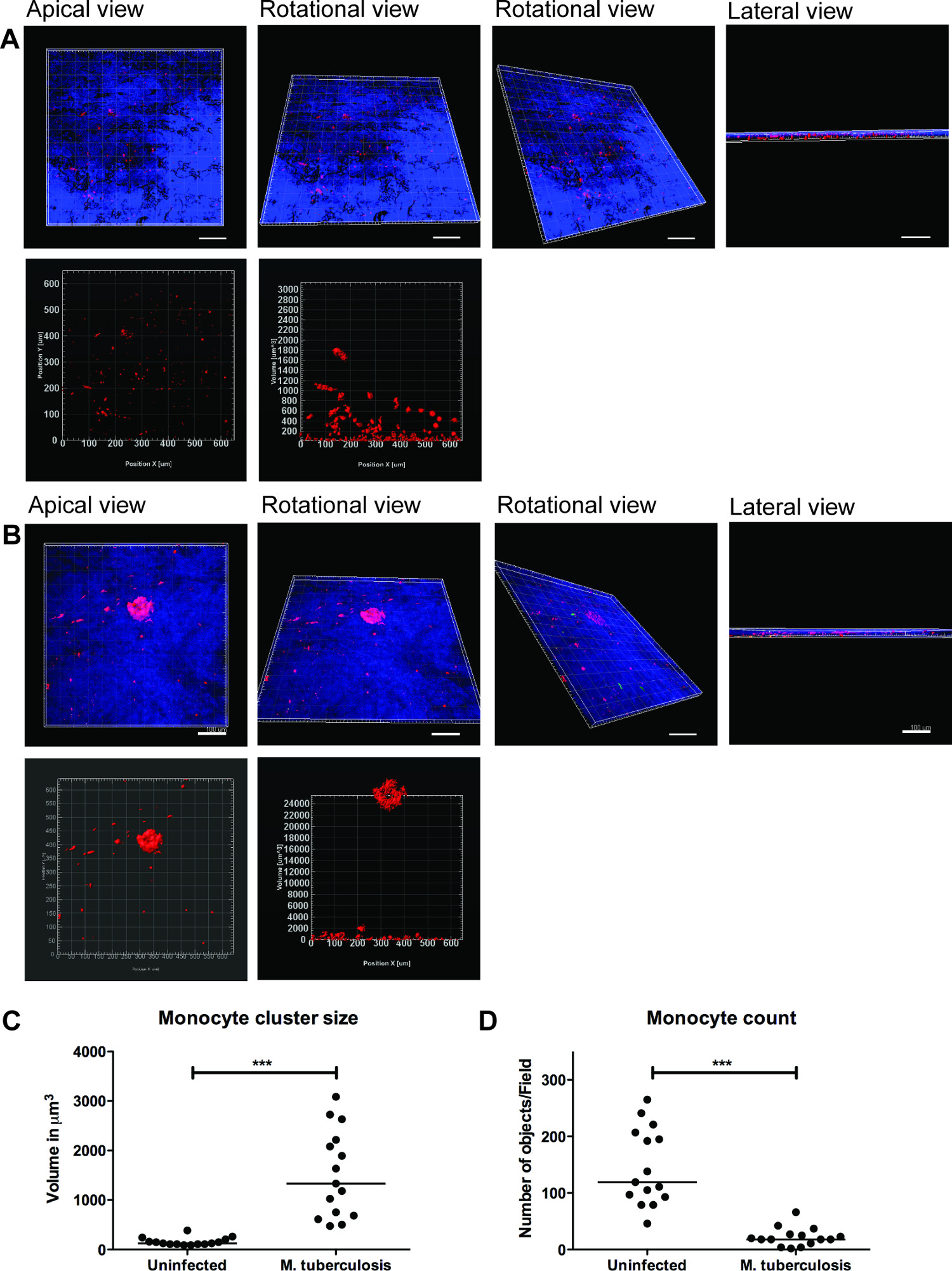
TB感染の監視に肺組織モデルの使用の例は、図2に示されている。M.を視覚化するため結核 -immune細胞遊走との相互作用は、我々 は 、Mに感染したマクロファージを導入しましたGFPを発現する結核組織モデル(核について染色ブルー、DAPI)に合わせて新たに単離したPKH26標識単球(赤)と(緑)。 Mの 7日後の追加について組織モデルに結核に感染した細胞は、共焦点顕微鏡は、人間の結核9の特徴的な病変を模倣感染(緑)のサイト( 図2)、で赤単球のクラスターを明らかにしています。
Mの 3次元可視化のための代表的な一連の画像結核は、細胞クラスターの組織モデルと定量化は、図3に示されているに感染し。3Dビジュアライゼーションは、3D画像にいくつかの機能を、対話調べ、定量化するため、ユーザーの柔軟性を提供します。 M.部位で単球のクラスターを明らかに図3Bに示すように、緑と赤の細菌単球クラスタの空間的配置は、頂端の回転及び側面図から分かるよう結核。クラスター OBませんでした感染していない組織 ( 図3A)で提供しています。個々の単球の数がMに(p <0.01)減少し、我々は、単球細胞集団のサイズと数を定量した (p <0.001)、細胞塊の大きさ(体積)が増強されることを見出しました結核は感染していない組織モデル( 図3Cおよび3D)と比較して組織を感染させました。このデータは、Mの初期の肉芽腫形成の我々の以前の発見を検証します結核感染は、2D組織切片9で分析肺組織モデルで観察しました。
我々のデータは、組織モデルは、M細胞-複雑なホストを調査するために自然な立体生息地を提供していることを示唆しています結核の通信ネットワーク。また、3次元可視化と定量分析は、組織モデル( 図3)の機能を研究するための優れたツールであることがわかりました。細胞塊の定量化(例えば、肉芽腫)、多くの場合、ストレッチいくつかの細胞層のHESとは完全に3次元定量分析によって捕捉することができます。また、モデル内の個々の細胞や細菌の正確な空間的および時間的特徴の可視化は、指定された実験室でのライブイメージング、移動および追跡調査を可能にします。

病原性M.周囲の組織モデルクラスター図2.単球結核。感染していないと、Mの代表的な共焦点画像結核に感染した組織モデルが提示されています。感染していない組織と比較して、パネルの緑色から(M. tuberculosis- GFP)、赤(PKH26標識単球)、青(DAPI染色核)と合併チャネルが感染組織における単球の動員を示します。スケール - 100μmです。large.jpg "スタイル="フォントサイズ:14px;行の高さ:28px; "ターゲット=" _空白 ">この図の拡大版をご覧になるにはこちらをクリックしてください。

図3. 3次元可視化と組織モデルの定量分析は、有用な情報を提供しています。全体の組織モデル(A)非感染組織の3次元可視化の代表的な画像、(B)に感染したM.結核、IMARIS画像処理ソフトウェア(バージョン7.6.8)によりツァイスLSM700共焦点顕微鏡および定量分析を用いた光学セクショニングを介し。これらの画像は20X倍率で取得された、頂端、回転水平、回転、垂直および側面図(AとB)からの可視化を可能にする、1.5μmの間隔で19.5ミクロンの組織の厚さをカバーする14のzスタック。(C)QUA単球細胞クラスターのntitative分析はM.後早期肉芽腫クラスタの増強した(p <0.0001)のサイズを明らかに感染組織で複数のクラスタを繰り返し強調、感染していない組織と比較して、感染の不在に比べ結核感染 。(D)単球数の定量は、感染組織の減少した(p <0.01)を示しました。グリーン- M.結核 -GFP、レッド- PKH26標識単球、ブルー-細胞核、スケール- 。100ミクロンこの図の拡大版をご覧になるにはこちらをクリックしてください。
ディスカッション
The ability to recruit and form organized cell clusters at the site of infection is the hallmark of human TB 11. These dynamic structures known as tubercle granulomas primarily consist of immune cells (macrophages, monocytes, T-cells and B-cells) and multi-nucleated giant cells surrounding M. tuberculosis. The role of the granuloma has long been considered to wall off the infection, preventing local spread of bacteria. However, more recent studies show that granuloma formation is critical for early bacterial survival, growth and dissemination 12. A strategy of new studies is to identify molecules or pathways that could efficiently be targeted to inhibit the cellular migration in granuloma formation and/or TB dissemination.
A caveat for novel studies on TB is the lack of models that recapitulate human TB. The most widely used experimental animals do not form true granuloma upon M. tuberculosis infection, and are therefore not appropriate choices for studies of TB 13-16. Non-human primates have the closest resemblance to human TB 17, but are not the preferred choice owing to high operational costs and ethical issues. Human TB is a complex immunological process and is difficult to model in vitro. Cell cultures of monolayers or co-cultures lack the 3D environment and tissue responses. Therefore, we have developed an innovative lung tissue model based on human primary immune cells and human lung-specific cell lines 8,9. The model displays characteristic features of human lung tissue, including epithelia with evenly integrated macrophages, formation of extracellular matrix, stratified epithelia and mucus secretion 9.
The 3D human lung tissue model has several benefits over the in vitro single or co-cultures seeded on tissue culture plates or transwell inserts. First, the human lung-specific cells (fibroblasts and epithelial cells) are not commonly included in the in vitro single or co-cultures. Second, the immune cells and lung-specific cells are embedded in a 3D physiological context (collagen rich extra-cellular matrix products). The response of cells to a stimulus/infection and the migratory behaviour of cells, for instance formation of a granuloma, differ significantly between a 2D and 3D environment. Furthermore, the described method enables the 3D visualization and robust 3D quantitative analysis that provides pivotal information on spatial distribution and intricate cellular interactions.
Experimental infection in the model tissue with M. tuberculosis resulted in clustering of macrophages at the site of infection, reminiscent of early TB granuloma (Figure 2 and 3). We have recently demonstrated that mutant strains defective in the ability to secrete the virulence factor ESAT-6 or Mycobacterium bovis BCG that lacks ESAT-6 did not induce the clustering of monocytes (no early granuloma), in contrast to the virulent M. tuberculosis 9. These data are consistent with the observations made from Mycobacterium marinum-infected zebrafish embryos, whose transparency allows for elegant live imaging of granuloma formation 12. As there is no gold-standard model for TB, we took advantage of the surgically resected tissue biopsies from TB patients for validation of the method 9. Our in vitro tissue model shares several characteristics with the lung and lymph node biopsies from TB patients, including the aggregation of macrophages in granuloma, the presence of both intra- and extracellular bacteria 18 and induction of necrosis 11.
Although the described model has physiological relevance to human TB and has several advantages over other in vitro models, it has some limitations. For instance, out of more than 20 collagen proteins identified in humans, only type I is included to the model to mimic the extra-cellular matrix. However, type I collagen is a complex mixture of extra-cellular matrix products and is the most abundant collagen in the human body. Further, we have demonstrated the presence of collagen IV and several extra-cellular matrix proteins such as tropoelastin, vimentin and laminin, which are produced by the epithelial cells and fibroblasts in the tissue model, indicating the synthesis of new collagen 8. Presently, the lung tissue model only has monocytes and macrophages, besides lung-specific cells. It lacks neutrophils and lymphocytes that are also known to be present in the granuloma. Remarkably the model is not limited to the introduction of additional immune cells and is of interest to explore how they contribute to the complex cellular interactions in human TB. Implantation of primary alveolar macrophages, skin-specific cells and lung carcinoma cells has already been tested in the model. Since our objective was to use a model that closely resembles human TB, introduction of mouse cells have not been attempted.
In summary, the lung tissue model has implications for both basic mechanistic and applied studies. Potential applications of the lung model include the study of innate immunity, investigating mechanistic aspects of host defences such as phagosomal maturation, autophagy, production of cytokines, chemokines and anti-microbial peptides, and functional characterization of individual cell types. Strikingly, the in vitro tissue model allows manipulation of one or more cells types and provides a relevant tissue micro-environment, not only for studies on TB, but for a variety of infectious and non-infectious diseases that affect the lungs.
開示事項
The authors declare no competing financial interests.
謝辞
The authors acknowledge the Microscopy core facility at the Faculty of Health Sciences, Linköping University for providing access to advanced imaging systems; Karl-Eric Magnusson (Emeritus Scientist) at the Dept. of Clinical and Experimental Medicine, Linköping University for providing access to Imaris 3D/4D image processing software (Bitplane, Switzerland); and S. Braian for his help with the lung model cartoon. This work was supported by funds from the Swedish Research Council (Alternatives to animal research, 2012-1951) and Swedish Research Council (2012-3349) to M.L. and Swedish Foundation for Strategic Research to S.B. S.B. receive grants from the Karolinska Institutet, Swedish Research Council, the Swedish International Development Cooperation Agency (Sida) and the Swedish Civil Contingencies Agency (MSB), and the Swedish Heart and Lung Foundation (HLF). M.S. received grants from the Karolinska Institutet and Stockholm County Council.
資料
| Name | Company | Catalog Number | Comments |
| Cell culture inserts | BD Falcon | 353092 | |
| 6-well culture plates | BD Falcon | 353046 | |
| MRC-5 cells, lung fibroblasts | ATCC#CCL-171 | ||
| 16HBE cells, lung epithelial cells | Gift from Dr. Dieter Gruenert, Mt. Zion Cancer Center, University of California, San Fransisco, USA | ||
| 5 x Dulbecco’s modified Eagle’s medium (5 x DMEM) | Gibco | 12800-082 | Made from powder but add 5 times less water. Adjust pH to 7.3 and filter it using a 0.2 µm filter. |
| Dulbecco’s modified Eagle’s medium with glucose (DMEM) 1x | Gibco | 41965-039 | |
| Minimum Essential Medium (MEM) 1x with Earle’s salts | Sigma | M4655 | |
| Non-Essential Amino Acids Solution, 100x | Life Technologies | 11140-035 | |
| L-glutamine 200 mM (100x) | Gibco | 25030-024 | |
| Sodium Pyruvate | Life Technologies | 11360-039 | |
| NaHCO3 (71.2 mg/ml) | Prepared in house | ||
| Heat inactivated Fetal Bovine Serum (FBS) | Gibco | 10270-106 | Heat inactivated for 30 min, 56 °C |
| Gentamicin (50 mg/ml) | Gibco | 15750-060 | |
| Hepes buffer solution 1M | Gibco | 15630-056 | |
| Penicillin Streptomycin (Pen Strep) | Gibco | 15140-122 | |
| Lymphoprep | Axis-Shield | 7801 | |
| Ultrapure 0.5 M EDTA | Gibco | 15575 | |
| Bovine Collagen PA treated (500 ml) | Organogenesis | 200-055 | |
| Pure col purified Bovine Collagen solution (100 ml) | Advanced biomatrix | 5005-B | |
| Extracellular matrix protein, Fibronectin (1 mg) | BD | 354008 | |
| Primary human monocytes/macrophages | Isolated from human whole blood or buffy coats. | ||
| PKH26 Red fluorescent cell linker | Sigma | MINI26 | |
| Mycobacterium tuberculosis H37Rv expressing green fluorescent protein | M. tuberculosis H37Rv wild type was transformed with the pFPV2 plasmid constitutively expressing GFP. | ||
| Middlebrook 7H9 medium | Difco | 271310 | |
| BBL Middlebrook ADC Enrichment | BBL | 211887 | |
| Tween-80 | |||
| Glycerol | |||
| Kanamycin B sulfate (20 µg/ml) | Sigma | B5264 | |
| Prolong Gold anti=-fade reagent with DAPI | Invitrogen | P36935 | |
| Trypsin -EDTA | |||
| Bovine serum albumin | |||
| Paraformaldehyde | |||
| DAPI | |||
| LSM700 Confocal microscope | Zeiss | ||
| iMaris Scientific 3D/4D image processing software, version 7.6.8 | Bitplane AG |
参考文献
- Sakamoto, K. The pathology of Mycobacterium tuberculosis infection. Veterinary pathology. 49, 423-439 (2012).
- Saunders, B. M., Britton, W. J. Life and death in the granuloma: immunopathology of tuberculosis. Immunol Cell Biol. 85, 103-111 (2007).
- Kaufmann, S. H. New issues in tuberculosis. Ann Rheum Dis. Ann Rheum Dis. 63, ii50-ii56 (2004).
- Morrison, J., Pai, M., Hopewell, P. C. Tuberculosis and latent tuberculosis infection in close contacts of people with pulmonary tuberculosis in low-income and middle-income countries: a systematic review and meta-analysis. Lancet Infect Dis. 8, 359-368 (2008).
- Barry 3rd, C. E., et al. The spectrum of latent tuberculosis: rethinking the biology and intervention strategies. Nat Rev Microbiol. 7, 845-855 (2009).
- Puissegur, M. P., et al. An in vitro dual model of mycobacterial granulomas to investigate the molecular interactions between mycobacteria and human host cells. Cell Microbiol. 6, 423-433 (2004).
- Kapoor, N., et al. Human Granuloma In Vitro Model, for TB Dormancy and Resuscitation. PLoS One. 8, e53657 (2013).
- Nguyen Hoang, ., T, A., et al. Dendritic cell functional properties in a three-dimensional tissue model of human lung mucosa. Am J Physiol Lung Cell Mol Physiol. 302, L226-L237 (2012).
- Parasa, V. R., et al. Modeling Mycobacterium tuberculosis early granuloma formation in experimental human lung tissue. Dis Model Mech. 7, 281-288 (2014).
- Cozens, A. L., et al. CFTR expression and chloride secretion in polarized immortal human bronchial epithelial cells. Am J Respir Cell Mol Biol. 10, 38-47 (1994).
- Brighenti, S., Andersson, J. Local immune responses in human tuberculosis: learning from the site of infection. J Infect Dis. 205, S316-S324 (2012).
- Davis, J. M., Ramakrishnan, L. The role of the granuloma in expansion and dissemination of early tuberculous infection. Cell. 136, 37-49 (2009).
- Gupta, U. D., Katoch, V. M. Animal models of tuberculosis. Tuberculosis (Edinb). 85, 277-293 (2005).
- Kashino, S. S., Napolitano, D. R., Skobe, Z., Campos-Neto, A. Guinea pig model of Mycobacterium tuberculosis latent/dormant infection. Microbes Infect. 10, 1469-1476 (2008).
- Singhal, A., et al. Experimental tuberculosis in the Wistar rat: a model for protective immunity and control of infection. PLoS One. 6, e18632 (2011).
- Subbian, S., et al. Phosphodiesterase-4 inhibition alters gene expression and improves isoniazid-mediated clearance of Mycobacterium tuberculosis in rabbit lungs. PLoS Pathog. 7, e1002262 (2011).
- Lin, P. L., et al. Tumor necrosis factor neutralization results in disseminated disease in acute and latent Mycobacterium tuberculosis infection with normal granuloma structure in a cynomolgus macaque model. Arthritis Rheum. 62, 340-350 (2010).
- Rahman, S., et al. Compartmentalization of immune responses in human tuberculosis: few CD8+ effector T cells but elevated levels of FoxP3+ regulatory t cells in the granulomatous lesions. Am J Pathol. 174, 2211-2224 (2009).
転載および許可
このJoVE論文のテキスト又は図を再利用するための許可を申請します
許可を申請さらに記事を探す
This article has been published
Video Coming Soon
Copyright © 2023 MyJoVE Corporation. All rights reserved