完全に再構成されたCMGの動きを画像化するためのハイブリッドアンサンブルおよび単一分子アッセイ
要約
蛍光標識され、完全に再構成されたCdc45/Mcm2-7/GINS(CMG)ヘリカーゼの動きを光学トラップに保持された直鎖状DNA分子上で直接画像化および定量化するためのハイブリッドアンサンブルおよび単一分子アッセイを報告します。
要約
真核生物には、CMGとして知られる複製ヘリカーゼが1つあり、これがレプリソームを中央に整理して駆動し、複製フォークの先頭で道をリードしています。CMGのダイナミクスを深くメカニズム的に理解することは、細胞が細胞サイクルごとに全ゲノムを効率的かつ正確に複製するという膨大なタスクをどのように達成するかを解明するために重要です。単一分子技術は、その比類のない時間的および空間的分解能により、CMGのダイナミクスを定量化するのに特に適しています。それにもかかわらず、CMG運動の単一分子研究は、これまで複合体として細胞から精製された事前に形成されたCMGに依存しており、その活性化に至るまでのステップの研究が不可能でした。ここでは、36種類の精製された 出芽酵母 ポリペプチドからの合成と活性化を完全に再構成した後、蛍光標識CMGの運動を1分子レベルでイメージングすることを可能にしたハイブリッドアンサンブルおよび単一分子アッセイについて説明します。このアッセイは、直交する2つの結合部分を持つ直鎖状DNA基質の末端の二重官能基化に依存しており、1分子レベルで同様に複雑なDNAプロセッシングメカニズムの研究に適合させることができます。
概要
真核生物のDNA複製は、レプリソーム1として知られる動的タンパク質複合体によって行われます。この複合体の主要な構成要素は、真核生物の複製ヘリカーゼCdc45/Mcm2-7/GINS(CMG)であり、これはレプリソームを駆動して中央に組織化し、レプリケーションフォーク1,2の先頭をリードしています。したがって、CMGのダイナミクスを深く定量的に理解することは、レプリソームのダイナミクスを理解するために重要です。このような理解は、比類のない空間的および時間的分解能により、CMGなどの分子モーターの研究に特に適した単一分子技術で獲得でき、その機能、確率性、およびダイナミクスの比類のない定量的理解を提供することができます2,3,4,5,6,7,8,9。
In vivoでは、CMGは時間的に分離された方法でロードされ、活性化され、複製が細胞周期1,10,11ごとに1回だけ起こることを確実にします。まず、細胞周期のG1期において、ローディングファクターとして知られる一組のタンパク質が、CMGの第1の構成要素であるMcm2-7ヘキサマー複合体をdsDNA12,13,14,15,16に、ヘッド・トゥ・ヘッド構成の二重ヘキサマーの形でロードする15,17,18 .酵母の特定のケースでは、この初期プロセスは、複製の起源1として知られる特定のDNA配列で発生します。Mcm2-7は複製ヘリカーゼの運動コアであるが、それ自体では、完全に活性なCMG11,19,20,21を生じさせるためにロードされたMcm2-7に動員する必要がある2つのヘリカーゼ活性化因子Cdc45およびGINSなしではDNA19を巻き戻すことができない。ヘリカーゼ活性化のプロセスは、細胞周期のS期に起こり、細胞周期調節キナーゼDDK22,23,24によるMcm2-7ダブルヘキサマーの選択的リン酸化から始まります。これらのリン酸化事象は、発火因子10,11,26として知られるタンパク質の第2のセットによって、Cdc45およびGINSのMcm2-7二重六量体10,22,23,24,25,26への動員を促進する.Cdc45とGINSの結合は、2つの姉妹CMGヘリカーゼを生じさせ、これらは最初に親DNAの両方の鎖を囲み、依然として直接対向する構成で位置している11,27。最終活性化ステップでは、発火因子Mcm10が、各姉妹CMG11からの1本のDNA鎖のATP加水分解依存的な押し出しを触媒する。鎖の押し出し後、姉妹のCMGヘリカーゼは、ATP加水分解依存的な方法でssDNAに沿って転座することにより互いに迂回して分離し(11,20,21,28)、非転座鎖29を立体的に排除してDNAを巻き戻す。この全過程は、精製された出芽酵母ポリペプチド10,11の36個の最小セットからin vitroで完全に再構成されている。
上述のCMGの集合および活性化のin vivoでの精巧な調節にもかかわらず、CMG2,30,31,32,33,34のin vitro再構成単一分子運動研究は、これまで細胞20,21から複合体として精製された活性化済みCMGに依存してきた、そのアクティブ化前のすべてのステップとそのモーションの双方向性が欠落しています。この事前に活性化されたCMGアプローチは、完全に再構成されたCMGアセンブリ反応の生化学的複雑さに一部起因して、単一分子分野のゴールドスタンダードとなっています10,11。この生化学反応は、いくつかの理由から、バルク生化学レベルから単一分子レベルに変換するのが困難でした。まず、反応効率を最大化するために、CMGの組み立てと活性化に必要なローディングファクターと燃焼ファクターが、10〜200 nM 10,11,27の範囲の濃度で必要になります。これらの濃度範囲は、特に蛍光標識された成分35を使用する場合に、ほとんどの単一分子技術が許容できる上限に対応する。最後に、CMGはセル36,37,38,39内の数千の塩基対を巡航するように進化しました。したがって、生物学的に関連性のある空間スケールでその運動を研究するためには、長いDNA基質(典型的には数十キロベースのオーダーの長さ)30,31,34,40,41,42が必要である。このような長いDNA基質を用いると、DNA基質が長ければ長いほど、タンパク質やタンパク質凝集体の非特異的結合部位が有する可能性が高くなるという課題が生じます。CMGの場合、後者の点が特に重要であり、なぜなら、CMGの集合および活性化に関与するローディングおよびファイアリング因子のいくつかは、本質的に無秩序な領域43を含み、凝集しやすいからである。
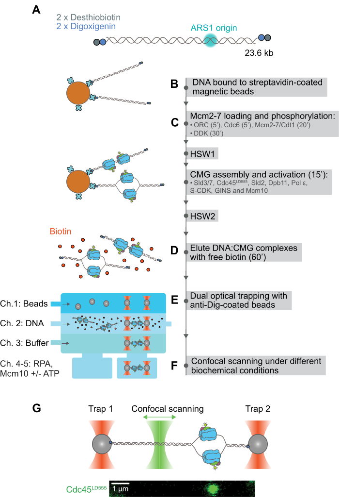
ここでは、36精製された 出芽酵母 ポリペプチド28からのCMGのアセンブリと活性化を完全に再構成した後のCMGの動きの観察と定量を可能にしたハイブリッドアンサンブルと単一分子アッセイを報告する。このアッセイは、デスチオビオチンとジゴキシゲニン2 の2つの直交結合部分を持つDNA基質の両端の二重機能化に依存しています(図1A)。第1の部分であるデスチオビオチンは、DNA基質をストレプトアビジン被覆磁気ビーズ44 (図1B)に可逆的に結合するために使用される。これに続いて、蛍光標識されたCMGをビーズ結合DNA上に組み立てて活性化し、得られた磁気ビーズ結合DNA:CMG複合体を磁気ラックで精製および洗浄します(図1C)。そうすることで、そうでなければDNA基質上に凝集する余分なタンパク質が除去されます。これにより、実質的に凝集しないDNA:CMG複合体が得られます。その後、モル酸の過剰の遊離ビオチンを添加することにより、磁気ビーズから無傷の錯体が溶出され、デスチオビオチン-ストレプトアビジン相互作用に対抗することができます(図1D)。次に、個々のDNA:CMG複合体を、抗ジゴキシゲニン抗体(抗Dig)でコーティングした2つの光学的にトラップされたポリスチレンビーズの間に結合します。このステップでは、DNAの2番目の部分であるジゴキシゲニンが、遊離ビオチンを過剰に含む緩衝液でも抗Digに結合できるため、使用します(図1E)。DNA:CMG複合体が光学トラップの所定の位置に保持されると、蛍光標識されたCMGの動きが共焦点走査レーザーで画像化されます(図1F)。このアッセイは、同様に複雑なDNA:タンパク質相互作用の単一分子レベルでの研究に容易に適応できると期待しています。
プロトコル
1. 二重官能基化直鎖状DNA基質の合成と磁気ビーズへの結合
- デスチオビオチンおよびジゴキシゲニン部分を有するDNA基質の二重官能基化
- 20 μg の 23.6 kb プラスミド pGL50-ARS1 (天然の ARS1 複製起点を含み、社内でクローニングされ、ご要望に応じて入手可能) を 200 μg の制限酵素 AflII で 37 °C で 16 時間、最終容量 200 μL の 1x バッファー (50 mM 酢酸カリウム、20 mM トリス酢酸、10 mM 酢酸マグネシウム) を直鎖化します。 100 μg/ml BSA、pH 7.9)。
注:このステップは、より便利な場合は、歩留まりを低下させることなく4時間に短縮できます。 - 反応液を65°Cで20分間インキュベートすることにより、AflIIを不活性化します。得られた 4-ヌクレオチド TTAA は、200 μL の直鎖化反応に 60 ユニットの Klenow Fragment (3' to 5' exo-) ポリメラーゼ、17 μL の 10x バッファー (500 mM NaCl、100 mM Tris-HCl、100 mM MgCl2、10 mM DTT、pH 7.9)、33 μM D-デスチオビオチン-7-dATP、33 μM ジゴキシゲニン-11-dUTP、33 μM dCTP、33 μM dGTP、100 μL を添加して、オーバーハングを平滑化します。 超純水の最終容量370 μMまで、反応液を37°Cで30分間インキュベートします。
注:このステップでは、Klenow Fragmentは、各末端に2つのD-デスチオビオチン-7-dATPヌクレオチドと2つのジゴキシゲニン-11-dUTPヌクレオチドを組み込むことにより、直鎖状DNAの両端のオーバーハングを鈍化します。 - 反応液に10 mM EDTAを添加し、反応液を75°Cで20分間インキュベートしてKlenow Fragmentを不活性化します。超純水で反応量を400μLにします。
- 4本のS-400スピンカラムをボルテックスし、少なくとも30秒間ボルテックスして樹脂を再懸濁した後、735 x g で1分間遠心分離して保存バッファーを除去します。カラムを洗浄した1.5 mLチューブに移します。
- 直ちに、各カラムに100 μLのDNA溶液を加え、735 x gで2分間遠心分離します。DNAはフロースルー中にあり、カラムは廃棄できます。
- 4本のカラムのフロースルーをプールし、ピペットで容量を測定し、分光光度計でDNA濃度を測定します。これは、ビーズに結合したDNAの量を計算するために使用されます。
- 20 μg の 23.6 kb プラスミド pGL50-ARS1 (天然の ARS1 複製起点を含み、社内でクローニングされ、ご要望に応じて入手可能) を 200 μg の制限酵素 AflII で 37 °C で 16 時間、最終容量 200 μL の 1x バッファー (50 mM 酢酸カリウム、20 mM トリス酢酸、10 mM 酢酸マグネシウム) を直鎖化します。 100 μg/ml BSA、pH 7.9)。
- 二重官能基化直鎖DNAのストレプトアビジン被覆磁気ビーズへの結合
- Vortex M-280ストレプトアビジンコーティングされた磁気ビーズを30秒間使用して再懸濁します。
- 再懸濁したM-280ストレプトアビジン被覆磁気ビーズ4 mgを清潔な1.5 mLチューブに移します。チューブを磁気ラックに置き、ビーズが収集されるまで1分間待ってから、保存バッファーを取り外します。
- 1xバッファーA(5 mM Tris-HCl pH 7.5、0.5 mM EDTA、および1 M NaCl)1 mLをビーズに加え、5秒間ボルテックスして再懸濁します。ビーズを室温で5分間インキュベートします。
注:すべてのバッファーは滅菌ろ過する必要があります。時間を節約するために、事前に2xバッファA、1xバッファB(下記参照)、および1xバッファC(下記参照)を大量に作成してください(推奨)。これらのバッファーは、磁気ビーズのメーカーが推奨するもので、-20°Cで保存できます。 - チューブをマグネットホルダーに入れ、ビーズが収集されるまで1分間待ってから、バッファーAを取り出します。
- ピペッティングによりビーズを400 μLの2倍バッファーAに再懸濁し、次に400 μLの官能基化DNA溶液を加えます(これにより、2倍バッファーAがビーズへのDNA結合に最適な最終濃度1倍に希釈されることに注意してください)。ピペッティングで穏やかに混合します。
- ビーズ/DNA混合物を4°Cで一晩インキュベートし、エンドオーバーエンド回転させて、機能化されたDNAがビーズに結合できるようにします。
- マグネティックラックを使用して上清を除去し(ピペットでその体積を測定し、分光光度計で未結合DNAの濃度を測定する前に廃棄しないでください)、ビーズを500μLのバッファーB(10 mM HEPES-KOH pH 7.6、1 mM EDTA、および1 M KOAc)で2回洗浄して、非特異的に結合したDNAを除去します。メーカーが推奨する保存バッファーであるバッファーC(10 mM HEPES-KOH pH 7.6および1 mM EDTA)500 μLでビーズを2回洗浄します。
- ビーズに添加されたDNAの総量と上清に残ったDNAの量を比較することにより、磁気ビーズに結合したDNAの総量を計算します。収率は、磁気ビーズ1mgあたり2.3-2.9mgのDNA(~150-190fmol)の範囲である必要があります。収量が低い場合は、S400カラムが乾燥していないことを確認してから使用してください。ゲル電気泳動によりDNAの完全性を常に監視し、初期のプラスミドDNA基質が分解されていないことを確認します。
- ビーズを300 μLのバッファーCに再懸濁し、4°Cで保存します。 ニッキングを防ぐために、1 mgの磁気ビーズの使い捨てアリコートを4つ作成し、DNAを2週間以上保存しないでください。
2. アンサンブルと1分子のハイブリッドアッセイにより、相関デュアルビーム光ピンセットと共焦点顕微鏡法を用いて、完全に再構成されたCMGの動きを画像化し、定量化します。
- 磁気ビーズ結合DNA(図1C).
注:CMGの組み立てと活性化反応は、Mcm2-7のローディングとリン酸化、およびCMGの組み立てと活性化の2段階で行われました。特に指定がない限り、すべてのバッファー交換ステップは、ビーズを磁石で1分間収集し、その後上清を除去することにより、磁気ラックの助けを借りて行われました。すべてのインキュベーションは、蛍光タンパク質の光退色を防ぐために、蓋付きの温度制御されたヒートブロックで行われました。蓋が利用できない場合は、チューブを錫箔で覆う必要があります。このプロトコルで使用されるすべてのタンパク質の精製および蛍光標識は、前述のように実施されています10,11,28.- Mcm2-7のローディングとリン酸化
- DNA結合ストレプトアビジン被覆磁気ビーズ1mgを取り、保存バッファー(バッファーC)を取り出します。
- ビーズを200 μLのローディングバッファー(25 mM HEPES-KOH pH 7.6、100 mM Kグルタミン酸、10 mM MgOAc、0.02% NP40代替品、10%グリセロール、2 mM DTT、100 μg/mL BSA、および5 mM ATP)で洗浄します。
- ローディングバッファーを取り外し、ビーズを75 μLのローディングバッファーに再懸濁します。ピペッティングで穏やかに混合します。
- ビーズ結合DNAに最終濃度37.5 nMのORCを添加し、800 rpmの攪拌を行いながら30°Cで5分間反応をインキュベートします。
- Cdc6を最終濃度50 nMで添加し、800 rpmの攪拌を行いながら30°Cで5分間反応をインキュベートします。
- 最終濃度100 nMでMcm2-7/Cdt1(または蛍光標識されたMcm2-7JF646-Halo-Mcm3/Cdt1)を添加し、800 rpmの攪拌で30°Cで20分間反応をインキュベートします。
- 最終濃度100 nMでDDKを添加し、800 rpmの攪拌を行いながら30°Cで反応を30分間インキュベートします。
- 上清を取り除き、ビーズ結合DNA(リン酸化Mcm2-7ヘキサマーを含む)を200 μLの高塩洗浄(HSW)バッファー(25 mM HEPES-KOH pH 7.6、300 mM KCl、10 mM MgOAc、0.02% NP40代替物、10%グリセロール、1 mM DTT、および400 μg/mL BSA)で洗浄します。ピペッティングで混合し、ビーズがバッファー内に完全に再懸濁され、凝集物が見えないことを確認します。
- HSWバッファーを取り外し、ビーズを200 μLのCMGバッファー(25 mM HEPES-KOH pH 7.6、250 mM Kグルタミン酸、10 mM MgOAc、0.02% NP40代替品、10%グリセロール、1 mM DTT、および400 μg/mL BSA)で1回洗浄します。
- 蛍光標識CMGの組み立てと活性化
- CMGバッファーを取り出し、DNA結合ビーズを5 mM ATPを添加した50 μLのCMGバッファーに再懸濁します。
- ビーズ結合DNAに50 nM Dpb11、200 nM GINS、30 nM Polɛ、20 nM S-CDK、50 nM Cdc45LD555、30 nM Sld3/7、55 nM Sld2、および10 nM Mcm10を添加します。このステップでは、DNAにタンパク質を添加する直前に、すべてのタンパク質を1つのチューブで混合し、氷の上に置きます。再懸濁したビーズ結合DNAをタンパク質ミックスに加えます。反応を30°Cで15分間インキュベートし、800rpmの撹拌を行います。
- ビーズを200μLのHSWバッファーで3回洗浄します。ビーズを200 μLのCMGバッファーで1回洗浄します。
- 磁気ビーズからのインタクトDNA:CMG複合体の溶出(図1D)
- CMGバッファーを磁気ビーズから取り出し、CMG含有DNA結合磁気ビーズを200 μLの溶出バッファー(10 mMビオチンを添加したCMGバッファー)に再懸濁して、磁気ビーズからDNA:CMG複合体を溶出します。800 rpmの撹拌を行いながら、室温で1時間インキュベートします。
- チューブを磁気ラックに入れ、ビーズを5分間収集します。上清(溶出されたDNA:CMG複合体を含む)を沈殿したビーズを乱さずに慎重に収集し、新しいチューブに移します。
- 溶液中にビーズが残らないようにするため(溶液中に残った磁気ビーズはアッセイの光トラップ部分に干渉する可能性があるため)、回収した上清を再び磁気ラックに置き、残ったビーズをさらに5分間回収します。上清を慎重に収集し、新しいチューブに移します。
- 200 μLの上清に1400 μLのCMGバッファーを加えます。これで、サンプルを1分子イメージングする準備が整いました。
- Mcm2-7のローディングとリン酸化
- 相関デュアルビーム光ピンセットと共焦点顕微鏡法(図1E-G).
注:アッセイの単一分子部分は、デュアルビーム光ピンセットと共焦点顕微鏡およびマイクロ流体工学を組み合わせた商用セットアップで実施されます45,46 (図1E-G)ですが、自作のセットアップで実施することもできます。ここで使用される商用セットアップには、5つの入口(それぞれチャネル1〜5と呼ばれる)と1つの出口(図1E).以下の手順では、すべてのボタンやパネルの名前は、この商用セットアップで提供されるソフトウェアに固有のものです。- 市販のセットアップの5つのインレットを次のように使用します:左からチャネル1〜3を注入し、ビーズトラップ(チャネル1)、DNA-タンパク質複合体の結合(チャネル2)、およびCMGの存在の確認(チャネル3)に使用します。チャネル4および5は、タンパク質リザーバーおよびバッファー交換位置として使用します。各実験の前に、1 mg/mL のウシ血清アルブミンでマイクロ流体フローセルを少なくとも 30 分間不動態化し、続いて 0.5% プルロニック F-127 を超純水に溶解します。
- 各実験の各チャネルの内容が説明どおりであることを確認します (図 1E)。
チャンネル1:抗ジゴキシゲニン被覆ポリスチレンビーズ(直径2.06μm)をPBSで1:50に希釈。
チャンネル2:DNA:磁気ビーズから溶出するCMG複合体。
チャンネル3:イメージングバッファー(CMGバッファーに2 mM 1,3,5,7シクロオクタテトラエン、2 mM 4-ニトロベンジルアルホホール、および2 mM Troloxを添加);
チャンネル4および5:25 nM RPA、10 nM Mcm10、および5 mM ATP、5 mM ATPγS、またはヌクレオチドなしのいずれかを添加したイメージングバッファー。
- 各実験の各チャネルの内容が説明どおりであることを確認します (図 1E)。
- 実験の前に、ソフトウェアのパワーコントロールパネルにあるRe-calibrateボタンをクリックして、両方のトラップ33,46で0.3 pN / nmの剛性を達成するためにトラッピングレーザー出力を調整します。ソフトウェアのImage Scanパネルで、共焦点ピクセルサイズを50 x 50 nm、画像サイズを160 x 18ピクセル、ピクセルあたりの照明時間を0.2 ms、フレームレートを5秒に設定します。[温度]パネルで温度制御を30°Cに設定します。
注:さらに、最大ステージ速度(チャネル間の移動に使用)を0.2 mm/sに設定して、抗力によるDNAからのCMGの解離を最小限に抑えることをお勧めします。 - 注入ポートで0.5 barの一定圧力ですべての溶液をフローセルに流します(これはソフトウェアの圧力バーパネルで設定できます)。次に、チャネル 4 と 5 のフローをオフにします。最初にすべての溶液を流した後、圧力を0.2バールに下げます。
- トラップレーザーをチャネル1に移動し、各光トラップに1つのビーズが引っかかるまで移動させます。トリガーを押しながらジョイスティックを動かして、トラップされたビーズをチャンネル2に移動します。次いで、DNAテザーが捕捉されるまで、引き金を押さずにジョイスティックを使用して右のビーズを左のビーズに近づけたり遠ざけたりすることにより、DNA:CMG複合体を釣る(これは、ソフトウェアのFDビューアパネルでトラップされたDNA47 の力-伸長曲線を監視することによって知られている)。
- DNAテザリングの場合は、DNAの長さと正しい温度値をReference Modelパネルに入力すると、FD ViewerパネルにDNAコンストラクトの理論的な拡張可能なワーム様鎖モデルが自動的にプロットされます。1つのDNA分子が2つのビーズの間に挟まれている場合、DNAの力-伸長挙動は、プロットされた拡張可能なワーム様鎖モデルと密接に一致するはずです。これが当てはまることを確認するには、実験的な力-伸長曲線を監視し、ソフトウェアが理論上の曲線の上に自動的にプロットします。
注:理論モデルからの逸脱は、ビーズ間に結合した複数のDNA分子の存在、および/またはDNAに結合して圧縮するタンパク質凝集体の存在を示している可能性があります。力によるタンパク質のDNAからの解離を防ぐために、この初期試験では10pNの張力を超えないようにしてください。
- DNAテザリングの場合は、DNAの長さと正しい温度値をReference Modelパネルに入力すると、FD ViewerパネルにDNAコンストラクトの理論的な拡張可能なワーム様鎖モデルが自動的にプロットされます。1つのDNA分子が2つのビーズの間に挟まれている場合、DNAの力-伸長挙動は、プロットされた拡張可能なワーム様鎖モデルと密接に一致するはずです。これが当てはまることを確認するには、実験的な力-伸長曲線を監視し、ソフトウェアが理論上の曲線の上に自動的にプロットします。
- ビーズをチャネル3に移動し、すべてのチャネルで流れをすぐに停止します。チャンネル3で1フレームのテストスキャンを行い、CMGの存在を確認し、対物レンズで測定した4μWのパワーで561nmレーザーで照らします。Excitation lasersパネルでイメージングレーザーの出力を調整します。存在する場合、CMG は 、図 1G と 図 2A に示すように、2 次元回折限界スポットとして表示されます。
- DNAを捕捉できない場合は、チャネル2に気泡がないか確認し、チャネル2の流れを遅くする可能性があります(チャネル1および3と比較して)。気泡が存在する場合は、チャネル2の圧力を1バールに上げて、気泡を洗い流してみてください。
- CMGが存在する場合は、DNAテザーをチャネル4または5に移動してイメージングします。それ以外の場合は、フローを再びオンにし、ビーズを破棄して、ステップ 2.2.4 に戻ります。
- チャネル4または5で、ビーズ間の距離を調整して、DNAテザーの初期張力を2pNにします。このためには、Force Spectroscopyパネルに2 pNを入力し、Enableボタンをクリックしてフォースクランプを開始します。初期張力が2 pNに達したら、イメージング前にフォーススペクトロスコープパネルの[Enable]ボタンをクリックして、フォースクランプを無効にします。
- 対物レンズで測定した4μWの出力で561nmレーザーを使用して、CMGCdc45-LD555 を5秒ごとに画像化します(図1F)。これを行うには、イメージスキャンパネルの[スキャンの開始]ボタンをクリックします。このようなスキャンでは、CMGは 図1Gの例のように、2次元の回折限界スポットとして表示されます。
- CMGが観察されたが、漂白前に動かないように見える場合は、DNA上の傷がCMGをDNA41から解離させ、その見かけの処理能力を大幅に低下させるため、ヌクレアーゼ活性に使用されるすべてのタンパク質をテストしてください。
注:ここで使用する市販の顕微鏡を使用する場合、4μWのレーザー出力で一貫した結果が得られるはずです。自作のセットアップを使用する場合は、最適なレーザー出力を経験的に見積もることをお勧めします。最適なレーザー出力は、蛍光色素分子から目的の精度で局在化するのに十分な信号を提供し、蛍光色素分子の寿命が実験の目的の時間範囲に近いものでなければなりません。
注:この出版物は、ハイブリッドアンサンブルおよび単一分子アッセイに焦点を当てているが、我々は以前に、このアッセイ48で生成されたデータの分析の包括的な説明を発表している。
- CMGが観察されたが、漂白前に動かないように見える場合は、DNA上の傷がCMGをDNA41から解離させ、その見かけの処理能力を大幅に低下させるため、ヌクレアーゼ活性に使用されるすべてのタンパク質をテストしてください。
- 市販のセットアップの5つのインレットを次のように使用します:左からチャネル1〜3を注入し、ビーズトラップ(チャネル1)、DNA-タンパク質複合体の結合(チャネル2)、およびCMGの存在の確認(チャネル3)に使用します。チャネル4および5は、タンパク質リザーバーおよびバッファー交換位置として使用します。各実験の前に、1 mg/mL のウシ血清アルブミンでマイクロ流体フローセルを少なくとも 30 分間不動態化し、続いて 0.5% プルロニック F-127 を超純水に溶解します。
結果
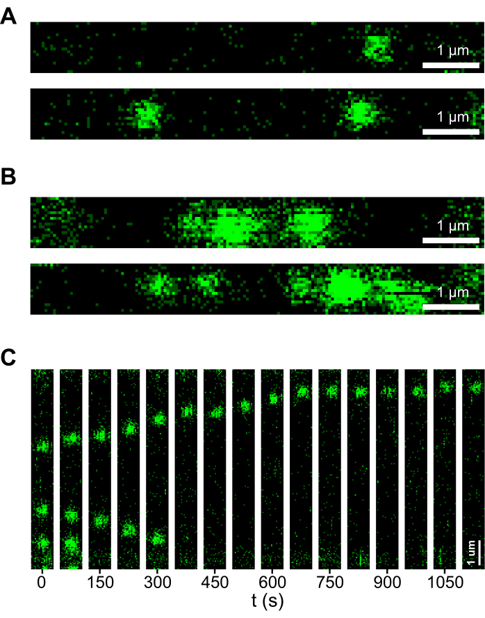
正しく実施された場合、ここで説明するプロトコルは、実質的に凝集しないDNA:CMG複合体をもたらすはずです。凝集のない反応は、マイクロ流体フローセルのチャネルを詰まらせてはならず、DNAを壊すことなく、トラップされたDNA分子をその輪郭長の10%以内でエンドツーエンドの伸長まで引き伸ばすことができるはずです。逆に、反応に凝集があると、DNA分子が凝集体によって圧縮されることがあり、引き伸ばすとDNAが切断されることがよくあります。タンパク質凝集体を認識する良い方法は、DNAの2Dスキャンを見ることです。凝集のない反応では、CMGは、 図2Aに示すスキャンのように、DNAにまばらに密集した離散的で対称的な回折限界スポットとして現れます。それどころか、凝集体は離散性が低く、 図2Bに示すスキャンのように、DNAのより長い長さに密集する非対称のブロブが発生することがあります。さらに、アッセイが成功裏に実施され、精製されたタンパク質の高純度が達成された場合、 図2Cに示すキモグラフで観察されるように、ATPの存在下でのCMGの長距離運動が見られます。

図1:完全に再構成されたCMGの動きを画像化および定量するためのハイブリッドアンサンブルおよび単一分子アッセイの図解的説明(A)自然に発生するARS1複製起点を含む23.6 kbの直鎖DNAは、デスチオビオチンおよびジゴキシゲニン部分で両端が二重に官能化されています。(B)二重官能基化されたDNAは、そのデスチオビオチン部分を通じてストレプトアビジン被覆磁気ビーズに結合している。(C)CMGは、磁気ビーズ結合DNA上に段階的に組み立てられ、活性化され、過剰な未結合タンパク質およびタンパク質凝集体を除去するために異なる洗浄ステップが含まれています。(D)インタクトDNA:CMG複合体は、過剰な遊離ビオチンを添加することにより磁気ビーズから溶出され、デスチオビオチン-ストレプトアビジン相互作用を凌駕します。(E)個々のDNA:CMG複合体は、マイクロ流体フローセルの助けを借りて、2つの抗ジゴキシゲニン被覆光学トラップポリスチレンビーズの間に結合しています。なお、Dig-anti-Dig相互作用はビオチン-アビジン相互作用と直交しているため、遊離ビオチンの存在による影響を受けません。(F)光ピンセットで所定の位置に保持されると、DNA:CMG複合体を異なる緩衝条件に移し、DNA平面を共焦点走査レーザーでスキャンして、DNAに沿ったCMGの動きを経時的に画像化します。(G)トップパネルは、共焦点走査レーザーによってスキャンされている2つの光学的にトラップされた抗Digコーティングポリスチレンビーズの間に保持されているDNA:CMG複合体の図を示しています。下部のパネルは、光学トラップで所定の位置に保持されたDNAに結合したCMGの2Dスキャンの例を示しています。これらの実験ではDNAは標識されていませんが、画像の中央を通る水平線と考えることができます。この数値は平成28年度から修正されています。この図の拡大版を表示するには、ここをクリックしてください。

図2:成功した実験と失敗した実験のデータの例。 (A)例:アグリゲーションフリーサンプル中のCMG含有DNAの2Dスキャン。どちらのスキャンでも、CMGはDNAに沿ってまばらに分布した対称的で離散的な回折限界スポットを示しています。(B)例:凝集体を含むサンプル中のCMG含有DNAの2Dスキャン。どちらのスキャンでも、CMGはDNAを密集させる対称性の低いブロブを形成します。(C)ATPの存在下でCMG回折限界スポットのDNA上の位置を経時的に示し、CMG複合体の長距離運動を示すキモグラフ。 この図の拡大版を表示するには、ここをクリックしてください。
ディスカッション
重要なステップと重要な試薬品質チェック
ここでは、アッセイにおける重要なステップと生物学的試薬の品質チェックに焦点を当てています。まず、タンパク質サンプル中の小さなヌクレアーゼ汚染物質によって引き起こされるDNA分解でさえデータに悪影響を与えるため、使用するタンパク質の純度が重要です。これは、デュアルビーム光ピンセットに閉じ込められるのは、無傷の(または部分的に傷のある)DNA分子だけであるためです。さらに重要なことに、DNA上の傷はCMGを解離させ、CMGの長距離運動の観察を複雑にします41。精製された各タンパク質のヌクレアーゼ活性を試験するとともに、開始プラスミド基質の完全性を常に監視して、ニッキングが最小限に抑えられていることを確認することを強くお勧めします。2つ目の重要なステップは、DNA:CMG複合体の溶出後に磁気ビーズを慎重に除去することです。このステップでの上清の除去は、収集したビーズを乱さないようにゆっくりと行う必要があります。光ピンセットに飛ばされたサンプルに磁気ビーズが残っていると、光学的にトラップされたポリスチレンビーズにぶつかることが多く、光トラップから逃れてデータ取得が複雑になります。最後に、DNA:CMG複合体は光ピンセットで慎重に取り扱う必要があります。この目的のために、力を加えるとDNAからCMGが解離する可能性があるため、DNAの張力を10pN以上に増加させないことをお勧めします。さらに、マイクロ流体フローセル内のチャネル間の移動は、結果として生じる抗力がDNAからCMGを解離するのを防ぐために、できるだけゆっくりと(~0.2 mm/s)行う必要があります。
メソッドの変更
アッセイには、変更可能ないくつかのステップがあります。例えば、溶出時間を60分から30分に短縮しても、溶出収率に大きな影響を与えることはないことを示しました。さらに、CMGがDNA末端から拡散するのを防ぎ、CMG28を一般的に安定化させるために、低濃度(1 mM未満)のATPまたはATPγSを溶出バッファーに添加することをお勧めします。さらに、ここで報告する緩衝液組成物およびタンパク質濃度は、以前のアンサンブル生化学的および単一分子研究11,18で採用されたものに基づいているが、我々が説明するアッセイは、CMG26,27を組み立てるための他のプロトコルと完全に互換性がある。したがって、CMGのアセンブルまたは活性化の効率を向上させると報告されている生化学的進歩は、収量を増やすためにアッセイのバルク部分に実施することができ、また実施すべきです。最後に、フレーム間の時間を長くすると、CMGを画像化できる合計時間が長くなり、蛍光色素の漂白前の長距離CMG運動の観察が容易になります。
方法の制限
ここで説明するハイブリッド法は、CMGを活性化した後に一括でしか画像化できないという点で制限されています。CMGの活性化をリアルタイムで観察するには、さらなる研究が必要になります。もう1つの重要な制限は、CMGがペア17,26,27に組み立てられると予想される一方で、観察されるDNAあたりのCMG複合体の総数は大部分が1つである28であり、CMG、または少なくともCdc45が敏感なDNA:CMG複合体の取り扱い中にDNAから解離していることを示唆している。単一分子イメージングの前の取り扱いステップ数を減らし、マイクロ流体フローセルのプラスチックチューブとガラスのより良いパッシベーション戦略を開発することで、この収率を向上させることができます。
この方法の意義
CMGの単一分子運動研究では、これまで細胞から複合体として精製された活性化済みCMGが用いられてきました。比較的単純ですが、この事前に活性化されたCMGアプローチは、CMGの活性化に至るまでのステップを見逃すだけでなく、CMGの双方向性やレプリソームモーションを見逃すという点で制限されています。一方、CMGの組み立てと活性化の完全な再構成は、活性化前のステップを研究するだけでなく、CMGの動きを双方向で研究する可能性があります。それにもかかわらず、このアプローチは、より多くの精製されたタンパク質因子とステップを含むため、バルク生化学レベルから単一分子レベルに変換するのが困難です。ここで説明するアッセイは、完全に再構成されたCMGの動きを単一分子レベルで画像化することを可能にし、以前に見逃されていた活性化前のダイナミクス28にアクセスできるようにすることで、これらの課題を克服するのに役立ちました。さらに、DNAごとに1つのCMGが見られることがほとんどですが、2つのCMG複合体が反対方向に動いているいくつかのインスタンスを観察することができ、姉妹CMGの互いにの初期分離を捉えることさえできました28、双方向複製の確立に関するいくつかの洞察を提供しました。
以前のCMGモーションと比較したこのアッセイのもう一つの利点は、私たちが採用するDNA基質の完全な二本鎖の性質にあります(図1A)。以前の活性化CMG研究では、CMGをDNA基質に結合する最も一般的な方法は、3' ssDNAフラップを使用することです。これにより、ねじれ制約が容易になり得ないDNA構築物が得られ、したがって、レプリソーム進行におけるスーパーコイルの役割の研究が禁止されます。逆に、ここで説明する新しいアプローチは、使用されるDNA基質が完全に二本鎖であるため、このプロセスにおけるトルクの役割を研究するために適応できる可能性があります。
このメソッドの幅広いアプリケーション
記載されているハイブリッドアッセイは、完全な真核生物のレプリソームの完全な再構成への道を開き、私たちや他の人々がレプリソームがすべての異なるタスクで成功できるようにする重要なダイナミクスを観察し、定量化することを可能にします。DNA複製はさておき、私たちが報告するアッセイは、複雑な生化学反応をバルク生化学から単一分子レベルに変換する上での重要な進歩を表しています。このアッセイは、さまざまなDNAプロセッシングメカニズムに関与する同様に複雑なDNA:タンパク質相互作用を研究するために簡単に変更できると予想しています。
開示事項
著者は何も開示していません。
謝辞
著者らは、非標識タンパク質の過剰発現のための酵母株を提供してくれたAnne Early氏、Lucy Drury氏、Max Douglas氏、およびローディングファクターとDDKの精製を支援してくれたN.D.ラボのメンバーであるAnuj Kumar氏、Katinka Ligthart氏、Julien Gros氏に感謝します。また、著者らは、有益な科学的議論をしてくれたKaley McCluskey氏、Dorian Mikolajczak氏、Joseph Yeeles氏、Jacob Lewis氏、Alessandro Costa氏、Hasan Yardimci氏、Taekjip Ha氏にも感謝しています。D.R.M.は、ベーリンガーインゲルハイム財団博士フェローシップからの資金提供を認めています。 NDは、オランダ科学研究機構(NWO)からのトップ助成金714.017.002を通じて、および欧州研究会議からのアドバンスドグラント(REPLICHROMA、助成金番号789267)を通じて資金提供を受けていることを認めています。
資料
| Name | Company | Catalog Number | Comments |
| AflII | NEB | R0520L | |
| Anti-digoxigenin coated polystyrene beads | Spheroteck | DIGP-20-2 | |
| ATP solution | Thermo Fisher | R0441 | |
| ATPγS | Roche | 11162306001 | |
| BSA | NEB | B9000S | |
| C-Trap | Lumicks | ||
| CutSmart Buffer | NEB | B6004S | Provided with AflII |
| dCTP | Promega | U122B | |
| D-Desthiobiotin-7-dATP | Jena Bioscience | NU-835-Desthiobio | |
| dGTP | Thermo Fisher | 10218014 | |
| Digoxigenin-11-dUTP | Jena Bioscience | NU-803-DIGXL | |
| Dynabeads M-280 Streptavidin magnetic beads | Invitrogen | 11205D | |
| Klenow Fragment (3'→5' exo-) | NEB | M0212L | |
| Microspin S-400 HR spin columns | GE Healthcare | GE27-5140-01 | |
| NEBuffer2 | NEB | B7002S | Provided with Klenow Fragment |
| Nonidet P 40 Substitute | Sigma | 74385 | |
| Pluronic F-127 | Sigma | P2443 |
参考文献
- Bell, S. P., Labib, K. Chromosome duplication in Saccharomyces cerevisiae. Genetics. 203 (3), 1027-1067 (2016).
- Burnham, D. R., Kose, H. B., Hoyle, R. B., Yardimci, H. The mechanism of DNA unwinding by the eukaryotic replicative helicase. Nat Commun. 10 (1), 1-14 (2019).
- Ticau, S., Friedman, L. J., Ivica, N. A., Gelles, J., Bell, S. P. Single-molecule studies of origin licensing reveal mechanisms ensuring bidirectional helicase loading. Cell. 161 (3), 513-525 (2015).
- Ticau, S., et al. Mechanism and timing of Mcm2-7 ring closure during DNA replication origin licensing. Nat Str Mol Biol. 24 (3), 309-315 (2017).
- Gupta, S., Friedman, L. J., Gelles, J., Bell, S. P. A helicase- tethered ORC flip enables bidirectional helicase loading. eLife. 10, e74282 (2021).
- Lewis, J. S., et al. Single-molecule visualization of Saccharomyces cerevisiae leading-strand synthesis reveals dynamic interaction between MTC and the replisome. Proc Natl Acad Sci U S A. 114 (40), 10630-10635 (2017).
- Dulin, D., Berghuis, B. A., Depken, M., Dekker, N. H. Untangling reaction pathways through modern approaches to high-throughput single-molecule force-spectroscopy experiments. Curr Opinion Str Biol. 34, 116-122 (2015).
- Lewis, J. S., van Oijen, A. M., Spenkelink, L. M. Embracing heterogeneity: Challenging the paradigm of replisomes as deterministic machines. Chem Rev. 123 (23), 13419-13440 (2023).
- Abbondanzieri, E. A., Greenleaf, W. J., Shaevitz, J. W., Landick, R., Block, S. M. Direct observation of base-pair stepping by RNA polymerase. Nature. 438 (7067), 460-465 (2005).
- Yeeles, J. T. P., Deegan, T. D., Janska, A., Early, A., Diffley, J. F. X. Regulated eukaryotic DNA replication origin firing with purified proteins. Nature. 519 (7544), 431-435 (2015).
- Douglas, M. E., Ali, F. A., Costa, A., Diffley, J. F. X. The mechanism of eukaryotic CMG helicase activation. Nature. 555 (7695), 265-268 (2018).
- Remus, D., et al. Concerted loading of Mcm2-7 double hexamers around DNA during DNA replication origin licensing. Cell. 139 (4), 719-730 (2009).
- Frigola, J., Remus, D., Mehanna, A., Diffley, J. F. X. ATPase-dependent quality control of DNA replication origin licensing. Nature. 495 (7441), 339-343 (2013).
- Coster, G., Frigola, J., Beuron, F., Morris, E. P., Diffley, J. F. X. Origin licensing requires ATP binding and hydrolysis by the MCM replicative helicase. Mol Cell. 55 (5), 666-677 (2014).
- Coster, G., Diffley, J. F. X. Bidirectional eukaryotic DNA replication is established by quasi-symmetrical helicase loading. Science. 357 (6348), 314-318 (2017).
- Miller, T. C. R., Locke, J., Greiwe, J. F., Diffley, J. F. X., Costa, A. Mechanism of head-to-head MCM double-hexamer formation revealed by cryo-EM. Nature. 575 (7784), 704-710 (2019).
- Abid Ali, F., et al. Cryo-EM structure of a licensed DNA replication origin. Nat Commun. 8 (1), 1-10 (2017).
- Sánchez, H., et al. DNA replication origins retain mobile licensing proteins. Nat Commun. 12 (1), 1908 (2021).
- Ilves, I., Petojevic, T., Pesavento, J. J., Botchan, M. R. Activation of the MCM2-7 helicase by association with Cdc45 and GINS proteins. Mol Cell. 37 (2), 247-258 (2010).
- Moyer, S. E., Lewis, P. W., Botchan, M. R. Isolation of the Cdc45/Mcm2-7/GINS (CMG) complex, a candidate for the eukaryotic DNA replication fork helicase. Proc Natl Acad Sci U S A. 103 (27), 10236-10241 (2006).
- Langston, L. D., et al. CMG helicase and DNA polymerase ε form a functional 15-subunit holoenzyme for eukaryotic leading-strand DNA replication. Proc Natl Acad Sci U S A. 111 (43), 15390-15395 (2014).
- Sheu, Y. J., Stillman, B. Cdc7-Dbf4 phosphorylates MCM proteins via a docking site-mediated mechanism to promote S phase progression. Mol Cell. 24 (1), 101-113 (2006).
- Sheu, Y. J., Stillman, B. The Dbf4-Cdc7 kinase promotes S phase by alleviating an inhibitory activity in Mcm4. Nature. 463 (7277), 113-117 (2010).
- Randell, J. C. W., et al. Mec1 is one of multiple kinases that prime the Mcm2-7 helicase for phosphorylation by Cdc7. Mol Cell. 40 (3), 353-363 (2010).
- Greiwe, J. F., et al. Structural mechanism for the selective phosphorylation of DNA-loaded MCM double hexamers by the Dbf4-dependent kinase. Nat Str Mol Biol. 29 (1), 10-20 (2021).
- de Jesús-Kim, L., Friedman, L. J., Ramsoomair, C., Gelles, J., Bell, S. P. DDK regulates replication initiation by controlling the multiplicity of Cdc45-GINS binding to Mcm2-7. eLife. 10, 65471 (2021).
- Lewis, J. S., et al. Mechanism of replication origin melting nucleated by CMG helicase assembly. Nature. 606 (7916), 1007-1014 (2022).
- Ramírez Montero, D., et al. Nucleotide binding halts diffusion of the eukaryotic replicative helicase during activation. Nat Commun. 14 (1), 2082 (2023).
- Kose, H. B., Larsen, N. B., Duxin, J. P., Yardimci, H. Dynamics of the eukaryotic replicative helicase at lagging-strand protein barriers support the steric exclusion model. Cell Rep. 26 (8), 2113-2125 (2019).
- Lewis, J. S., et al. Single-molecule visualization of Saccharomyces cerevisiae leading-strand synthesis reveals dynamic interaction between MTC and the replisome. Proc Natl Acad Sci U S A. 114 (40), 10630-10635 (2017).
- Lewis, J. S., et al. Tunability of DNA polymerase stability during eukaryotic DNA replication. Mol Cell. 77, 1-9 (2020).
- Schauer, G. D., et al. Replisome bypass of a protein-based R-loop block by Pif1. Proc Natl Acad Sci U S A. 117 (48), 30354-30361 (2020).
- Wasserman, M. R., Schauer, G. D., O'Donnell, M. E., Liu, S. Replication fork activation is enabled by a single-stranded DNA gate in CMG helicase. Cell. 178 (3), 600-611 (2019).
- Kose, H. B., Xie, S., Cameron, G., Strycharska, M. S., Yardimci, H. Duplex DNA engagement and RPA oppositely regulate the DNA-unwinding rate of CMG helicase. Nat Commun. 11 (1), 1-15 (2020).
- White, D. S., Smith, M. A., Chanda, B., Goldsmith, R. H. Strategies for overcoming the single-molecule concentration barrier. ACS Meas Sci Au. 3 (4), 239-257 (2023).
- Liachko, I., et al. A comprehensive genome-wide map of autonomously replicating sequences in a naive genome. PLoS Genet. 6 (5), 22 (2010).
- Kapadia, N., et al. Processive activity of replicative DNA polymerases in the replisome of live eukaryotic cells. Mol Cell. 80 (1), 114-126 (2020).
- Claussin, C., Vazquez, J., Whitehouse, I. Single-molecule mapping of replisome progression. Mol Cell. 82 (7), 1372-1382 (2022).
- Polo Rivera, C., Deegan, T. D. Replicon-seq: seeing is believing. Trends Genet. 38 (10), 987-988 (2022).
- Sparks, J. L., et al. The CMG helicase bypasses DNA-protein cross-links to facilitate their repair. Cell. 176 (1-2), 167-181 (2019).
- Vrtis, K. B., et al. Single-strand DNA breaks cause replisome disassembly. Mol Cell. 81 (6), 1309-1318 (2021).
- Low, E., Chistol, G., Zaher, M. S., Kochenova, O. V., Walter, J. C. The DNA replication fork suppresses CMG unloading from chromatin before termination. Genes Dev. 34 (21), 1534-1545 (2020).
- Parker, M. W., et al. A new class of disordered elements controls DNA replication through initiator self-assembly. eLife. 8, 1-35 (2019).
- Hirsch, J. D., et al. Easily reversible desthiobiotin binding to streptavidin, avidin, and other biotin-binding proteins: Uses for protein labeling, detection, and isolation. Anal Biochem. 308 (2), 343-357 (2002).
- Candelli, A., Wuite, G. J. L., Peterman, E. J. G. Combining optical trapping, fluorescence microscopy and micro-fluidics for single molecule studies of DNA-protein interactions. Phys Chem Chem Phys. 13 (16), 7263-7272 (2011).
- Candelli, A., et al. Visualization and quantification of nascent RAD51 filament formation at single-monomer resolution. Proc Natl Acad Sci U S A. 111 (42), 15090-15095 (2014).
- Bustamante, C., Marko, J. F., Siggia, E. D., Smith, S. Entropic elasticity of lambda-phage DNA. Science. 265 (5178), 1599-1600 (1994).
- Liu, Z., et al. A biophysics toolbox for reliable data acquisition and processing in integrated force-confocal fluorescence microscopy. ACS Photonics. 11 (4), 1592-1603 (2024).
転載および許可
このJoVE論文のテキスト又は図を再利用するための許可を申請します
許可を申請さらに記事を探す
This article has been published
Video Coming Soon
Copyright © 2023 MyJoVE Corporation. All rights reserved