Aby wyświetlić tę treść, wymagana jest subskrypcja JoVE. Zaloguj się lub rozpocznij bezpłatny okres próbny.
Method Article
Transfection of Mouse Retinal Ganglion Cells by in vivo Electroporation
W tym Artykule
Podsumowanie
We demonstrate an in vivo electroporation protocol for transfecting single or small clusters of retinal ganglion cells (RGCs) and other retinal cell types in postnatal mice over a wide range of ages. The ability to label and genetically manipulate postnatal RGCs in vivo is a powerful tool for developmental studies.
Streszczenie
The targeting and refinement of RGC projections to the midbrain is a popular and powerful model system for studying how precise patterns of neural connectivity form during development. In mice, retinofugal projections are arranged in a topographic manner and form eye-specific layers in the Lateral Geniculate Nucleus (dLGN) of the thalamus and the Superior Colliculus (SC). The development of these precise patterns of retinofugal projections has typically been studied by labeling populations of RGCs with fluorescent dyes and tracers, such as horseradish peroxidase1-4. However, these methods are too coarse to provide insight into developmental changes in individual RGC axonal arbor morphology that are the basis of retinotopic map formation. They also do not allow for the genetic manipulation of RGCs.
Recently, electroporation has become an effective method for providing precise spatial and temporal control for delivery of charged molecules into the retina5-11. Current retinal electroporation protocols do not allow for genetic manipulation and tracing of retinofugal projections of a single or small cluster of RGCs in postnatal mice. It has been argued that postnatal in vivo electroporation is not a viable method for transfecting RGCs since the labeling efficiency is extremely low and hence requires targeting at embryonic ages when RGC progenitors are undergoing differentiation and proliferation6.
In this video we describe an in vivo electroporation protocol for targeted delivery of genes, shRNA, and fluorescent dextrans to murine RGCs postnatally. This technique provides a cost effective, fast and relatively easy platform for efficient screening of candidate genes involved in several aspects of neural development including axon retraction, branching, lamination, regeneration and synapse formation at various stages of circuit development. In summary we describe here a valuable tool which will provide further insights into the molecular mechanisms underlying sensory map development.
Protokół
1. Equipment Set-up for Electroporation
- Electrodes: We modified Dumont #5 forceps to use as electrodes.
- Separate and break apart the forceps.
- Solder a wire at the wider end of each prong. Wrap the wire attached and prongs with insulation tape leaving approximately 25-30 mm of the tip of the prongs exposed.
- Put the modified forceps back together with any suitable plastic spacer (e.g a button) between the two prongs to provide spring action.
- Electrical Equipment: We use an electrical stimulator to deliver current pulses for electroporation and an oscilloscope and audio monitor to confirm the waveform and number of pulses being delivered.
- Connect the wire from one prong of the forceps to a foot pedal which is also connected in series with the stimulator. The foot pedal acts as a switch. When depressed it completes the circuit for electroporation.
- Connect the wire from the other prong to the electrical stimulator.
- Connect the stimulator to the oscilloscope and audio monitor.
- Micropipette and injector set-up: We use sharp pulled glass pipettes and the Nanoinject II injector system to inject very small volumes of dye or DNA solution directly into the retina.
- Mount injector system on a 3-axis micromanipulator.
- Pull glass pipettes with a long taper and small tip.
- Back fill the pulled pipette with mineral oil and secure to injector (for details see Nanoinect II instruction manual).
- Using sharp microdissecting scissorsa cut the tip of the pipette, creating a small opening (~2-3 μm).
- Fill the pipette with desired amount of injection solution through the tip of the pipette. As long as the integrity of the tip holds, it can be used for multiple injections and animals.
Note: i) Ensure enough injection solution is loaded in the tips such that backfilled mineral oil is never injected into the eye. ii) The Nanoinject II system and the specific electronic equipment used in this video are not critical for achieving successful labeling of RGCs. Injections made with other devices such as a picospritzer and electroporation with other common stimulators can also be used to label RGCs.
2. Plasmid Solutions for RGC Labeling
- For labeling small clusters of RGCs: We use a construct encoding EGFP (~2-3 μg/μL) enhanced green fluorescent protein) under the control of a CAG promoter (chicken β-actin promoter with a CMV immediate early enhancer). EGFP is tagged with the palmitoylation sequence of GAP-43 (growth associated protein-43) targeting it to the cell membrane (mut4EGFP)12. This construct is referred to as pCAG-gapEGFP.
- For single cell labeling: We use a combination of two constructs. The first vector is a CAG promoter driving Cre recombinase (pCAG-Cre, Addgene [Cambridge, MA] plasmid 13775)13 and the second vector contains a floxed STOP cassette followed by membrane targeted EGFP (pCAG-LNL-gapEGFP). pCAG-Cre (~0.15-ng/μL) is used at approximately 1,000-10,000-fold lower concentration than pCAG-LNL-gapEGFP (~1-2 μgμL), confining strong EGFP expression to a small number of cells (by virtue of the relatively low pCAG-Cre concentration).
3. Retinal Injection and Electroporation Protocol
- Postnatal day 0 (P0) to P5 pups are anesthetized by hypothermia, while mice older than P5 are anesthetized with an intraperitoneal injection (0.7 ml/kg) of a cocktail of ketamine (4.28 mg/mL), xylazine (0.82 mg/ml), and acepromazine (0.07mg/ml)14.
- Sterilize all surgical instruments and electrode tips in a hot bead sterilizer. Subsequently cool both in sterile saline prior to use to avoid thermal damage to the eye.
- Place mouse under a dissection scope and position injector.
- For mice younger than P14 (before eye opening), surgically open the eyelid by cutting along the entire length of future eyelid opening with micro dissecting spring scissorsb.
- Make the eyeball partially protrude by gently applying pressure around the eye with the tips of forcepsc.
- Hold the eye in place by gently pinching the skin around the eyeball.
- Adjust micromanipulator and bring pipette close to the eyeball.
- Press foot pedal, which is connected to the Nanoinject II system, to expel a few drops of injection solution and thus verify that the pipette is not clogged.
- With one hand stabilizing the eyeball, move the micromanipulator such that the tip of the glass pipette pierces through the retinal pigment epithelium and enters the retina.
- Inject desired amount of solution by pressing the foot pedal for the Nanoinject II system. For example, to label single RGCs we make a single injection of 2.3-4.6nL.
- Retract the pipette and carefully place electrode tips directly over the injection site and depress foot pedal for stimulator to complete the circuit and electroporate the eye. We typically use square pulses with settings of 25V strength, 50msec duration, 1sec apart. 10 pulses (5 pulses of each polarity) are applied for mice older than P4 and 6 pulses (3 pulses of each polarity) are applied for P0 to P3 mice.
- Gently push eyeball back into socket and apply sterile ophthalmic ointment over the cut eyelids.
- Place animals on a temperature controlled heat pad and on complete recovery, including adequate rewarming and mobility, return animal to their mother.
- Monitor animals every 12 hours for any sign of pain, distress or discomfort, such as loss of mobility, abnormal posture or failure to groom. Repeated rounds of anesthesia, injections and electroporation should not be performed on the same animal.
4. Notes
- The method of injection described in this video is a 'blind' injection. It is possible to visualize the amount and location of injection when working with albino mice and colored solutions (DiI or plasmid solution with trace amounts of fast blue). However, with pigmented mice strains the injection location cannot be directly visualized and hence it is difficult to verbally describe whether one has reached the intended retinal location or how far one should move the pipette to target the RGC layer specifically. Therefore, we strongly advise new users to first start with DiI injections in albino mice since these injections can be immediately visualized in the retina and the labeling of RGC projections to the retina and brain can be used to assess quality of injection. Prepare 10% DiI in N,N-Dimethylformamide (100%) for focal injections. Once this procedure is mastered users should be able to consistently target RGCs with clear DNA plasmid solutions in pigmented mice.
- The method of injection described in this video can be used to make focal DiI injections to label small populations of cells to study retinotopy (4.6nL labels a few hundred RGCS)14 and to bulk label RGCs with fluorophores (Alexa555, 488 etc) conjugated to cholera toxin subunit B to study eye-specific segregation, excluding the electroporation step in #10 above. A maximal total injection volume of 2-3uL for mice older than P14 and 1-2uL for mice younger than P14 of any solution is recommended.
- Pipettes can be backfilled with solutions to be injected followed by backfilling with mineral oil before mounting the pipette on the injector and breaking the tip. This is especially useful when using high concentration DNA solutions (~6ug/uL), which are general very viscous and difficult to load from the tip of the pipette.
- If the glass pipette becomes clogged, carefully wipe the pipette tip with a cotton swab soaked in water for DNA solutions and ethanol (100%) for dye (DiI) solutions.
- After injecting in the eye, retract the pipette tip from the eyeball and press the foot pedal to inject again. This is to confirm that the tip did not get clogged during the injection process. If no solution is dispensed, it is quite likely that the injection was not successful. However, this should not be taken as an absolute sign that the injection was not successful. The experimenter may inject the same eye again if multiple injection sites and labeling of larger number of cells is desired.
- For labeling single RGCs, set the injection speed to slow on the Nanoinject control box. This further reduces the amount of solution injected into the retina as the system takes more time at this setting to dispense the solution while simultaneously the tip of the glass pipettes is being retracted quickly from the eyeball.
- Electroporation of postnatal day 0 to 3 (P0 to P3) mice requires additional care since it is very easy to damage the eye (this becomes obvious after a few days of recovery, when the eye is demonstrably smaller than normal). Reducing the number of pulses and voltage applied is advisable for early neonatal mice. After P4, the eyeballs are more resilient to damage from the electroporation.
- In our experience membrane targeted versions of EGFP or RFP ('gapEGFP', see12) are far superior to unmodified GFP for examining retinofugal projections to the brain. However, pCAG-tdTomato, which lacks a membrane targeting modification, also works well (Figure 1E). In addition, dextran conjugated fluorescent dyes can also be used to label RGCs using the electroporation protocol described above.
- Typically, 9 out of 10 injections will lead to labeled RGCs, though only about 15% of injections with the dual plasmid approach will result in just a single RGC being labeled. The number of successful cases can be increased by injecting DNA in multiple locations in one eye or by injecting plasmids encoding different fluorescent proteins into each eye.
5. Representative Results
RGC labeling was observed at all ages, ranging from P2 to P25, with EGFP labeling in RGCs by 24hrs after electroporation and maintained expression for at least three weeks after transfection.
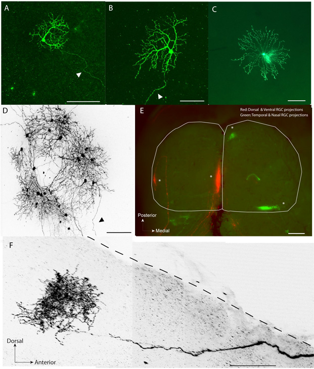
Fluorescently labeled dendrites (Figure 1A, B) and axonal arbors (Figure 1F) of single RGCs can be clearly visualized and reconstructed.
Apart from RGCs, this technique can be used to label other retinal cell types such as horizontal cells, bipolar cells and various amacrine cell subtypes (Figure 1C)
This method does not interfere with the normal time course of visual map refinement as demonstrated by normal retinotopy in the SC (Figure 1E).
At all ages, in about 90% of cases, a small volume injection (~2.3-4.6nL) of pCAG-gapEGFP led to expression in a few RGCs (Figure 1D).
In approximately 15% of the trials using a single injection of the pCAG-Cre and pCAG-LNL-gapEGFP combination plasmids per animal, led to single retinal neuron labeling including RGCs (Figure 1A,B,D) and other cell types such as amacrine cells (Figure 1C).

Figure 1 - A, B. Examples of EGFP labeled single retinal ganglion cells (arrow head pointing to axon) in a flat-mount retina at post-natal day 14 (P14). C. Example of single starburst amacrine cell at P8. D. A cluster of EGFP labeled retinal neurons including RGCs and amacrine cells in a flat-mount retina at P14. E. Dorsal and ventral RGCs in the right eye were electroporated and labeled with EGFP and temporal and ventral RGCs in the left eye were electroplated and labeled with tdTomato at P1. The target zones (asterisks) formed by the labeled RGCs can be seen in their topographically correct location in the SC at P9 (whole-mount, white outline). F. Example of a single EGFP labeled RGC arbor (2-D projection) in a sagittal section (250 μm thick) of the SC (dotted line). For clarity, images in (D) and (F) have been converted to grayscale and inverted. Scale bars (μm) : (A) - (D), (F): 100; (E): 500
Dyskusje
In this video we demonstrate an in vivo electroporation protocol that results in labeling of single or small clusters of retinal neurons in postnatal mice with DNA constructs encoding fluorescent proteins. Small clusters of fluorescently labeled RGC projections to the dLGN and SC reproduced similar projection patterns as previous studies using RGC labeling with lipophilic dyes, indicating that electroporation did not interfere with normal RGC axon arbor refinement. We have utilized this protocol to analyze the r...
Ujawnienia
No conflicts of interest declared.
Podziękowania
The pCAG-gapEGFP plasmid was a gift from Dr. S. McConnell (Stanford, CA). pCAG-tdTomato plasmid was a gift from Dr. M. Feller (Berkeley, CA). We thank Dr. Edward Ruthazer for suggesting the use of a two-plasmid strategy for single cell labeling and Anne Schohl (Montreal, QC) for validating the two-plasmid Cre/loxP strategy in pilot studies and Crair lab members for technical support. Supported by R01 MH62639 (MC), NIH R01 EY015788 (MC) and NIH P30 EY000785 (MC).
Materiały
| Name | Company | Catalog Number | Comments |
| Dumont #5 Forceps | Fine Science Tools | 11252-20 | |
| Electrical Stimulator | Grass Technologies | Model S4 | |
| Oscilloscope | Agilent Technologies | Model 54621A | |
| Audio monitor | Grass Technologies | Model AM8B | |
| Puller | Sutter Instrument Co. | Model P-97 | |
| Vannas Scissors a | World Precision Instruments, Inc. | 14003 | |
| Micro Scissors b | Ted Pella, Inc. | 1347 | |
| Dumont AA Forceps c | Fine Science Tools | 11210-20 | |
| Nanoinject II System | Drummond Scientific | 3-000-204 | |
| Glass Pipettes | Drummond Scientific | 3-000-203-G/X | |
| Foot pedal | Drummond Scientific | 3-000-026 | |
| Mineral Oil | Sigma-Aldrich | M3516 | |
| DiI | Invitrogen | D-383 | |
| N,N-Dimethylformamide | Sigma-Aldrich | D4551 |
Odniesienia
- Huberman, A. D., Feller, M. B., Chapman, B. Mechanisms underlying development of visual maps and receptive fields. Annu Rev Neurosci. 31, 479-509 (2008).
- McLaughlin, T., Torborg, C. L., Feller, M. B., O'Leary, D. D. Retinotopic map refinement requires spontaneous retinal waves during a brief critical period of development. Neuron. 40, 1147-1160 (2003).
- Godement, P., Salaun, J., Imbert, M. Prenatal and postnatal development of retinogeniculate and retinocollicular projections in the mouse. J Comp Neurol. 230, 552-575 (1984).
- Jaubert-Miazza, L. Structural and functional composition of the developing retinogeniculate pathway in the mouse. Vis Neurosci. 22, 661-676 (2005).
- Garcia-Frigola, C., Carreres, M. I., Vegar, C., Herrera, E. Gene delivery into mouse retinal ganglion cells by in utero electroporation. BMC Dev Biol. 7, 103-103 (2007).
- Matsuda, T., Cepko, C. L. Analysis of gene function in the retina. Methods Mol Biol. 423, 259-278 (2008).
- Petros, T. J., Rebsam, A., Mason, C. A. In utero and ex vivo electroporation for gene expression in mouse retinal ganglion cells. J Vis Exp. , (2009).
- Ishikawa, H. Effect of GDNF gene transfer into axotomized retinal ganglion cells using in vivo electroporation with a contact lens-type electrode. Gene Ther. 12, 289-298 (2005).
- Hewapathirane, D. S., Haas, K. Single cell electroporation in vivo within the intact developing brain. J Vis Exp. , (2008).
- Ruthazer, E. S., Haas, K., Javaherian, A., Jensen, K., Sin, W. C., Cline, H. T., Yuste, R., Konnerth, A. In vivo time- lapse imaging of neuronal development. Imaging in Neuroscience and Development: A Laboratory Manual. , 191-204 .
- Kachi, S., Oshima, Y., Esumi, N., Kachi, M., Rogers, B., Zack, D. J., Campochiaro, P. A. Nonviral ocular gene transfer. Gene Ther. 12, 843-851 (2005).
- Okada, A., Lansford, R., Weimann, J. M., Fraser, S. E., McConnell, S. K. Imaging cells in the developing nervous system with retrovirus expressing modified green fluorescent protein. Exp Neurol. 156, 394-406 (1999).
- Matsuda, T., Cepko, C. L. Controlled expression of transgenes introduced by in vivo electroporation. Proc Natl Acad Sci U S A. 104, 1027-1032 (2007).
- Plas, D. T. morphogenetic proteins, eye patterning, and retinocollicular map formation in the mouse. J Neurosci. 28, 7057-7067 (2008).
Przedruki i uprawnienia
Zapytaj o uprawnienia na użycie tekstu lub obrazów z tego artykułu JoVE
Zapytaj o uprawnieniaPrzeglądaj więcej artyków
This article has been published
Video Coming Soon
Copyright © 2025 MyJoVE Corporation. Wszelkie prawa zastrzeżone