
Aby wyświetlić tę treść, wymagana jest subskrypcja JoVE. Zaloguj się lub rozpocznij bezpłatny okres próbny.
Method Article
Gene Transfection toward Spheroid Cells on Micropatterned Culture Plates for Genetically-modified Cell Transplantation
W tym Artykule
Podsumowanie
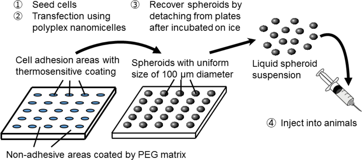
This protocol describes a cell transplantation system using genetically modified, injectable spheroids. Cell spheroids are cultured on micropatterned culture plates and recovered after gene introduction using polyplex nanomicelles. This system facilitates prolonged transgene expression from the transplanted cells in host animals while maintaining the innate function of the cells.
Streszczenie
To improve the therapeutic effectiveness of cell transplantation, a transplantation system of genetically modified, injectable spheroids was developed. The cell spheroids are prepared in a culture system on micropatterned plates coated with a thermosensitive polymer. A number of spheroids are formed on the plates, corresponding to the cell adhesion areas of 100 µm diameter that are regularly arrayed in a two-dimensional manner, surrounded by non-adhesive areas that are coated by a polyethylene glycol (PEG) matrix. The spheroids can be easily recovered as a liquid suspension by lowering the temperature of the plates, and their structure is well maintained by passing them through injection needles with a sufficiently large caliber (over 27 G). Genetic modification is achieved by gene transfection using the original non-viral gene carrier, polyplex nanomicelle, which is capable of introducing genes into cells without disrupting the spheroid structure. For primary hepatocyte spheroids transfected with a luciferase-expressing gene, the luciferase is sustainably obtained in transplanted animals, along with preserved hepatocyte function, as indicated by albumin expression. This system can be applied to a variety of cell types including mesenchymal stem cells.
Wprowadzenie
Cell transplantation therapy has attracted widespread attention for treating various intractable diseases. The activity and half-life of bioactive factors that are secreted by the transplanted cells are essential for improved therapeutic effectiveness of a cell transplantation system. Genetic modification of the cells prior to transplantation is a beneficial technique to regulate and manipulate cellular functions, including the secretion of the bioactive factors. It is also important to maintain a favorable microenvironment for the cells for avoiding cell death or loss of cell activity. Three-dimensional (3D) spheroid cell culture, in which cell-to-cell interactions are well preserved, is promising for this purpose, for example, for improving albumin secretion from primary hepatocytes and promoting multi-lineage differentiation from mesenchymal stem cells (MSCs) 1-7.
In this study, a novel combination system of spheroid culture and gene transfection is used to serve as a platform for genetically modified cell transplantation. For creating spheroid cells, a spheroid culture system on micropatterned culture plates is used. On these plates, cell adhesion areas of 100 µm diameter are regularly arrayed in a two-dimensional manner and are surrounded by non-adhesive areas coated by a PEG matrix3. By seeding an adequate number of cells, arrays of 3D spheroids of 100 µm in diameter are formed corresponding to the micropatterned culture bed.
The spheroids are recovered without disrupting their 3D structure by using thermosensitive cell-culture plates, that were coated with a thermosensitive polymer, poly(iso-propylacrylamide) (PIPAAm) 8-10. The micropatterned architecture is constructed on the thermosensitive plates (custom-built). By simply lowering the temperature of the plates, the spheroids are detached from the culture bed and dispersed in phosphate buffered saline (PBS). Thus, a large number of spheroids with a uniform size of 100 µm can be obtained in the form of an injectable suspension.

Figure 1. Schematic representation of the spheroid culture system on a micropatterned plate. Genetic modification is achieved by gene transfection using the original non-viral gene carrier, polyplex nanomicelle. It is composed of plasmid DNA (pDNA) and polyethylene glycol (PEG)-polycation block copolymers11. These have a characteristic core-shell structure consisting of a PEG shell and an inner core of condensed pDNA, allowing safe and effective gene introduction into cells for therapeutic purposes11. Please click here to view a larger version of this figure.

Figure 2. Structure of the polyplex nanomicelle formed by the complex of nucleic acids and PEG-block-polycation block copolymers. In this study, the primary advantage of this technique is that the spheroid structure is not disrupted during gene transfection by the nanomicelles. After nanomicelle-mediated transfections of rat primary hepatocyte spheroids, prolonged transgene expression is obtained for more than a month with continuous albumin secretion from the hepatocytes at a level comparable to that of untransfected spheroids12. The transgene expression and albumin secretion from the spheroids are also maintained after recovery from the thermosensitive plates. It is evident that nanomicelles can safely facilitate gene introduction without impairing the innate functions of the hepatocytes. Thus, the combination of spheroid cells cultured on thermosensitive micropatterned plates with gene introduction using nanomicelles is a promising platform for genetically modified cell transplantation. Please click here to view a larger version of this figure.
Access restricted. Please log in or start a trial to view this content.
Protokół
All the animal studies were conducted with the approval of the Animal Care and Use Committee of the University of Tokyo, Tokyo, Japan.
1. Cell Preparation
- For primary hepatocytes, follow the protocol for hepatocyte isolation from rat by a modified two-step collagenase digestion process13,14.
- Anesthetize Sprague Dawley (SD) rats (male, 5 weeks old) under inhalation anesthesia with isoflurane. Place a rat in a chamber connected to an anesthesia machine to provide isoflurane for the chamber. Take out the rat after falling asleep, and set it on the operating table with ventilation using a mask. Control the isoflurane flow approximately at 0.4 – 0.7 L/min by checking the conditions of the rat. Perfuse the livers of Sprague Dawley (SD) rats (male, 5 weeks old) from the hepatic portal vein with a special solution composed of 8 g/L sodium chloride (NaCl), 400 mg/L potassium chloride (KCl), 78 mg/L sodium dihydrogen phosphate dihydrate (NaH2PO4·2H2O), 151 mg/L disodium hydrogen phosphate dodecahydrate (Na2HPO4·12H2O), 2.38 g/L 2-[4-(2-hydroxyethyl)-1-piperazinyl] ethanesulfonic acid (HEPES), 190 mg/L ethylene glycol tetraacetic acid (EGTA), 350 mg/L sodium hydrogen carbonate (NaHCO3), and 900 mg/L glucose.
- Circulate collagenase solution through the liver.
NOTE: The solution is composed of 500 mg/L collagenase, 9.8 g/L Hank’s buffered salt, 2.38 g/ml HEPES, 556 mg/ml calcium chloride hydrate (CaCl2·H2O), 350 mg/L NaHCO3, and 50 mg/L trypsin inhibitor, with the pH adjusted to 7.2. - Remove the liver carefully, and mince it gently on a dish using a scalpel blade, add Dulbecco’s Modified Eagle’s Medium (DMEM) supplemented with 10% fetal bovine serum (FBS), and filter the cell suspension through a 100 μm nylon mesh. To remove additional debris, centrifuge the cell suspension at 20 × g for 1 min. At this step, hepatocytes are in the supernatant.
- Repeat the centrifugation step twice (a total of three centrifugations). Finally, centrifuge the hepatocytes at 50 × g for 3 min to recover them in the form of a pellet.
- Re-suspend the hepatocytes to a concentration of 4 × 10⁵ cells/ml in a special culture medium composed of DMEM supplemented with 10% FBS, 1% Pen-Strep-Glut (PSQ), 1% dimethyl sulfoxide (DMSO), 0.1 µmol/L dexamethasone, 0.5 µg/ml insulin, 10 mmol/L nicotinamide, 0.2 mmol/L phosphorylated ascorbate (Asc-2P), and 10 ng/ml human epidermal growth factor (hEGF)15. This special medium is mandatory for preserving the hepatic function under in vitro conditions.
- For obtaining rat MSCs, euthanize Sprague Dawley (SD) rats (male, 5 weeks old) by excessive administration of isoflurane. Resect the femurs and tibias, and collect the bone marrows by inserting a 22 G needle into the shaft of the bone to flush it out with 10 ml of DMEM supplemented with 10% FBS. Collect the cells by filtration through a 100 µm nylon mesh.
- Seed the cells onto 10 cm culture dishes using DMEM containing 10% FBS and 1% penicillin / streptomycin. For the spheroid experiments, use MSCs within 5 passages.
2. Preparation of 3D Cell Spheroids
- Commercially obtain micropatterned culture plates, on which the cell adhesive areas are regularly arrayed at 100 µm diameter in a two-dimensional manner, surrounded by non-adhesive areas coated by the PEG matrix.
NOTE: For cell transplantation, an additional coating with the thermosensitive polymer PIPAAm is necessary to allow cell detachment by cooling the plates (see step 5.1). Temperature fluctuations can induce changes in the chemistry of this polymer8-10. At 37 °C, PIPAAm is slightly hydrophobic, allowing the cells to be cultured under normal conditions. A decrease in the temperature below 32 °C results in rapid hydration of the polymer, leading to the spontaneous detachment of the cells. - Seed the hepatocytes or MSCs on 12-well micropatterned plates at a density of 4 × 10⁵ cells/well andincubate them at 37 °C in a humidified atmosphere containing 5% CO2. The cells will accumulate on the micropatterned adhesion areas and gradually form round spheroids in 2 days.
NOTE: For preparing control cells in a monolayer culture, use normal 12-well plates and seed the cells at an identical density, following a similar procedure as described above.
3. Preparation of Polyplex Nanomicelles
- Synthesize a block copolymer, PEG-PAsp(DET) (poly[N’-[N-(2-aminoethyl)-2-aminoethyl] aspartamide]) (PEG Mw = 12,000, polymerization degree (DP) of PAsp(DET) segment = 59), and a homopolymer that is composed of only the cationic segment [PAsp(DET)] (DP = 55), following the procedures previously described by the authors11,16,17.
- Prepare a mixed solution of the PEG-PAsp(DET) block copolymer and the PAsp(DET) homopolymer at an equal molar ratio of residual amino groups in 10 mM HEPES buffer (pH 7.3) by adjusting the polymer concentration to 33.3 µg/ml and 19.1 µg/ml, respectively.
NOTE: The polymer concentrations described above are representative values for preparing nanomicelles with a residual molar ratio of the total amino groups in the two polymers to the phosphate groups in the pDNA (N/P ratio) of 10. The N/P ratio can vary depending on the cell type and the purpose (for details, see Discussion). The combined use of the two polymers can achieve both effective PEG shielding and functioning of PAsp(DET) to enhance endosomal escape (for details, see Discussion)18. - Prepare the pDNA encoding Gaussia luciferase, GL4 luciferase, or erythropoietin by cloning the segment expressing the respective genes into the pCAG-GS plasmid (http://www.cdb.riken.jp/pcs/protocol/vector/map/m36.html) to obtain expression under the CAG promoter/enhancer using a commercial kit according to manufacturer’s protocol. Amplify the pDNA in a competent Escherichia coli strain and purify it using an endotoxin-free plasmid DNA purification system. Determine the pDNA concentration at an absorbance of 260 nm to obtain a 150 µg/ml solution in 10 mM HEPES buffer (pH 7.3).
- To prepare the polyplex nanomicelles, thoroughly mix the pDNA solution (150 µg/ml in 10 mM HEPES buffer) and the premixed solution of the two polymers at a ratio of 2:1 (by volume).
4. Gene Transfection into Spheroids
- Incubate the cells (hepatocytes or MSCs) for 72 hrs after seeding onto the micropatterned plates to allow for the formation of mature spheroids. For gene transfection, add 100 µl of the polyplex nanomicelle solution (containing 10 µg of pDNA) to each well after replacing the culture medium with 1 ml of fresh medium. Continue the incubation with the nanomicelle solution for 24 hrs.
- For a control using a lipid-based transfection reagent, mix pDNA solutions with the reagent at a weight ratio reagent / pDNA of 3. Adjust the final dose of pDNA to be equal for both the lipid-based reagent and nanomicelle methods.
5. Recovery and Transplantation of Cell Spheroids
- Replace the culture medium with 200 µl of chilled PBS and place the plates on ice.
NOTE: Generally, the spheroids can detach in about 15 min and can be recovered in the form of a suspension for transplantation. - Gently aspirate the cells in the 200 µl suspension using a syringe with a 23 G or 27 G needle for in vivo injections.
6. Evaluation of Transgene Expression
- For the in vitro evaluation of the expression of Gaussia luciferase secreted in the culture medium, collect 50 – 100 µl of the medium precisely 24 hrs after replacing with fresh medium. Estimate the luciferase expression using a commercial renilla luciferase assay system and a luminometer according to manufacturer’s protocol.
NOTE: The Gaussia luciferase remains stable in the culture medium for more than a week. Thus, to evaluate the real-time efficiency of the transgene expression, replace the medium with fresh one prior to collecting the sample medium. The timing of the medium change can be flexible. - For the in vivo evaluation of the transgene expression in host animals following cell transplantation, anesthetize BALB/c nude mice (female; 7 weeks old) under inhalation anesthesia with isoflurane.
- Place a mouse in a chamber connected to an anesthesia machine to provide isoflurane for the chamber. Take out the mouse after falling asleep, and set it on the operating table with ventilation using a mask. Control the isoflurane flow approximately at 0.2 – 0.5 L/min by checking the conditions of the mouse.
- Inject 200 µl of the cell suspension containing spheroids transfected with the GL4 luciferase-expressing pDNA (as described in 4.1) into the subcutaneous tissue of the abdominal region.
- Immediately after injecting D-luciferin (150 mg/kg; intravenous route), measure the luciferase expression using IVIS imaging system according to manufacturer’s protocol.
- For evaluating the therapeutic effects of the cell transplantation, inject 200 µl of the cell suspension containing spheroids transfected with the erythropoietin-encoding pDNA into the subcutaneous tissue of the abdominal region.
- Collect blood samples by submandibular bleeding to obtain approximately 200 µl of blood19. Measure the hemoglobin and hematocrit using a blood sample analyzer.
NOTE: The cell number for transplantation is regulated by the number seeded on the plates. Unfortunately, it is difficult to determine the exact cell number because the number inside spheroids cannot be measured.
- Collect blood samples by submandibular bleeding to obtain approximately 200 µl of blood19. Measure the hemoglobin and hematocrit using a blood sample analyzer.
- After experiments, place the mice on a heating pad connected with a temperature controller until awakening from anesthesia.
Access restricted. Please log in or start a trial to view this content.
Wyniki
Gene transfection of the Gaussia luciferase-expressing pDNA was performed in the spheroids formed by the hepatocytes or MSCs using polyplex nanomicelles or the control lipid-based transfection reagent12. The nanomicelles induced almost no change in the spheroid structure compared with non-transfected spheroids on the micropatterned plates, whereas the control reagent significantly disrupted the structure a day after the transfection (Figure 3). After transfection using the nanomicelles, consis...
Access restricted. Please log in or start a trial to view this content.
Dyskusje
In this protocol, it is critical to maintain the 3D structure of spheroids during the steps of gene introduction and spheroid recovery. It is essential to maintain a favorable microenvironments for the cells to avoid cell death or loss of cell activity. For example, albumin secretion, a representative innate function of hepatocytes, is well preserved in the hepatocyte spheroids, while the hepatocytes in the conventional monolayer culture rapidly lose their secretory capacity a few days after seeding12. For MSC...
Access restricted. Please log in or start a trial to view this content.
Ujawnienia
The authors declare that they have no competing financial interests.
Podziękowania
We deeply appreciate Dr. Takeshi Ikeya and technical staff in Toyo Gosei, Tokyo, Japan for providing thermosensitive micropatterned culture plates as well as scientific advice. We also thank Ms. Satomi Ogura, Ms. Sae Suzuki, Ms. Asuka Miyoshi and Ms. Katsue Morii for technical assistance with animal experiments. This work was financially supported in part by the JSPS KAKENHI Grant-in-Aid for Scientific Research, the Center of Innovation (COI) Program and the S- innovation program from the Japan Science and Technology Agency (JST), and the JSPS Core-to-Core Program, A. Advanced Research Networks.
Access restricted. Please log in or start a trial to view this content.
Materiały
| Name | Company | Catalog Number | Comments |
| Pen-Strep-Glut | GIBCO | ||
| Dexamethasone | Wako Pure Chemical Industries | 041-18861 | |
| Nicotinamide | Wako Pure Chemical Industries | 141-01202 | |
| Hank’s buffered salt and L-ascorbic acid 2-phosphate (Asc-2P) | Sigma-Aldrich | A8960 | |
| Human epidermal growth factor (hEGF) | Toyobo | PT10015 | |
| Cell-able multi-well plates | Toyo Gosei | PP-12 | |
| Thermosensitive cell culture plates (Upcell) | CellSeed Inc | The micropatterned architecture is constructed on the thermosensitive plates (custom-built by Toyo Gosei) | |
| Lipid-based transfection reagent (FuGENE HD) | Promega | E2311 | |
| Renilla Luciferase Assay System | Promega | E2810 | |
| pGL4 Luciferase Reporter Vector | Promega | E6651 | |
| pDNA expressing Gaussia luciferase | New England BioLabs | N8082S | |
| Mouse erhthropoietin-expressing vector | Origene | MC208445 | |
| pCAG-GS | Kindly provided by Laboratory for Pluripotent Cell Studies, Center for Developmental Biology, RIKEN | ||
| Escherichia coli DH5α competent cells | Takara | 9057 | |
| Endotoxin-free plasmid DNA purification system | Nippon Genetics | NucleoBond Xtra EF | |
| Collagenase | Wako Pure Chemical Industries | 639-00951 | |
| Trypsin inhibitor | GIBCO | R-007-100 | |
| Luminometer | Promega | GloMax™ 96 Microplate Luminometer | |
| IVIS Imaging System | Xenogen Corp. | Xenogen IVIS Spectrum in vivo imaging system | |
| Blood sample analyzer | Sysmex | pocH-100i Automated Hematology Analyzer |
Odniesienia
- Landry, J., Bernier, D., Ouellet, C., Goyette, R., Marceau, N. Spheroidal aggregate culture of rat liver cells: histotypic reorganization, biomatrix deposition, and maintenance of functional activities. J Cell Biol. 101 (3), 914-923 (1985).
- Yuasa, C., Tomita, Y., Shono, M., Ishimura, K., Ichihara, A. Importance of cell aggregation for expression of liver functions and regeneration demonstrated with primary cultured hepatocytes. J Cell Physiol. 156 (3), 522-530 (1993).
- Otsuka, H., et al. Two-dimensional multiarray formation of hepatocyte spheroids on a microfabricated PEG-brush surface. Chembiochem. 5 (6), 850-855 (2004).
- Wang, W., et al. 3D spheroid culture system on micropatterned substrates for improved differentiation efficiency of multipotent mesenchymal stem cells. Biomaterials. 30 (14), 2705-2715 (2009).
- Bartosh, T. J., et al. Aggregation of human mesenchymal stromal cells (MSCs) into 3D spheroids enhances their antiinflammatory properties. Proc Natl Acad Sci U S A. 107 (31), 13724-13729 (2010).
- Frith, J. E., Thomson, B., Genever, P. G. Dynamic three-dimensional culture methods enhance mesenchymal stem cell properties and increase therapeutic potential. Tissue Eng Part C Methods. 16 (4), 735-749 (2010).
- Nakasone, Y., Yamamoto, M., Tateishi, T., Otsuka, H. Hepatocyte spheroids underlayered with nonparenchymal cells for biomedical applications. IEICE Transactions on Electronics. E94, 176-180 (2011).
- Nishida, K., et al. Corneal reconstruction with tissue-engineered cell sheets composed of autologous oral mucosal epithelium. N Engl J Med. 351 (12), 1187-1196 (2004).
- Ohashi, K., et al. Engineering functional two- and three-dimensional liver systems in vivo using hepatic tissue sheets. Nat Med. 13 (7), 880-885 (2007).
- Sekine, H., et al. Cardiac cell sheet transplantation improves damaged heart function via superior cell survival in comparison with dissociated cell injection. Tissue Eng Part A. 17 (23-24), 2973-2980 (2011).
- Itaka, K., Kataoka, K. Progress and prospects of polyplex nanomicelles for plasmid DNA delivery. Curr Gene Ther. 11 (6), 457-465 (2011).
- Endo, T., Itaka, K., Shioyama, M., Uchida, S., Kataoka, K. Gene transfection to spheroid culture system on micropatterned culture plate by polyplex nanomicelle: a novel platform of genetically-modified cell transplantation. Drug Deliv and Transl Res. 2 (5), 398-405 (2012).
- Howard, R. B., Christensen, A. K., Gibbs, F. A., Pesch, L. A. The enzymatic preparation of isolated intact parenchymal cells from rat liver. J Cell Biol. 35 (3), 675-684 (1967).
- Berry, M. N., Friend, D. S. High-yield preparation of isolated rat liver parenchymal cells: a biochemical and fine structural study. J Cell Biol. 43 (3), 506-520 (1969).
- Tateno, C., Yoshizato, K. Long-term cultivation of adult rat hepatocytes that undergo multiple cell divisions and express normal parenchymal phenotypes. Am J Pathol. 148 (2), 383-392 (1996).
- Kanayama, N., et al. A PEG-based biocompatible block catiomer with high buffering capacity for the construction of polyplex micelles showing efficient gene transfer toward primary cells. ChemMedChem. 1 (4), 439-444 (2006).
- Itaka, K., Ishii, T., Hasegawa, Y., Kataoka, K. Biodegradable polyamino acid-based polycations as safe and effective gene carrier minimizing cumulative toxicity. Biomaterials. 31 (13), 3707-3714 (2010).
- Uchida, S., et al. PEGylated Polyplex With Optimized PEG Shielding Enhances Gene Introduction in Lungs by Minimizing Inflammatory Responses. Mol Ther. 20 (6), 1196-1203 (2012).
- Golde, W. T., Gollobin, P., Rodriguez, L. L. A rapid, simple, and humane method for submandibular bleeding of mice using a lancet. Lab Anim (NY). 34, 39-43 (2005).
- Uchida, S., et al. An injectable spheroid system with genetic modification for cell transplantation therapy. Biomaterials. 35 (8), 2499-2506 (2014).
Access restricted. Please log in or start a trial to view this content.
Przedruki i uprawnienia
Zapytaj o uprawnienia na użycie tekstu lub obrazów z tego artykułu JoVE
Zapytaj o uprawnieniaThis article has been published
Video Coming Soon
Copyright © 2025 MyJoVE Corporation. Wszelkie prawa zastrzeżone