
Aby wyświetlić tę treść, wymagana jest subskrypcja JoVE. Zaloguj się lub rozpocznij bezpłatny okres próbny.
Method Article
Utilizing the Ethylene-releasing Compound, 2-Chloroethylphosphonic Acid, as a Tool to Study Ethylene Response in Bacteria
W tym Artykule
Podsumowanie
The protocols outlined herein facilitate the convenient investigation of bacterial ethylene responses by utilizing 2-chloroethylphosphonic acid (CEPA). Ethylene is produced in situ through the decomposition of CEPA in an aqueous bacterial growth medium, circumventing the requirement for pure ethylene gas.
Streszczenie
Ethylene (C2H4) is a gaseous phytohormone that is involved in numerous aspects of plant development, playing a dominant role in senescence and fruit ripening. Exogenous ethylene applied during early plant development triggers the triple response phenotype; a shorter and thicker hypocotyl with an exaggerated apical hook. Despite the intimate relationship between plants and bacteria, the effect of exogenous ethylene on bacteria has been greatly overlooked. This is partly due to the difficulty of controlling gaseous ethylene within the laboratory without specialized equipment. 2-Chloroethylphosphonic acid (CEPA) is a compound that decomposes into ethylene, chlorine, and phosphate in a 1:1:1:1 molar ratio when dissolved in an aqueous medium of pH 3.5 or greater. Here we describe the use of CEPA to produce in situ ethylene for the investigation of ethylene response in bacteria using the fruit-associated, cellulose-producing bacterium Komagataeibacter xylinus as a model organism. The protocols described herein include both the verification of ethylene production from CEPA via the Arabidopsis thaliana triple response assay and the effects of exogenous ethylene on K. xylinus cellulose production, pellicle properties and colonial morphology. These protocols can be adapted to examine the effect of ethylene on other microbes using appropriate growth media and phenotype analyses. The use of CEPA provides researchers with a simple and efficient alternative to pure ethylene gas for the routine determination of bacterial ethylene response.
Wprowadzenie
The olefin ethylene (C2H4) was first discovered as a plant hormone in 1901 when it was observed that pea seedlings, grown in a laboratory that used coal gas lamps, exhibited an abnormal morphology in which stems (hypocotyls) were shorter, thicker and bent sideways compared to normal pea seedlings; a phenotype later termed the triple response1,2. Subsequent studies demonstrated that ethylene is a vital phytohormone that regulates numerous developmental processes such as growth, stress response, fruit ripening and senescence3. Arabidopsis thaliana, a model organism for plant biology research, has been well studied in regards to its response to ethylene. Several ethylene response mutants have been isolated by exploiting the triple response phenotype observed in dark-grown A. thaliana seedlings in the presence of ethylene1,4,5. The biosynthetic precursor for ethylene production in plants is 1-aminocyclopropane carboxylic acid (ACC)6 and is commonly used during the triple response assay to increase endogenous ethylene production that leads to the triple response phenotype1,4,5.
Although the ethylene response is widely studied in plants, the effect of exogenous ethylene on bacteria is vastly understudied despite the close association of bacteria with plants. One study reported that certain Pseudomonas strains can survive using ethylene as a sole source of carbon and energy7. However, only two studies have demonstrated that bacteria respond to ethylene. The first study showed that strains of Pseudomonas aeruginosa, P. fluorescens, P. putida, and P. syringae were chemotactic toward ethylene using an agarose plug assay in which molten agarose was mixed with a chemotaxis buffer equilibrated with pure ethylene gas8. However, to our knowledge, there have been no further reports using pure ethylene gas to characterize bacterial ethylene response, likely due to the difficultly of handling gases in the laboratory without specialized equipment. The second report of bacterial ethylene response demonstrated that ethylene increased bacterial cellulose production and influenced gene expression in the fruit-associated bacterium, Komagataeibacter (formerly Gluconacetobacter) xylinus9. In this case, the ethylene-releasing compound, 2-chloroethylphosphonic acid (CEPA) was used to produce ethylene in situ within the bacterial growth medium, bypassing the need for pure ethylene gas or specialized equipment.
CEPA produces ethylene at a 1:1 molar ratio above pH 3.510,11 through a base-catalyzed, first-order reaction12-14. The degradation of CEPA is positively correlated with pH and temperature13,14 and results in the production of ethylene, chloride and phosphate. CEPA provides researchers interested in studying bacterial responses to ethylene with a convenient alternative to gaseous ethylene.
The overall goal of the following protocols is to provide a simple and efficient method to study bacterial ethylene response and includes validation of physiologically relevant levels of ethylene production from CEPA decomposition in bacterial growth medium, analysis of culture pH to ensure CEPA decomposition is not impaired during bacterial growth, and assessment of the effect of ethylene on bacterial morphology and phenotype. We demonstrate these protocols using K. xylinus, however, these protocols can be adapted to study ethylene response in other bacteria by using the appropriate growth medium and phenotype analyses.
Access restricted. Please log in or start a trial to view this content.
Protokół
1. Chemicals
- Prepare a solution of 500 mM CEPA (144.49 g/mol), and a solution consisting of both 500 mM NaCl (58.44 g/mol) and 500 mM NaH2PO4·H2O (137.99 g/mol) in acidified (pH 2.5) ultra-pure water or 0.1 N HCl. Mix using a vortex until the solutions are clear.
- Serially dilute (10x) the 500 mM solutions in the same solvent to obtain 5 mM and 50 mM stocks.
- Prepare a 10 mM solution of 1-aminocyclopropane carboxylic acid (ACC; 101.1 g/mol) in ultra-pure water.
- Filter-sterilize stock solutions and store aliquots at -20 °C.
- Filter-sterilize cellulase. Store aliquots at 4 °C.
2. Verifying Ethylene Production from 2-Chloroethylphosphonic Acid Decomposition: Triple Response Assay
- Surface-sterilize Arabidopsis thaliana Ecotype Columbia Seeds Using the Vapor-phase Method:
CAUTION: The following step produces toxic chlorine gas. Conduct seed sterilization in a fume hood.- Obtain a sealable container deep enough to accommodate a 250 ml glass beaker for seed sterilization and place it in a fume hood.
- Add A. thaliana seeds to a microcentrifuge tube and place the tube in a rack. Place the rack containing the open tube into the sealable container.
Note: Do not fill individual tubes over half full to allow chlorine gas to penetrate the lower seeds. - Place a 250 ml beaker containing 100 ml of commercial bleach in the sealable container. Carefully add 3 ml of concentrated HCl to the bleach and immediately seal the container with its lid.
CAUTION: Bleach and HCl react to produce toxic chlorine gas which acts to surface-sterilize the seeds. - Incubate the seeds in the presence of chlorine gas for 4 hr in the fumehood.
Note: Longer sterilization times will reduce germination efficiency. - After sterilization, allow the chlorine gas to vent in the fume hood for at least 1 hr then seal the microcentrifuge tube. Leave the bleach and HCl mixture in the fumehood for at least 24 hr before discarding.
Note: Sterilized seeds may be stored at room temperature for immediate use. - Keep seeds at 4 °C for long-term storage. Bring seeds to room temperature before opening the microcentrifuge tube to prevent condensation. Store seeds in the dark.
- Prepare Agar Plates in 4-sector Petri Dishes (90 x 15 mm):
- Prepare 110 ml of growth medium for A. thaliana seedlings: 1x Murashige and Skoog (MS) basal medium15 (4.33 g/L) containing 1% (w/v) sucrose and 0.8% (w/v) agar. Adjust the MS medium to pH 6 with NaOH.
- Prepare 100 ml of bacterial growth medium for CEPA decomposition: Schramm and Hestrin (SH) medium16 containing 1.5% (w/v) agar. Adjust the SH medium to pH 7 with NaOH.
- Sterilize media by autoclaving and temper in a 55 °C water bath.
- Once the MS agar has tempered, add 40 ml to a sterile flask. To prepare the positive control growth medium for A. thaliana seedlings, supplement the 40 ml aliquot of MS agar with 40 µl of 10 mM ACC to obtain a final concentration of 10 µM ACC.
- Add 5 ml of medium to the appropriate quadrants of the sectored Petri dishes (Figure 1) and allow the agar to solidify. Prepare all plates in triplicate.
Note: CEPA is not added to the growth medium until later in the protocol.

Figure 1: Setup of agar plates used for the triple response assay with CEPA. A schematic illustrates the quadrants specific for the negative control (A), positive control (B), and experimental plates (C). This figure has been modified from Augimeri and Strap9. Please click here to view a larger version of this figure.
- Stratify A. thaliana Seeds to Ensure Synchronous Germination:
- Add approximately fifty A. thaliana seeds onto each quadrant containing MS or MS+ACC agar. Ensure seeds are evenly distributed to facilitate seedling removal and analysis.
- Incubate plates containing seeds in the dark at 4 °C for 3-4 days.
- Decompose CEPA on Bacterial Growth Medium (SH) and Perform Triple Response Assay:
- After stratification, expose the seeds to fluorescent light for 2 hr.
- Spread 10 µl of the 500 mM CEPA stock solution onto the quadrants of the experimental plates containing SH agar (pH 7; Figure 1) to obtain a final CEPA concentration of 1 mM.
- Seal the plates with laboratory film and cover with foil to create a dark environment for the seeds.
- Germinate seeds by incubating plates in the dark at 23 °C for 3 days with the agar side down.
Note: Plates can be stacked, but negative controls should be placed on the bottom since ethylene is lighter than air.
- Analyze Triple Response Assay Data:
- With flame-sterilized forceps, remove single seedlings from a plate corresponding to each treatment and view under a dissecting or digital USB microscope. Ensure the bottom of the seedlings are aligned and photograph.
- Remove 30 seedlings from each quadrant (60 seedlings per biological replicate and 180 seedlings per treatment). Align on a surface with a black background and photograph with a ruler.
- Measure the hypocotyl length (mm) of replicate seedlings using ImageJ software17. Set the scale by clicking and dragging the *Straight* tool to select a length of 10 mm. Select "Set Scale" under the "Analyze" tab and set the known distance to 10. Using the *Segmented* tool, click and drag to select the length of the hypocotyl and press M to measure distance. Compare the means of biological replicates using a one-way ANOVA with a Tukey's multiple comparison test. Differences are considered significant if p < 0.05.
3. Analysis of pH throughout Bacterial Growth
- Grow and Quantify Komagataeibacter xylinus Starter Cultures in Triplicate:
- Inoculate a single colony of K. xylinus into 5 ml of SH medium (pH 5) supplemented with 0.2% (v/v) filter-sterilized cellulase. Incubate cultures at 30 °C with agitation at 150 rpm until an OD600 of 0.3 to 0.4 is reached (about 72 hr).
- Harvest starter cultures by centrifugation (2,000 x g; 4 °C; 10 min). With 5 ml sterile 0.85% (w/v) NaCl solution, wash the cells twice and resuspend the cell pellet. Keep cells on ice.
- Quantify cells using a Petroff-Hausser counting chamber.
- Inoculate Cultures for Analysis of pH:
- Add 150 ml of SH broth (pH 7) supplemented with 0.2% (v/v) filter-sterilized cellulase to twelve 500 ml flasks with foil lids. Inoculate flasks with K. xylinus starter cultures at a concentration of 105 cells/ml. Using the three starter cultures, prepare three biological replicates per treatment.
- Supplement cultures with 300 µl of the 5, 50, or 500 mM CEPA stock solutions to obtain final CEPA concentrations of 0.01, 0.1, and 1.0 mM, respectively. Supplement the untreated control cultures with 300 µl of the solvent used to dissolve CEPA.
- Seal the flasks by tightly taping the foil lids to the flasks and incubate cultures for 14 days at 30 °C with agitation at 150 rpm.
- Each day, aseptically remove 5 ml samples from each flask and pellet cells by centrifugation (2,000 x g; 4 °C; 10 min). Transfer supernatants to a clean tube and measure the pH of each biological replicate using a pH meter.
- Analyze the time-course data by graphing the mean pH of the biological replicates.
Note: It is important that the culture pH does not drop below 3.5; a culture pH of 5 significantly reduces ethylene release from CEPA, and a pH below 3.5 completely inhibits ethylene release. - To control for chlorine and phosphate levels, perform an identical experiment using 0.01, 0.1, and 1.0 mM of the NaCl-NaH2PO4 solution using the 5, 50, and 500 mM stocks, respectively.
4. Colony Morphology
- Grow K. xylinus Starter Cultures in Triplicate:
- Inoculate a single colony of K. xylinus into 5 ml of SH medium (pH 5) supplemented with 0.2% (v/v) filter-sterilized cellulase. Incubate cultures at 30 °C with agitation at 150 rpm until an OD600 of 0.3 to 0.4 is reached (about 72 hr).
- Harvest starter cultures by centrifugation (2,000 x g; 4 °C; 10 min). With 5 ml of sterile saline, wash the cells and then resuspend the cell pellet. Keep cells on ice.
- Prepare 24 agar plates containing 25 ml of SH (pH 7) medium with 1.5% (w/v) agar.
- Once the agar has solidified, spread 50 µl of the 5, 50, and 500 mM CEPA stock solutions on the agar to obtain final CEPA concentrations of 0.01, 0.1, and 1.0 mM, respectively. Set up untreated and solvent control plates, which consist of no amendment and spreading of 50 µl of the solvent used to dissolve the test compounds.
- Streak plates for isolated colonies with a loop-full (~5 μl) of K. xylinus starter culture and seal them with paraffin film. Inoculate all plates in triplicate. Incubate plates for five days at 30 °C.
- Photograph colonies at 20X magnification using a digital USB microscope and qualitatively assess colonial morphology and cellulose production on solid medium.
Note: Cellulose appears as a hazy substance along the margin of the colony. - To control for chlorine and phosphate levels, perform an identical experiment using 0.01, 0.1, and 1.0 mM of the NaCl-NaH2PO4 solution using the 5, 50, and 500 mM stocks, respectively.
5. Pellicle Assays
- Grow and Quantify K. xylinus Starter Cultures in Triplicate:
- In triplicate, inoculate a single colony of K. xylinus into 5 ml of SH medium (pH 5) supplemented with 0.2% (v/v) filter-sterilized cellulase. Incubate cultures at 30 °C with agitation at 150 rpm until an OD600 of 0.3 to 0.4 is reached (about 72 hr).
- Harvest starter cultures by centrifugation (2,000 x g; 4 °C; 10 min). With 5 ml of sterile saline, wash the cells twice and resuspend the cell pellet. Keep cells on ice.
- Quantify cells using a Petroff-Hausser counting chamber.
- Prepare Master Mixes to Inoculate 24-well Plates:
- Supplement 60 ml of SH medium (pH 7) with 120 µl of the 5, 50, and 500 mM CEPA stocks to obtain final CEPA concentrations of 0.01, 0.1, and 1.0 mM, respectively. Supplement another 60 ml of SH medium (pH 7) with 120 µl of the solvent used to dissolve CEPA. Vortex to mix.
- Divide each 60 ml of CEPA-containing medium into four 14 ml aliquots. Inoculate three of the 14 ml aliquots with biological replicates of starter culture at a concentration of 105 cells/ml. Keep tubes with cells on ice to prevent cellulose production.
Note: The remaining 14 ml aliquot will be used for sterile control wells.
- Inoculate 24-well Plates:
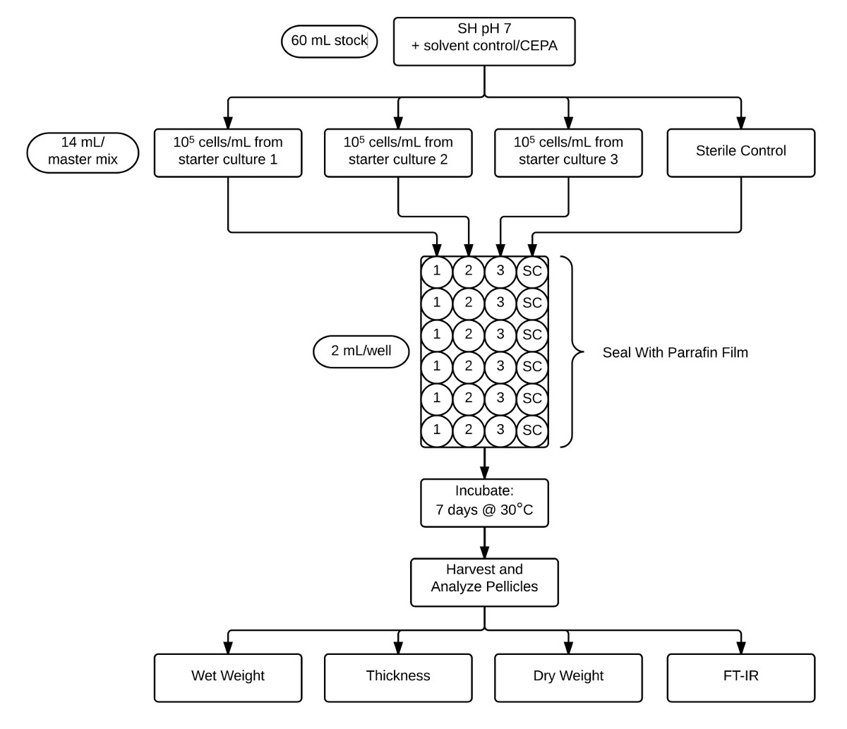
- Test each treatment in its own plate with three rows of biological replicates and a row of sterile controls (Figure 2).

Figure 2: Flow-chart illustrating the protocol used for pellicle assay and analysis. Stock CEPA-supplemented pH 7 SH medium (60 ml) is aliquoted for three separate biological replicate inoculations and a sterile control (14 ml each). These cultures are then aliquoted into six technical replicates (2 ml) into a 24 well plate and then sealed with paraffin film. After incubation for 7 days at 30 °C, pellicles are harvested and characterized by determining wet weight, thickness, dry weight, and crystallinity by FT-IR. Please click here to view a larger version of this figure
- Using the 14 ml master mix, add 2 ml into each of six wells of a sterile 24-well plate. Complete for the three biological replicates and sterile control (Figure 2). Repeat for each treatment.
- Seal plates with paraffin film and incubate statically for 7 days at 30 °C.
- Harvest and Measure Pellicle Wet Weight, Thickness, dry Weight (Cellulose Yield) and Crystallinity (Figure 2):
- Depress one side of the pellicle to elevate the opposing pellicle edge and remove individual pellicles with forceps. While retaining grip, place them on fresh paper towel for 3 sec to remove excess medium before weighing to determine their wet weights.
- Align pellicles adjacent to a ruler and photograph from the side using a high resolution digital camera.
- Using ImageJ software17, measure pellicle thickness on the left shoulder, right shoulder and the center of each pellicle. Average all technical replicates for each biological replicate.
- Individually transfer the pellicles into the wells of a 6-well plate. Treat pellicles with 12 ml of 0.1 N NaOH at 80 °C for 20 min to lyse cells.
- Remove the NaOH and neutralize pellicles by washing with ultra-pure water for 24 hr with agitation. Change water every 6 hr.
Note: Pellicles should be white upon completion of the washing step. - Place pellicles on silicon mats and dry at 50 °C for 48 hr to constant weight. Once dry, remove from mats and measure pellicle weights on an analytical scale to determine bacterial cellulose yield.
- Analyze pellicle crystallinity using Fourier-transform infrared spectroscopy (FT-IR) using 32 scans and a resolution of 4 cm-1 in the range of 4,000 to 650 cm-1. Calculate the crystallinity index, CI(IR), using A1437/A895; the absorbance ratio of the "crystalline band" and "amorphous band" as previously described18.
- To control for chlorine and phosphate levels, perform an identical experiment using 0.01, 0.1, and 1.0 mM of the NaCl-NaH2PO4 solution using the 5, 50, and 500 mM stocks, respectively.
- Analyze Pellicle Data:
- Calculate pellicle hydration by determining the difference between pellicle wet weight and dry weight.
- Average the values of all technical replicates to obtain a single value for each biological replicate for statistical analysis. Compare treatments using a one-way ANOVA with Tukey's multiple comparison test. Differences are significant if p <0.05.
- Normalize data as the percent of untreated controls and plot the means of biological replicates.
Access restricted. Please log in or start a trial to view this content.
Wyniki
A schematic plate setup for verification of ethylene liberation from CEPA in SH medium (pH 7) by the triple response assay is shown in Figure 1A-C. A flow-chart illustrating the pellicle protocol is shown in Figure 2. Dark-grown A. thaliana seedlings exhibit the triple response phenotype (shorter and thicker hypocotyl with an exaggerated apical hook) in the presence of ACC and in the presence of ethylene produced through the dec...
Access restricted. Please log in or start a trial to view this content.
Dyskusje
The methods described here outline the in situ production of ethylene from CEPA for the study of bacterial ethylene response using the model organism, K. xylinus. This method is very useful as ethylene can be produced by supplementing any aqueous medium that has a pH greater than 3.510,11 with CEPA negating the need for pure ethylene gas or specialized laboratory equipment. This method is not limited to studying the effects of CEPA-derived ethylene on bacteria but can be also be adapted to st...
Access restricted. Please log in or start a trial to view this content.
Ujawnienia
The authors have nothing to disclose.
Podziękowania
The authors thank Dr. Dario Bonetta for providing Arabidopsis thaliana seeds and for technical assistance in regards to the triple response assay, as well as Simone Quaranta for help with FT-IR. This work was supported by a Natural Sciences and Engineering Research Council of Canada Discovery Grant (NSERC-DG) to JLS, an Ontario Graduate Scholarship (OGS) to RVA, and a Queen Elizabeth II Graduate Scholarship in Science and Technology (QEII-GSST) to AJV.
Access restricted. Please log in or start a trial to view this content.
Materiały
| Name | Company | Catalog Number | Comments |
| 1-aminocyclopropane carboxylic acid (ACC) | Sigma | A3903 | Biosynthetic precursor of ethylene in plants |
| 4-sector Petri dish | Phoenix Biomedical | CA73370-022 | For testing triple response |
| Agar | BioShop | AGR001.1 | To solidify medium |
| Canon Rebel T1i DLSR camera | Canon | 3818B004 | For pictures of pellicles |
| Cellulase from Trichoderma reesei ATCC 26921 | Sigma | C2730 | Aqueous solution |
| Citric acid | BioShop | CIT002.500 | For SH medium |
| Commercial bleach | Life Brand | 57800861874 | Bleach for seed sterilization |
| Concentrated HCl | BioShop | HCL666.500 | Hydrochloric acid for pH adjustment |
| Digital USB microscope | Plugable | N/A | For pictures of colonies |
| Ethephon (≥96%; 2-chloroethylphosphonic acid) | Sigma | C0143 | Ethylene-releasing compound |
| Glucose | BioBasic | GB0219 | For SH medium |
| Komagataeibacter xylinus ATCC 53582 | ATCC | 53582 | Bacterial cellulose-producing alphaproteobacterium |
| Microcentrifuge tube | LifeGene | LMCT1.7B | 1.7 ml microcentrifuge tube |
| Murashige and Skoog (MS) basal medium | Sigma | M5519 | Arabidopsis thaliana growth medium |
| Na2HPO4·7H2O | BioShop | SPD579.500 | Sodium phosphate, dibasic heptahydrate for SH medium |
| NaCl | BioBasic | SOD001.1 | Sodium chloride for saline and control solution |
| NaH2PO4·H2O | BioShop | SPM306.500 | Sodium phosphate, monobasic monohydrate for control solution |
| NaOH | BioShop | SHY700.500 | Sodium hydroxide for pH adjustment |
| Paraffin film | Parafilm | PM996 | For sealing plates and flasks |
| Peptone (bacteriological) | BioShop | PEP403.1 | For SH medium |
| Petroff-Hausser counting chamber | Hausser scientific | 3900 | Bacterial cell counting chamber |
| Polyethersulfone sterilization filter 0.2 µm | VWR | 28145-501 | For sterilizing cellulase |
| Sucrose | BioShop | SUC600.1 | Sucrose for MS medium |
| Yeast extract | BioBasic | G0961 | For SH medium |
Odniesienia
- Guzmán, P., Ecker, J. R. Exploiting the triple response of Arabidopsis to identify ethylene-related mutants. Plant Cell. 2 (6), 513-523 (1990).
- Bakshi, A., Shemansky, J. M., Chang, C., Binder, B. M. History of research on the plant hormone ethylene. J. Plant Growth Regul. 34 (4), 809-827 (2015).
- Schaller, G. E. Ethylene and the regulation of plant development. BMC Biol. 10 (1), (2012).
- Hua, J., Sakai, H., et al. EIN4 and ERS2 are members of the putative ethylene receptor gene family in Arabidopsis. Plant Cell. 10 (8), 1321-1332 (1998).
- Bleecker, A. B., Estelle, M. A., Somerville, C., Kende, H. Insensitivity to ethylene conferred by a dominant Mutation in Arabidopsis thaliana. Science. 241 (4869), 1086-1089 (1988).
- Hamilton, A. J., Bouzayen, M., Grierson, D. Identification of a tomato gene for the ethylene-forming enzyme by expression in yeast. Proc. Natl. Acad. Sci. 88 (16), 7434-7437 (1991).
- Kim, J. Assessment of ethylene removal with Pseudomonas strains. J. Hazard. Mater. 131 (3), 131-136 (2006).
- Kim, H. E., Shitashiro, M., Kuroda, A., Takiguchi, N., Kato, J. Ethylene chemotaxis in Pseudomonas aeruginosa and other Pseudomonas species. Microbes Environ. 22 (2), 186-189 (2007).
- Augimeri, R. V., Strap, J. L. The phytohormone ethylene enhances bacterial cellulose production, regulates CRP/FNRKx transcription and causes differential gene expression within the cellulose synthesis operon of Komagataeibacter (Gluconacetobacter) xylinus ATCC 53582. Front. Microbiol. 6, 1459(2015).
- Zhang, W., Wen, C. K. Preparation of ethylene gas and comparison of ethylene responses induced by ethylene, ACC, and ethephon. Plant Physiol. Biochem. 48 (1), 45-53 (2010).
- Zhang, W., Hu, W., Wen, C. K. Ethylene preparation and its application to physiological experiments. Plant Signal. Behav. 5 (4), 453-457 (2010).
- Warner, H. L., Leopold, A. C. Ethylene evolution from 2-chloroethylphosphonic acid. Plant Physiol. 44 (1), 156-158 (1969).
- Biddle, E., Kerfoot, D. G. S., Kho, Y. H., Russell, K. E. Kinetic studies of the thermal decomposition of 2-chloroethylphosphonic acid in aqueous solution. Plant Physiol. 58 (5), 700-702 (1976).
- Klein, I., Lavee, S., Ben-Tal, Y. Effect of water vapor pressure on the thermal decomposition of 2-chloroethylphosphonic acid. Plant Physiol. 63 (3), 474-477 (1979).
- Murashige, T., Skoog, F. A revised medium for rapid growth and bio assays with tobacco tissue cultures. Physiol. Plant. 15 (3), 473-497 (1962).
- Schramm, M., Hestrin, S. Factors affecting production of cellulose at the air/liquid interface of a culture of Acetobacter xylinum. J. Gen. Microbiol. 11 (1), 123-129 (1954).
- Schneider, C. A., Rasband, W. S., Eliceiri, K. W. NIH Image to ImageJ: 25 years of image analysis. Nat. Methods. 9 (7), 671-675 (2012).
- Ciolacu, D., Ciolacu, F., Popa, V. I. Amorphous cellulose-structure and characterization. Cellul. Chem. Technol. 45 (1), 13-21 (2011).
Access restricted. Please log in or start a trial to view this content.
Przedruki i uprawnienia
Zapytaj o uprawnienia na użycie tekstu lub obrazów z tego artykułu JoVE
Zapytaj o uprawnieniaThis article has been published
Video Coming Soon
Copyright © 2025 MyJoVE Corporation. Wszelkie prawa zastrzeżone