
Aby wyświetlić tę treść, wymagana jest subskrypcja JoVE. Zaloguj się lub rozpocznij bezpłatny okres próbny.
Method Article
Zika Virus Infectious Cell Culture System and the In Vitro Prophylactic Effect of Interferons
W tym Artykule
Podsumowanie
Zika Virus (ZIKV), an emerging pathogen, is linked to fetal developmental abnormalities and microcephaly. The establishment of an effective infectious cell culture system is crucial for studies of ZIKV replication as well as vaccine and drug development. In this study, various virological assays pertaining to ZIKV are illustrated and discussed.
Streszczenie
Zika Virus (ZIKV) is an emerging pathogen that is linked to fetal developmental abnormalities such as microcephaly, eye defects, and impaired growth. ZIKV is an RNA virus of the Flaviviridae family. ZIKV is mainly transmitted by mosquitoes, but can also be spread by maternal to fetal vertical transmission as well as sexual contact. To date, there are no reliable treatment or vaccine options available to protect those infected by the virus. The development of a reproducible, effective Zika virus infectious cell culture system is critical for studying the molecular mechanisms of ZIKV replication as well as drug and vaccine development. In this regard, a protocol describing a mammalian cell-based in vitro Zika virus culture system for viral production and growth analysis is reported here. Details on the formation of plaques by Zika virus on a cell monolayer and plaque assay for measuring viral titer are presented. Viral genome replication kinetics and double-stranded RNA genome replicatory intermediates are determined. This culture platform was utilized to screen against a library of a small set of cytokines resulting in the identification of interferon-α (IFN-α), IFN-β and IFN-γ as potent inhibitors of Zika viral growth. In summary, an in vitro infectious Zika viral culture system and various virological assays are demonstrated in this study, which has the potential to greatly benefit the research community in elucidating further the mechanisms of viral pathogenesis and the evolution of viral virulence. Antiviral IFN-alpha can further be evaluated as a prophylactic, post-exposure prophylactic, and treatment option for Zika virus infections in high-risk populations, including infected pregnant women.
Wprowadzenie
Zika Virus (ZIKV) is an important human pathogen associated with microcephaly and poor pregnancy outcomes1,4. ZIKV belongs to the set of medically relevant flaviviruses that can cause neurological defects such as the Dengue, West Nile, and St. Louis encephalitis viruses. The main mode of viral transmission is by the mosquito vector Aedes aegypti, and, in addition, sexual transmission has also been reported5,6. ZIKV has become a major global health issue due to the expanding geographical distribution of the mosquito vector and its strong correlation with birth defects. ZIKV was first isolated in 1947 from a sentinel rhesus monkey in the Zika forest, Uganda and the first human case was reported in 19527,8. Individuals that become infected with ZIKV present with mild symptoms such as fever, rash, headache, conjunctivitis, and muscle/joint pain. Infected pregnant women can transmit ZIKV to the developing fetus1. ZIKV infection has also been linked to Guillain-Barre syndrome, a peripheral nerve auto-immune demyelination disorder9.
The Zika viral genome consists of a positive sense, single-stranded RNA molecule which is about 10.8 kilobases in length. The genome's structure is organized as 5'NCR-C-prM-E-NS1-NS2A-NS2B-NS3-NS4A-2K-NS4B-NS5-3'NCR, with non-coding regions (NCR) flanking a protein-coding region6. A single polyprotein (3,419 aa) is translated that is co- and post-translationally cleaved into 10 smaller peptides. Both the 5'NCR and 3'NCR RNA stem-loop structures play a critical part in the commencement of viral genome translation and replication. The structural components of the genome are comprised of the capsid, membrane, and envelope proteins. The non-structural proteins are critical for genome replication.
Currently, Zika viral strains are grouped into three main genotypes: West African, East African, and Asian6,10-13. It has been proposed that the East African lineage spread to West Africa and Asia, where it later further evolved12. The Asian genotype is responsible for the current outbreaks in the Americas. Zika virus can be cultured in both mosquito and mammalian cells. Primary dermal fibroblasts, immature dendritic cells, cortical neural progenitor cells, and Vero cells are susceptible to Zika viral infection10,14,15. Both type I and type II interferons have been shown to restrict ZIKV growth in skin fibroblasts15. The objectives of this study are to provide a step-wise, detailed protocol for the production and assaying of the Asian genotype ZIKA viral strain PRVABC59 in a mammalian cell culture system and to demonstrate the utility of this infectious culture system as a drug development platform. This resource has the potential to greatly benefit the Zika viral and neurological research community to further elucidate its mechanisms of viral pathogenesis and evolution of viral virulence.
Protokół
Note: A schematic outline of the work flow is presented in Figure 1.
1. Cells
- Use Vero cells for Zika virus production and analysis of viral replication cycle.
- Prepare complete growth media containing 10% fetal bovine serum (FBS), 2 mM L-glutamine, penicillin (100 units/ml), streptomycin (100 units/ml), and 10 mM HEPES.
- Culture Vero cells with the specified complete growth medium at 37 °C with 5% CO2.
2. Zika Virus Production
- Harvest the Vero cells at 80% cell density using 0.25% trypsin in T-75 flask and count the cells.
- Aspirate the media from the T-75 culture flask and rinse the cells with 2 ml phosphate buffered saline (PBS).
- Add 2 ml of 0.25% trypsin and incubate the flask at 37 °C for 5 min.
- Add 8 ml of serum containing growth medium to inactivate trypsin. Pipet up and down to suspend cells and transfer the cells to a 15 ml tube.
- Remove 10 µl of cells and mix with equal amount of 0.4% trypan blue. Load the prepared mix to the cell counting chamber slide and obtain viable cell counts using an automated cell counter.
- Seed a total of 7 million cells in a 30 ml volume into a T-160 flask.
- The next day, prepare an appropriate multiplicity of infection (0.01 to 0.1 MOI) of Zika virus inoculum in a 10 ml serum free culture media per flask.
- Remove the spent media from the T-160 flask and then add the freshly prepared viral inoculum (10 ml).
- Incubate the inoculated flasks in 37 °C with 5% CO2 for 4-6 hr. Spread the inoculum by gently tilting the flasks sideways at every hr.
- At the completion of incubation, replace the inoculum with warm serum-supplemented growth media (30 ml) for each flask. Then continue the viral culture for next 96 hr.
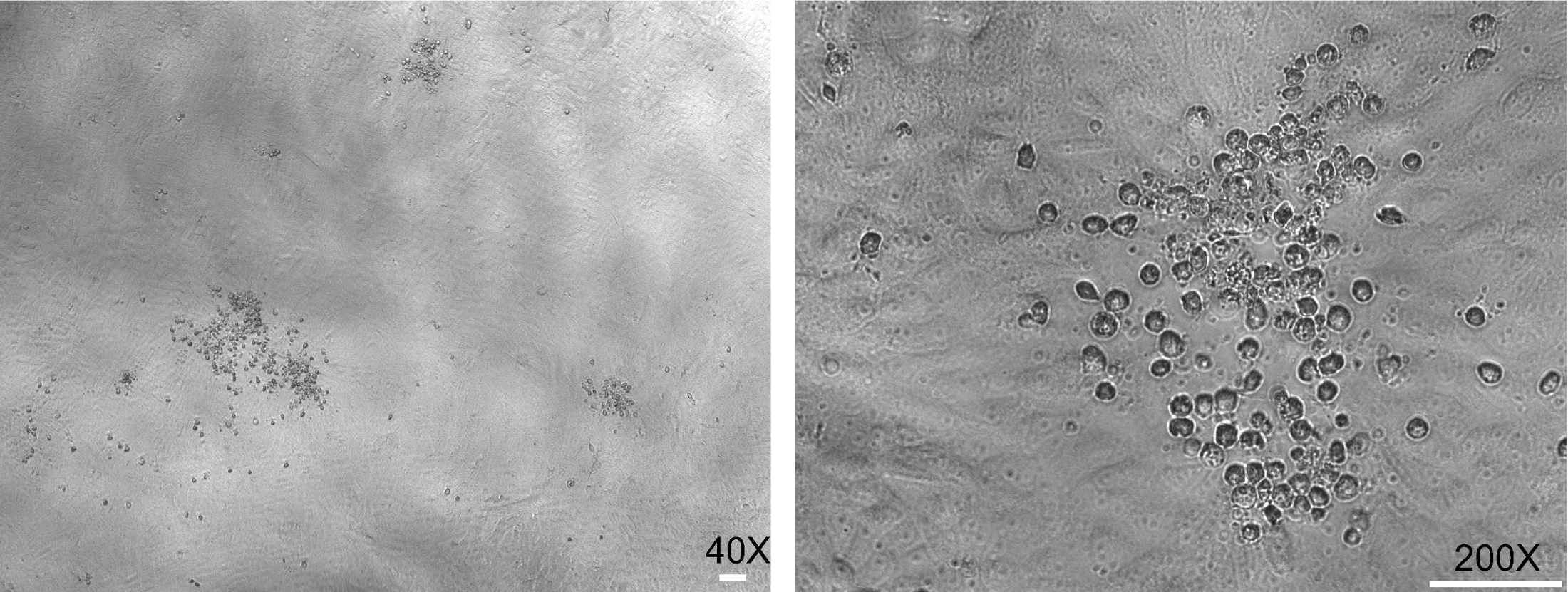
- Verify the progression of infection by observing the appearance of viral plaques on the cell monolayer using a phase contrast microscope and take images (Figure 2) as needed at 40X and 200X magnifications.

Figure 2: Plaques formed by Zika virus on a monolayer of Vero cells. Bright field images of various magnifications show Zika viral plaques at 48 hpi. Note the presence of rounded cell foci on the monolayer. (Scale bar = 50 µm) Please click here to view a larger version of this figure.
- Harvest cell culture supernatants at the 96 hr time point and centrifuge the supernatant at 300 x g for 10 min at 4 °C to remove cell debris.
- Carefully remove the supernatant without disturbing pelleted debris and transfer to 15 ml tubes. Ultracentrifugation at 24,000 x g can be performed to concentrate the viral particles (optional).
- Store the viral culture supernatants in multiple aliquots at -80 °C.
3. Measuring Zika Virus Titer by Plaque Assay
- Seed naive Vero cells at 1 x 10⁵ cells per well in 2 ml volume using a 12-well plate.
- The following day, prepare 10-fold serial dilutions of viral culture supernatants collected from the T-160 flask using serum-free media. Remove the spent media from each well and add 400 µl of prepared inoculum onto Vero cells in triplicate.
- Incubate the inoculated flasks in 37 °C with 5% CO2 for 4-6 hr. Spread the inoculum by gently tilting the plate sideways at every one hr.
- At the end of incubation, replace the inoculum with serum supplemented media (2 ml per well).
- At 48 hr post-inoculation, count the viral foci using a phase contrast microscope. Calculate the viral titer as plaque forming units (PFU) per ml. See Figure 3 for plaque assay.
- Fix and stain the cells in 4% formaldehyde and 0.1% crystal violet solution prepared in 20% ethanol for clear visualization of plaques.
4. Zika Viral Genome Replication Assay
- Seed naïve Vero cells at 1 x 10⁵ cells per well in 2 ml volumes using a 12-well plate. The following day, prepare viral inocula (MOI of 0.01 and 0.1; 400 µl/well) of low and high titer using serum free media in triplicate. For mock infection, use serum free media (400 µl/well) only.
- Repeat the steps 3.2 to 3.4.
- At the 48 and 96 hr time points, collect the samples for RNA and immunocytochemistry (ICC).
- For harvesting RNA samples, remove the media and then add 400 µl of lysis solution directly to each well and collect the lysates.
- For ICC, remove the media and then fix the cells by adding 1 ml of methanol to each well and incubate the plate at 4 °C for 30 min.
- From the cell lysate, isolate the total RNA using a RNA isolation kit per the manufacturer's instructions. Quantify the RNA using a spectrophotometer.
- Perform a two-step reverse transcription quantitative PCR (RT-qPCR) to determine the ZIKV genome content from harvested RNA.
- To reverse transcribe the RNA, first set up a 13 µl reaction mix comprised of 5 µg of isolated RNA, 1 µl of dNTPs (10 mM), and 1 µl random hexamer (250 ng) in a 0.2 ml tube. Incubate the tube at 65 °C for 5 min and then place it on ice for one minute. Subsequently, add 1 µl of reverse transcriptase, 4 µl of 5x strand synthesis buffer, 1 µl of 0.1 M dithiothreitol, and 1 µl of RNase inhibitor to the reaction mix. Incubate the tube for an additional 5 min at 25 °C, followed by 60 min at 50 °C and inactivate the reaction by heating to 70 °C for 15 min.
- Perform qPCR using 1 µl of the resulting cDNA with 12.5 µl of 2x green dye super mix containing DNA polymerase and 1 µl of 10 mM individual primers specific for Zika virus [Zika virus pan-genotype primers (For: 5'-AARTACACATACCARAACAAAGTGGT-3'; Rev: 5'-TCCRCTCCCYCTYTGGTCTTG-3'), Zika virus Asian genotype PRVABC59 strain primers (5'-AAGTACACATACCAAAACAAAGTGGT-3'; Rev: 5'-TCCGCTCCCCCTTTGGTCTTG-3')], or hepatitis C virus (JFH RTQ F: 5'CTGGGTCCTTTCTTGGATAA-3; JFH RTQ R: 5'CCTATCAGGCAGTACCACA-3'), or cellular housekeeping gene GAPDH (For: 5'-CCACCTTTGACGCTGGG-3'; Rev: 5'-CATACCAGGAAATGAGCTTGACA-3') in a 25 µl reaction volume.
- Perform PCR using the run condition 95 °C for 15 sec and 60 °C for 30 sec (40 cycles) in a real-time PCR system.
- Use GAPDH expression level based on cycle threshold (Ct) value to normalize the Zika viral genome measurement. Calculate the delta Ct (ΔCt) value of Zika genome compared to that of GAPDH and obtain the 2ΔCt value. Then, calculate the fold change by taking the ratio of normalized Zika genome contents between infected and uninfected cells at indicated time points. See Figure 4 for ZIKV genome replication results.
- Perform ICC using the methanol fixed cells.
- Wash the fixed cells three times with 1x PBS and block with ICC blocking buffer (3% goat serum, 3% BSA, 0.1% Triton-x 100 in PBS).
- Use mouse monoclonal anti-dsRNA antibody J2 (1 µg/ml) at a dilution of 1:100 in blocking buffer and incubate overnight at 4 °C.
- Wash the cells with 1x PBS and add secondary antibody goat anti-mouse IgG-594 (1 µg/ml) at a 1:1,000 dilution in blocking buffer and incubate for one hour at room temperature.
- Wash cells with 1x PBS and stain for nuclei using Hoechst dye and observe the cells using a fluorescent microscope at 100X magnification (Figure 4).
5. Screening Cytokine Library against Zika Virus Infection
- Seed naive Vero cells at 1 x 10⁵ cells per well using a 12-well plate. Once the cells are attached (6 hr post-seeding), add each cytokine at indicated concentrations in biological duplicates (2 ml volume/well). Include vehicle (PBS) alone control.
- At 12 hr post-treatment, perform Zika viral infection (MOI of 0.1 in 400 µl per well). Include negative control wells without infection (mock).
- At 4 hr post-infection, replace the viral inoculum with cytokine treated media (2 ml). Incubate the cells at 37 °C for additional 44 hr.
- At 48 hr post-infection, count viral plaques using a phase contrast microscope and acquire representative images of infected cells at 40X magnification (Figure 5).
Wyniki
A Zika viral strain (PRVABC59; GenBank accession number KU501215) of the Asian genotype was utilized in this study12. Vero cells at 80% confluency were used for investigating de novo Zika viral infection. For viral production and subsequent virological characterization, an early passage (P3) Zika virus was employed. The viral plaques were observed on the second day of infection. Zika viral progenies released from the initially infected cell can spread to neighboring ce...
Dyskusje
Here, a streamlined protocol for culturing Zika virus in vitro is presented. Critical steps including, identifying optimum end points for expanding virus culture, measuring titer, and quantifying genome replication were provided. Zika virus is a human pathogen, so, while handling infectious agents, biosafety procedures are to be strictly followed. A monkey kidney cell line, Vero, was used for demonstrating various virological assays. Zika viral replication kinetics may differ in cells of various tissues and spec...
Ujawnienia
The authors have nothing to disclose.
Podziękowania
We would like to thank Dr. Aaron Brault and Dr. Brandy Russell of the Centers for Disease Control and Prevention (CDC), USA for providing Zika viral strain PRVABC59. We thank Nicholas Ten of Yale University for copy-editing this manuscript. This work was supported by the Cedars-Sinai Medical Center Institutional Programmatic Research Award to V.A.
Materiały
| Name | Company | Catalog Number | Comments |
| Dulbecco’s modified Eagle’s medium (DMEM) | Sigma Life Science | D5796 | |
| HEPES | Life Technologies | 15630080 | |
| Glutamax | Life Technologies | 35050061 | |
| 2.5% Trypsin, 10x [-] Phenol Red | Corning | 25-054-C1 | |
| Trypan Blue Stain 0.4% | Life Technologies | T10282 | |
| Countess – Automated Cell Counter | ThermoFisher Scientific | C10227 | |
| Countess-cell counting chamber slides | ThermoFisher Scientific | C10283 | |
| Rneasy Mini Kit | Qiagen | 74104 | |
| Nanodrop 2000 | Thermo Scientific | Nanodrop 2000 | |
| mouse monoclonal anti-dsRNA antibody J2 | English & Scientific Consulting Kft. | 10010200 | |
| Goat anti-rabbit IgG Alexa Fluor 594 | Life Technologies | A11020 | |
| SUPERSCRIPT III RT | Life Technologies | 18080085 | |
| SYBR QPCR SUPERMIX W/ROX | Life Technologies | 11744500 | |
| QuantStudio12K Flex Real-Time PCR System | Thermo Fischer | 4471088 | |
| RNase-Free DNase | Promega | M6101 | |
| Vero Cell Line | ATCC | CCL-81 | |
| Zika viral strain PRVABC59 | Centers for Disease Control and Prevention (CDC) | ||
| IL-6 | Peprotech | 200-06 | |
| IL-1 alpha | Peprotech | 200-01A | |
| TNF-alpha | Peprotech | 300-01A | |
| Interferon alpha A | R & D Systems | 11100-1 | |
| Interferon beta | Peprotech | 300-02BC | |
| Interferon gamma | Peprotech | 300-02 | |
| Centrifuge 5415R | Eppendorf | 5415R | |
| Centrifuge 5810R | Eppendorf | 5810R | |
| Nikon Eclipse Ti Immunofluorescence Microscope with Nikon Intenselight C-HGFI | Nikon | Visit Nikon for Request |
Odniesienia
- Brasil, P., et al. Zika Virus Infection in Pregnant Women in Rio de Janeiro - Preliminary Report. N Engl J Med. , 1-11 (2016).
- Lucey, D. R., Gostin, L. O. The Emerging Zika Pandemic: Enhancing Preparedness. JAMA. 315 (9), 865-866 (2016).
- Mlakar, J., et al. Zika Virus Associated with Microcephaly. N Engl J Med. 374 (10), 951-958 (2016).
- Schuler-Faccini, L., et al. Possible Association Between Zika Virus Infection and Microcephaly. MMWR Morb Mortal Wkly Rep. 65 (3), 59-62 (2015).
- Foy, B. D., et al. Probable non-vector-borne transmission of Zika virus, Colorado, USA. Emerg Infect Dis. 17 (5), 880-882 (2011).
- Kuno, G., Chang, G. J. Full-length sequencing and genomic characterization of Bagaza, Kedougou, and Zika viruses. Arch Virol. 152 (4), 687-696 (2007).
- Dick, G. W. Zika virus. II. Pathogenicity and physical properties. Trans R Soc Trop Med Hyg. 46 (5), 521-534 (1952).
- Dick, G. W., Kitchen, S. F., Haddow, A. J. Zika virus. I. Isolations and serological specificity. Trans R Soc Trop Med Hyg. 46 (5), 509-520 (1952).
- Oehler, E., et al. Zika virus infection complicated by Guillain-Barre syndrome--case report, French Polynesia. Euro Surveill. 19 (9), 1-3 (2013).
- Baronti, C., et al. Complete coding sequence of zika virus from a French polynesia outbreak in 2013. Genome Announc. 2 (3), e00500-e00514 (2014).
- Lanciotti, R. S., et al. Genetic and serologic properties of Zika virus associated with an epidemic, Yap State, Micronesia, 2007. Emerg Infect Dis. 14 (8), 1232-1239 (2008).
- Lanciotti, R. S., et al. Phylogeny of Zika virus in Western Hemisphere, 2015 [Letter]. Emerg Infect Dis. 22 (5), (2016).
- Musso, D., Nilles, E. J., Cao-Lormeau, V. M. Rapid spread of emerging Zika virus in the Pacific area. Clin Microbiol Infect. 20 (10), O595-O596 (2014).
- Tang, H., et al. Zika Virus Infects Human Cortical Neural Progenitors and Attenuates Their Growth. Cell Stem Cell. , 1-5 (2016).
- Hamel, R., et al. Biology of Zika Virus Infection in Human Skin Cells. J Virol. 89 (17), 8880-8896 (2015).
- Faye, O., et al. Quantitative real-time PCR detection of Zika virus and evaluation with field-caught mosquitoes. Virol J. 10, 311 (2013).
- Chu, D., et al. Systematic analysis of enhancer and critical cis-acting RNA elements in the protein-encoding region of the hepatitis C virus genome. J Virol. 87 (10), 5678-5696 (2013).
- Hiratsuka, M., et al. Administration of interferon-alpha during pregnancy: effects on fetus. J Perinat Med. 28 (5), 372-376 (2000).
- Ozaslan, E., et al. Interferon therapy for acute hepatitis C during pregnancy. Ann Pharmacother. 36 (11), 1715-1718 (2002).
Przedruki i uprawnienia
Zapytaj o uprawnienia na użycie tekstu lub obrazów z tego artykułu JoVE
Zapytaj o uprawnieniaThis article has been published
Video Coming Soon
Copyright © 2025 MyJoVE Corporation. Wszelkie prawa zastrzeżone