The Split Brain
Source: Laboratories of Jonas T. Kaplan and Sarah I. Gimbel—University of Southern California
The study of how damage to the brain affects cognitive functioning has historically been one of the most important tools for cognitive neuroscience. While the brain is one of the most well protected parts of the body, there are many events that can affect the functioning of the brain. Vascular issues, tumors, degenerative diseases, infections, blunt force traumas, and neurosurgery are just some of the underlying causes of brain damage, all of which may produce different patterns of tissue damage that affect brain functioning in different ways.
The history of neuropsychology is marked by several well-known cases that led to advances in the understanding of the brain. For instance, in 1861 Paul Broca observed how damage to the left frontal lobe resulted in aphasia, an acquired language disorder. As another example, a great deal about memory has been learned from patients with amnesia, such as the famous case of Henry Molaison, known for many years in the neuropsychology literature as "H.M.," whose temporal lobe surgery led to a profound deficit in forming certain kinds of new memories.
While the observation and testing of patients with focal brain damage has provided neuroscience with insight into the functioning of the brain, great care must be taken in designing tests to reveal the specific nature of the deficit. Also, because the brain is a complex network of interconnected neurons, damage to one brain region can affect functioning in regions far away from the damage. To demonstrate how brain damage can affect connections among brain regions, this video examines the case of the so-called split brain.
The corpus callosum is a large bundle of fibers that connects the left and right hemispheres of the brain. It is one of the largest white matter tracts in the brain and can be easily recognized on a sagittal view of the midline of the brain. In the 1960s, neurosurgeons discovered that cutting the corpus callosum could be a successful treatment for certain kinds of epilepsy, which involves uncontrollable neural activity spreading through the brain. People who underwent the split-brain operation had their two hemispheres surgically separated, such that the left and right hemispheres were no longer able to communicate. This condition allowed experimenters to probe the functions of the left and right hemisphere independently, to learn about the relative abilities, and about the nature of communication between them.
This video demonstrates how to test a split-brain patient to reveal some of the differences between the two hemispheres of the brain and to see some dramatic consequences of such a disconnection. The original versions of these experiments were developed by Michael Gazzaniga and colleagues1, 2 and later were elaborated upon by others;3 the version presented here incorporates more recent modernizations of the methodology.
1. Patient and control recruitment
- There are a variety of patients with disconnection syndromes, including complete and partial surgical callosotomies and congenital conditions, such as agenesis of the corpus callosum (ACC), in which the corpus callosum does not fully develop. There are multiple tracts that connect the two hemispheres; the largest is the corpus callosum, but some fibers cross at the anterior commissure, hippocampal commissure, and posterior commissure.
Note that these different varieties of disconnection may lead to different behavioral outcomes in this testing. - For the purposes of this experiment, preselect the patient through the use of neuroimaging to confirm the absence of connecting fibers.
- Standard MRI and diffusion imaging, which can be used to image white matter tracts, are particularly useful. Knowing which connecting fibers are present in the patient helps with the interpretation of the results. In this demonstration, a patient with a complete callosotomy has been selected.
- Make sure that the patient has been fully informed of the research procedures and has signed all the appropriate consent forms.
- Recruit 20 participants of the same age and gender as the patient, matched for intelligence, using scores on the Wechsler Adult Intelligence Scale (WAIS).
2. Data collection
- In order to present visual stimuli to the left or right hemisphere alone, the stimuli must be properly presented to one visual field. Note that this is not equivalent to presenting the stimuli to one eye. Each eye projects to both hemispheres of the brain; for example, the part of the left eye that sees the left visual field is processed by the right hemisphere, but the part of the left eye that processes the right visual field is seen by the left hemisphere. Therefore, to present an image to the left hemisphere, present it entirely within the right visual field, which is to the right of where the patient is looking.
- To achieve this lateralization, use a chinrest to maintain the eyes approximately 22 in. from the computer screen. Place the patient's chin comfortably within the chinrest, facing the screen.
- Have a small cross remain in the center of the screen to provide a location for the patient to fixate their eyes.
- Instruct the patient to maintain their fixation on this cross throughout the experiment, even as images appear to the left or right side of it.
- Explain to the patient that when an image appears, they should say the name of the object out loud.
- Present images of well-known objects briefly on the left or right side of the screen to project them to the right or left hemispheres of the brain, respectively. 50 images are presented in random order from a set of objects that include easily recognizable drawings, such as an apple, a ball, a broom, and a chicken.
- Present the images for less than 150 ms to ensure proper lateralization. This is enough time to see the stimulus, but fast enough so the patient isn't able to move their eyes to see the stimulus in central vision.
- Ask the patient to name the objects presented on the screen out loud, and record their responses. This is a test of verbal linguistic capability and should reveal the differences in speaking ability between the hemispheres.
- If the patient is unable to name any of the objects, ask the patient to draw the object, without looking at the paper, with the hand ipsilateral to (on the same side as) the stimulus. This serves as a non-linguistic measure of knowledge of the stimulus.
- The hand ipsilateral to the stimulus is controlled by the hemisphere that saw the stimulus. For example, when the stimulus is presented in the left visual field, it is processed by the right hemisphere. The right hemisphere is largely responsible for the control of the left hand.
- Make sure the patient does not look at their hand while it is drawing to maintain the isolation of the stimulus to one hemisphere.
- When the patient finishes drawing an object, ask them to look at the object and say what it is out loud. This confirms that the patient knows the name of the object when it is presented in central vision, even if they are unable to name it when it is presented to a single hemisphere.
- Repeat the procedure for each control participant.
3. Data Analysis
- To analyze the patient's performance, compare the data from the left and right visual half-field with each other. To do this, tabulate the number of correct and incorrect responses in each visual field, and test the likelihood of obtaining a difference as large as the one observed using a chi-square test of independence.
- Compare the data from the patient with the data from the age, gender, and intelligence-matched control population to determine deficits in patient behavior. To do this, compile the average score for each person's left visual field and right visual field separately, and compare the distributions using a repeated-measures analysis of variance test (ANOVA).
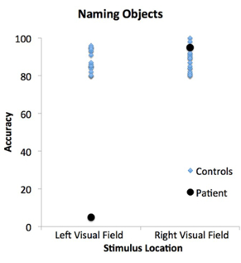
Typically, callosotomy patients exhibit an anomia for objects presented in the left visual half-field. Anomia is the inability to name objects. Objects presented to the right visual field, however, are named with high accuracy (Figure 1).

Figure 1: Patient and control performance in the naming objects task for stimuli presented in the left and right visual fields. The patient (black circles) is not able to verbally name objects presented in the left visual field, but is able to name objects in the right visual field. In contrast, the control population (blue diamonds) can name objects presented in both the left and right visual fields.
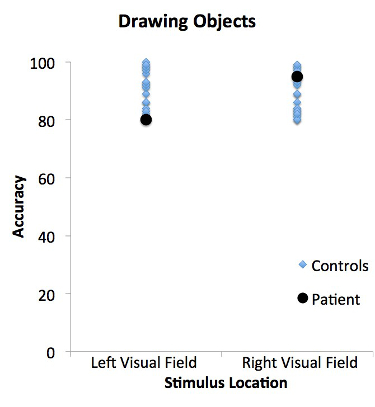
Some patients may be able to successfully draw objects presented to the left visual field, even though they cannot verbally name them (Figure 2).

Figure 2: Patient and control performance in the drawing objects task for stimuli presented in the left and right visual fields. The patient (black circles) and control population (blue diamonds) are able to draw objects presented in both the left and right visual fields. The patient's performance does not differ from matched controls.
In this case, the patient usually says they haven't seen anything. This is because the left hemisphere, which is controlling speech, has not seen the visual image. However, the right hemisphere, which has seen the object, can recognize it but is unable to generate speech. Since the right hemisphere is largely in control of the left hand, the patient is able to draw the object with the left hand. This result demonstrates a dissociation between the ability to recognize an object and the ability to verbally name an object.
The control population, with intact corpora callosa, can both name and draw objects presented in the left or right visual fields. This is because information can freely pass from one hemisphere to the other, allowing for the sharing of information between the brain regions.
The case of the split-brain patient reveals the relative specialization of the two cerebral hemispheres. Many of these specializations can also be demonstrated in healthy people with intact commissures using similar techniques. For example, people tend to recognize words faster when they are presented briefly in the right visual field compared to when they are presented in the left visual field. This experiment also shows that even when two brain regions are healthy, damage to the connections between different regions can affect behavior.
However, it is important to remember that while testing the split brain demonstrates the differences between the two cerebral hemispheres, in the intact brain, the two hemispheres are continually interacting with each other and working in concert. To isolate a stimulus to one visual field requires specialized equipment that can present stimuli very briefly and away from central fixation. Since central vision is processed by both hemispheres, and the eyes typically scan an environment, this is not a situation that is likely to be encountered in everyday life.
- Gazzaniga, M. S., Bogen, J. E., & Sperry, R. W. (1962). Some functional effects of sectioning the cerebral commissures in man. Proc Natl Acad Sci U S A, 48, 1765-1769.
- Gazzaniga, M. S., Bogen, J. E., & Sperry, R. W. (1965). Observations on visual perception after disconnexion of the cerebral hemispheres in man. Brain, 88(2), 221-236.
- Zaidel, E., Zaidel, D., & Bogen, J. E. (1990). Testing the commussurotomy patient. In A. Boulton, G. Baker, & M. Hiscock (Eds.), Neuromethods (pp. 147-201). Clifton, NJ: Humana Press.
Skip to...
Videos from this collection:

Now Playing
The Split Brain
Neuropsychology
68.1K Views

Motor Maps
Neuropsychology
27.4K Views

Perspectives on Neuropsychology
Neuropsychology
12.0K Views

Decision-making and the Iowa Gambling Task
Neuropsychology
32.1K Views

Executive Function in Autism Spectrum Disorder
Neuropsychology
17.5K Views

Anterograde Amnesia
Neuropsychology
30.2K Views

Physiological Correlates of Emotion Recognition
Neuropsychology
16.2K Views

Event-related Potentials and the Oddball Task
Neuropsychology
27.4K Views

Language: The N400 in Semantic Incongruity
Neuropsychology
19.5K Views

Learning and Memory: The Remember-Know Task
Neuropsychology
17.1K Views

Measuring Grey Matter Differences with Voxel-based Morphometry: The Musical Brain
Neuropsychology
17.2K Views

Decoding Auditory Imagery with Multivoxel Pattern Analysis
Neuropsychology
6.4K Views

Visual Attention: fMRI Investigation of Object-based Attentional Control
Neuropsychology
40.7K Views

Using Diffusion Tensor Imaging in Traumatic Brain Injury
Neuropsychology
16.7K Views

Using TMS to Measure Motor Excitability During Action Observation
Neuropsychology
10.1K Views
Copyright © 2025 MyJoVE Corporation. All rights reserved