A subscription to JoVE is required to view this content. Sign in or start your free trial.
Embedding Adult Drosophila Heads in Paraffin: Preparing Tissue for Histology
Overview
This video describes how to embed Drosophila heads in paraffin using the collar method, a technique that prepares fly heads for serial sectioning. Paraffin sections are used for histological examinations of the brain's anatomy and for the assessment of neurodegeneration. The featured protocol demonstrates how multiple flies can be processed in one preparation, thereby increasing time-efficiency and minimizing experimental artifacts.
Protocol
This protocol is an excerpt from Sunderhaus and Kretzschmar, Mass Histology to Quantify Neurodegeneration in Drosophila, J. Vis. Exp. (2016).
1. Fixing the Head on Collars and Embedding in Paraffin
NOTE: All of the steps in the fixation process should be done in a fume hood. Methylbenzoate, while not posing a health risk, has a highly distinct odor, whic.......
Representative Results

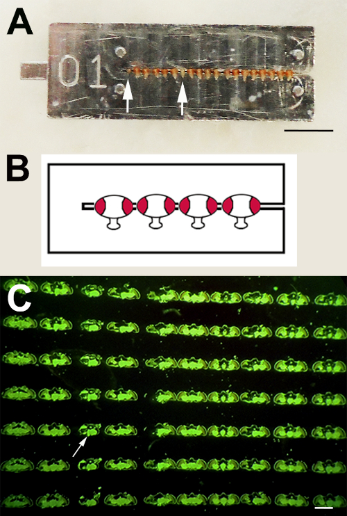
Figure 1: Paraffin Serial Sections. A) Using the collar method, experimental and control flies can be processed as one sample by threading them onto one collar. Eyeless sine oculis flies are inserted for orientation (arrows). B) Schematic showing the orientation of the fly heads in the collar. C) In this image, sections from different fly heads are oriented .......
This article has been published
Video Coming Soon
Source: Sunderhaus, E. R., Kretzschmar, D. Mass Histology to Quantify Neurodegeneration in Drosophila .J. Vis. Exp. (2016).
ABOUT JoVE
Copyright © 2025 MyJoVE Corporation. All rights reserved