A subscription to JoVE is required to view this content. Sign in or start your free trial.
Immunofluorescent Staining of Spheroids: A Technique to Analyze Spheroids for Expression of Intra- and Extracellular Antigen Expression
Overview
This video describes the technique of immunofluorescent staining of spheroids to understand the differentiation of the fibroblasts or CAFs. This protocol helps detect expression of intra- and extracellular antigens, including deposition of ECM proteins within the spheroids.
Protocol
1. Immunofluorescent staining of spheroids
- Collect the spheroids after about 24 h into one 1.5 mL reaction tube per experimental condition or per protein to be analyzed. For example, place the spheroids of iNFs stimulated with TGF-β1 in a tube different from the control iNFs, which were not treated. To avoid rupture of spheroids due to too strong shear forces, cut the end of the pipette tip to enlarge the orifice. Include at least 5-10 spheroids in one reaction tube, to allow replicates and take into acc.......
Representative Results

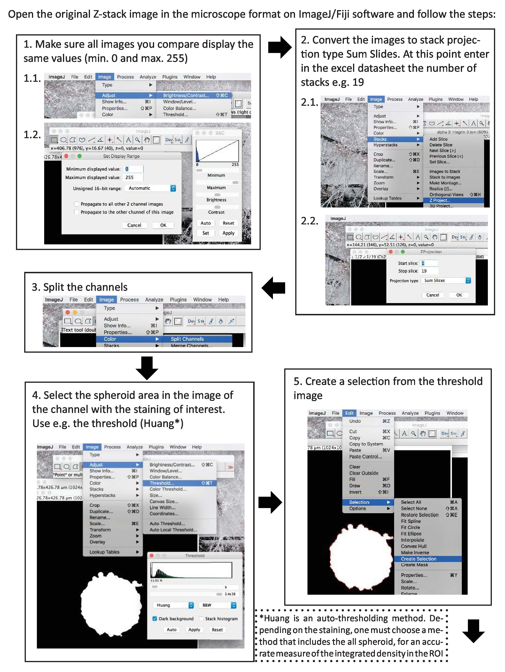
Figure 1. Sequential steps for quantification of the immunofluorescent staining of a protein of interest in spheroids, using ImageJ. Please click here to view a larger version of this figure.
Reprints and Permissions
Request permission to reuse the text or figures of this JoVE article
Request PermissionThis article has been published
Video Coming Soon
Source: Cavaco, A. C. M. et al. A 3D Spheroid Model as a More Physiological System for Cancer-Associated Fibroblasts Differentiation and Invasion In Vitro Studies. J. Vis. Exp. (2019).
ABOUT JoVE
Copyright © 2025 MyJoVE Corporation. All rights reserved