A subscription to JoVE is required to view this content. Sign in or start your free trial.
Spontaneous Metastasis Mouse Models: A Platform to Study the Metastatic Potential of Orthotopically Injected Colorectal Cancer Cells
In This Article
Overview
This video describes the protocol to establish a spontaneous metastasis mouse model of colorectal cancer patient-derived xenografts or PDXs. These xenograft models partially mimic the features of human carcinomas and understand the critical aspects of human malignancies, including invasion and metastasis.
Protocol
All procedures involving animal models have been reviewed by the local institutional animal care committee and the JoVE veterinary review board.
1. Dissection of CRC PDXs from Mice
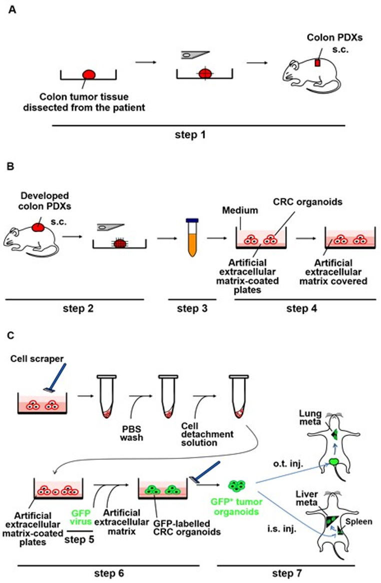
Experimental procedures for dissection of CRC PDXs are outlined in Figure 1B (step 2).
- Allow the tumor to grow subcutaneously to ~1 cm3 in size, which usually takes approximately 1-3 months.
- Anesthetize the mouse with isoflurane inhalation using the small animal inhalation anesthesia device and disinfect the skin with 70% ethanol.
- Place the mouse in ....
Representative Results

Figure 1: Schematic representation of the generation of metastases by the PDX-derived CRC organoids labeled with GFP lentivirus in NOG mice. (A) Implantation of small pieces of the CRC tissue subcutaneously into NOG mice (step 1). The CRC tissue surgically dissected from the patient was cut into pieces and implanted subcutaneously into NOG mice. s.c.: subcut.......
This article has been published
Video Coming Soon
Source: Okazawa, Y., et al. High-sensitivity Detection of Micrometastases Generated by GFP Lentivirus-transduced Organoids Cultured from a Patient-derived Colon Tumor. J. Vis. Exp. (2018).
Copyright © 2025 MyJoVE Corporation. All rights reserved