A subscription to JoVE is required to view this content. Sign in or start your free trial.
Patient-derived Xenograft Modeling: A Technique to Generate Melanoma Mouse Models
Overview
In this video, human-derived melanoma cells are implanted subcutaneously into the flank region of an immunocompromised mouse. The PDX model allows preclinical investigation of melanoma cells that better recapitulate the tumor heterogeneity and melanoma aggressiveness observed in in vivo conditions.
Protocol
All procedures involving animal models have been reviewed by the local institutional animal care committee and the JoVE veterinary review board.
1. Tumor tissue processing for mouse implantation
- Surgical excision or surgical biopsy tissue processing
- Transfer the tissue to a sterile Petri dish and separate the tumor tissue from the surrounding normal tissue as much as possible.
- Remove necrotic tissue (usually identified as pale-whitish ti.......
Representative Results

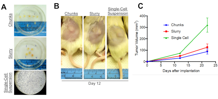
Figure 1: Alternative implantation methods. (A) Tumors can be processed into either chunks, a slurry suspension, or as a single-cell suspension. (B) All three methods will allow for the growth of tumors in vivo. Shown here are mice subcutaneously implanted with tumor and imaged 12 days after implantation. (C) Shown are tumor growth curves fo.......
Reprints and Permissions
Request permission to reuse the text or figures of this JoVE article
Request PermissionThis article has been published
Video Coming Soon
Source: Min Xiao et al. A Melanoma Patient-Derived Xenograft Model. J. Vis. Exp. (2018)
ABOUT JoVE
Copyright © 2025 MyJoVE Corporation. All rights reserved