A subscription to JoVE is required to view this content. Sign in or start your free trial.
Extracting Intact Kidney from Mouse: A Technique to Obtain Kidney Without Renal Capsule from Murine Models
Overview
In this video, we describe the procedure for kidney extraction from the retroperitoneal space of the mouse. The extracted kidney can further be microdissected to obtain an intact kidney without a renal capsule and the surrounding fat layer.
Protocol
All procedures involving animal models have been reviewed by the local institutional animal care committee and the JoVE veterinary review board.
1. Kidney Dissection
- Anaesthetize mice by inhalation of 3–4% isoflurane in a ventilated hood. Confirm the induction of deep anesthesia by loss of toe and/or tail pinch reflex and then euthanize the mice by cervical dislocation.
- Apply 70% ethanol to the chest to dampen the fur. Using.......
Representative Results

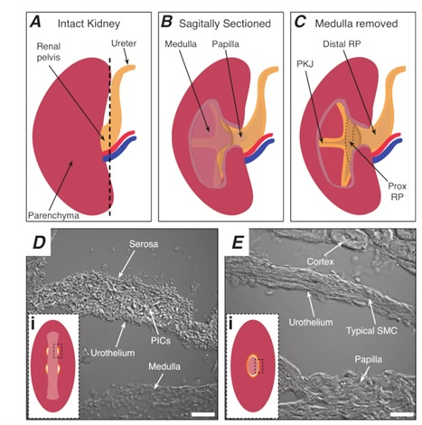
Figure 1: Basic kidney anatomy and location of PKJ pacemaker region. (A) Diagram of the intact kidney showing the orientation of the RP and ureter. The renal artery and renal vein are displayed in red and blue, respectively. (B) The intact kidney can be cut along a sagittal plane to expose the inner aspect of the kidney, including the medulla, papilla (dista.......
This article has been published
Video Coming Soon
Source: Grainger, N. et al. Isolating and Imaging Live, Intact Pacemaker Regions of Mouse Renal Pelvis by Vibratome Sectioning. J. Vis. Exp. (2021)
ABOUT JoVE
Copyright © 2025 MyJoVE Corporation. All rights reserved