A subscription to JoVE is required to view this content. Sign in or start your free trial.
Ex Vivo Electroporation of Chick Embryo Cerebellar Slices: A Method to Introduce Plasmid DNA Encoding Green Fluorescent Protein to Visualize Granular Cell Development
Overview
In this video, we perform electroporation of chick embryo cerebellum slices with reporter gene plasmids. The technique facilitates visualization of the development and migration of granule cell precursors to form granular layer.
Protocol
1. Electroporation of Slices
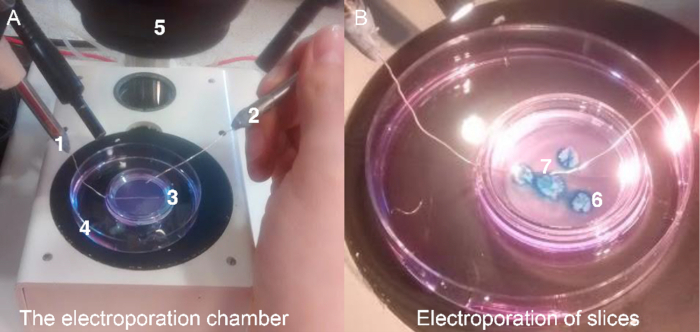
- Construct an electroporation chamber by fixing the anode of an electroporator to the base of a 60 mm Petri dish with insulation tape. Add approximately 1 mL of HBSS to cover the electrode.
- Place a 0.4 µm culture insert on top of the electrode covered in HBSS. Allow the culture insert to rest on the electrode making sure there is always contact between the insert and the electrode.
NOTE: In this setup, the culture insert with .......
Representative Results

Figure 1. The electroporation chamber set up. (A) A picture of the custom-made electroporation chamber. The chamber consists of an anode of an electroporator placed securely on the base of a 60 mm Petri dish. The dish contains approximately 1 mL of HBSS to cover the electrode. The culture insert should rest on the electrode with constant contact between.......
Reprints and Permissions
Request permission to reuse the text or figures of this JoVE article
Request PermissionThis article has been published
Video Coming Soon
Source: Hanzel, M. et al. Ex Vivo Culture of Chick Cerebellar Slices and Spatially Targeted Electroporation of Granule Cell Precursors. J. Vis. Exp. (2015)
ABOUT JoVE
Copyright © 2025 MyJoVE Corporation. All rights reserved