A subscription to JoVE is required to view this content. Sign in or start your free trial.
Three-Chambered Array-Based Impedance Assay: A Real-Time Analysis Technique to Assess Invasive Potential of Cancer Cells by Measuring Electrical Impedance
Overview
This video describes an invasion assay using a three-chambered array based on electrical impedance. This technique measures the invasive potential of cancer cells under the influence of soluble factors secreted by the resident stromal cells in tumor microenvironments.
Protocol
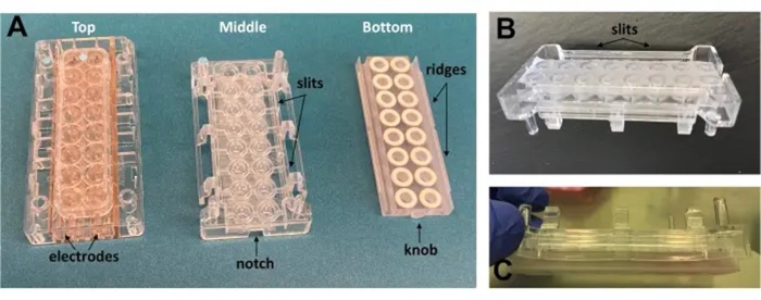
1. New chamber design (Figure 1)
- Open a new dual-chamber cell analyzer plate. Set aside the top chamber with electrodes.
- Using a milling machine, shave off 2 mm of the U-shaped bottom wells of the cell analyzer plate.
- Attach a 2 cm x 7 cm polyethersulfone (PES) membrane with 0.2 μm pore size to the bottom of the shaved wells using UV-curated adhesive. Allow 30 min curation time to ensure the glue is completely cured and inert.
- Using a milling machine, cut out two longitudina.......
Representative Results

Figure 1: Images of the array chambers and modifications.
(A) The three chambers used to build the array. No modification was made on the top chamber harboring the electrodes. (B) From the middle chamber wells, a height of 2 mm has been shaved off and a membrane attached to the open bottom; longitudinal slits (1.5 mm x 5.6 mm) were add.......
Reprints and Permissions
Request permission to reuse the text or figures of this JoVE article
Request PermissionThis article has been published
Video Coming Soon
Source: Sharif, G. M. et al., Real-Time Detection and Capture of Invasive Cell Subpopulations from Co-Cultures. J. Vis. Exp. (2022).
ABOUT JoVE
Copyright © 2025 MyJoVE Corporation. All rights reserved