A subscription to JoVE is required to view this content. Sign in or start your free trial.
Protein Aggregate Formation Assay: A Method to Detect and Quantify Protein Aggregation in Cultured Cells upon Induction by Proteasome Inhibitor
In This Article
Overview
In this video, we demonstrate a cell-based protein aggregation assay using proteasome inhibitors, which block proteasome activity, preventing misfolded, mutant proteins, fused to a fluorescent label, from undergoing ubiquitin-dependent proteasomal degradation, leading to their accumulation within the cell cytoplasm. The protein aggregates are then visualized and quantified by fluorescence microscopy.
Protocol
1. Lentivirus production
NOTE: The production and manipulation of lentiviral vectors was carried out according to the National Institutes of Health (NIH) guidelines for research involving recombinant DNA. The plasmid encoding the wild-type and A4V mutant SOD1 tagged with enhanced YFP (SOD1WT-YFP and SOD1A4V-YFP) are used. Both gene fusion products were amplified using the PCR primer pair 5′-ATCGTCTAGACACCATGGCGACGAAGGTCGTGTGC-3′ and 5′-TAGCGG CCGCTACTTGTACAGCTCGTCCATGCC-3′and inserted into the pTRIP-delta U3 CMV plasmid using XhoI and BsrGI restriction sites. Avoidance of more than 20 passages and main....
Results

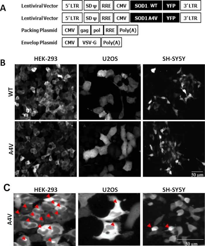
Figure 1. SOD1 WT and A4V stable line generation. (A) Schematic diagram of lentiviral and packaging vectors (Packing and Envelop plasmids) for wild type and mutant SOD1 A4V lentivirus generation. (B) Selected confocal images of three cells (HEK-293, U2OS, and SH-SY5Y) transduced with the lentivirus wild type SOD1 (WT) and mutant SOD1 (A4V). (C) Representative ima.......
Disclosures
Materials
| Name | Company | Catalog Number | Comments |
| ALLN (C20H37N3O4) | Millipore | 208719 | |
| MG132 (C26H41N3O5) | Sigma-Aldrich | C2211 | |
| Epoxomicin (C28H50N4O7) | Sigma-Aldrich | E3652 | |
| Hoechst 33342 | Invitrogen | H-3570 | |
| Opera | Perkin Elmer | OP-QEHS-01 | |
| Opera EvoShell software | Perkin Elmer | Ver 1.8.1 | |
| Operetta | Perkin Elmer | OPRT1288 | |
| Harmony Imaging software | Perkin Elmer | Ver 3.0.0 | |
| Columbus Image analysis software | Perkin Elmer | Ver 2.3.2 | |
| CyBi Hummingwell liquid handling | CyBio AG | OL 3387 3 0110 |
This article has been published
Video Coming Soon
Source: Lee, H. et al. Assay Development for High Content Quantification of Sod1 Mutant Protein Aggregate Formation in Living Cells. J. Vis. Exp. (2017)
Copyright © 2025 MyJoVE Corporation. All rights reserved