A subscription to JoVE is required to view this content. Sign in or start your free trial.
Fluorescence Microplate-Based Cycloheximide Chase Assay: A Technique to Monitor the Degradation Kinetics of Fluorescent Nuclear Misfolded Proteins
In This Article
Overview
This video describes an in vitro fluorescence-based microplate assay to study the degradation kinetics of a fluorescently-labeled misfolded luciferase mutant protein expressed in the nuclei of transfected mammalian cells. The assay involves the treatment of cells expressing the fluorescent mutant protein with a translation inhibitor and a proteasome inhibitor to assess the proteasome-mediated degradation of the misfolded protein.
Protocol
1. Preparation of Reagent
- Prepare low-fluorescence DMEM medium for assays using a microplate fluorescence reader. Mix 25 mM glucose, 0.4 mM glycine, 0.4 mM arginine, 0.2 mM cysteine, 4.0 mM glutamine, 0.2 mM histidine, 0.8 mM isoleucine, 0.8 mM leucine, 0.8 mM lysine, 0.2 mM methionine, 0.4 mM phenylalanine, 0.4 mM serine, 0.8 mM threonine, 0.078 mM tryptophan, 0.4 mM tyrosine, 0.8 mM valine, 1.8 mM CaCl2, 0.81 mM MgSO4, 5.33 mM KCl, 44.0 mM NaHCO3, 110 mM NaCl, 0.9 mM NaH2PO4. Adjust the pH of the solution using HCl or NaOH to pH 7.4. Sterilize the medium through filtration.
NOTE....
Results

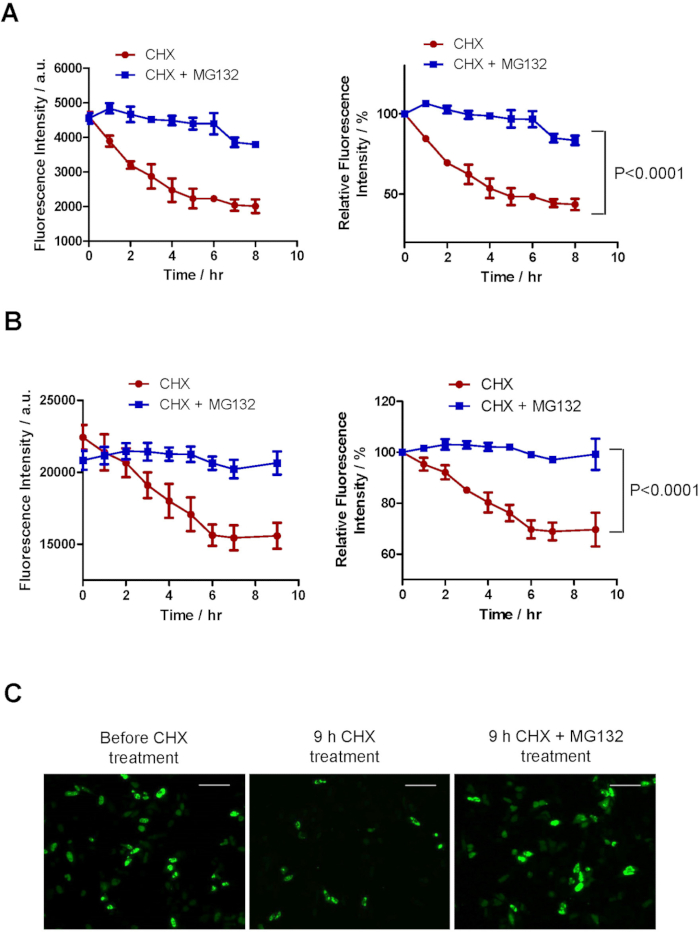
Figure 1: Fluorescent microplate-based assay for NLS-LucDM-GFP degradation. HeLa cells seeded on 96 well plates were transfected with 0.05 µg (A) or 0.1 µg (B) NLS-LucDM-GFP and treated with CHX in the presence or absence of MG132. (A and B) Fluorescence intensities of wells were measured at the indicated time points (M.......
Disclosures
Materials
| Name | Company | Catalog Number | Comments |
| Dulbecco's Modified Eagle Medium | Life Technologies | 11995-092 | |
| Fetal Bovine Serum | Life Technologies | 10082147 | |
| Lipofectamine 2000 | Life Technologies | 11668019 | |
| MG132 | Sigma-Aldrich | M8699 | |
| Amino acids | Sigma-Aldrich | Amino acids are used for making low fluorecence culturing medium | |
| Cycloheximide | Sigma-Aldrich | C7698 | |
| Olympus IX-81 Inverted Fluorescence Microscope | Olympus | IX71/IX81 | |
| 96 Well Black TC Plate w/ Transluscent Clear Bottom | Sigma-Greiner | 89135-048 | |
| Fluorescence Bottom Plate Reader Infinite 200® PRO | TECAN | Infinite 200® PRO | |
| Prism 5 | GraphPad | Statistical analysis software |
This article has been published
Video Coming Soon
Source: Guo, L. et. al., Assays for the Degradation of Misfolded Proteins in Cells. J. Vis. Exp. (2016)
Copyright © 2025 MyJoVE Corporation. All rights reserved