A subscription to JoVE is required to view this content. Sign in or start your free trial.
Neuronal Death Assay to Evaluate Cell Death in Ex Vivo Epilepsy Model
Overview
This video demonstrates the technique of assessing neuronal death in rhinal cortex-hippocampus organotypic slices. Neuronal death occurs via evolving epileptic seizures — resembling in vivo epilepsy — and is detected through dual-staining of the dead cells using propidium iodide and immunofluorescence.
Protocol
All procedures involving animal models have been reviewed by the local institutional animal care committee and the JoVE veterinary review board.
1. Preparation of rhinal cortex-hippocampus slices
NOTE: The preparation of rhinal cortex-hippocampus slices uses P6-7 Sprague-Dawley rats.
- Culture setup and medium preparation
- On the day before the culture, prepare the required media and place them at 4 °C.
- Prepare d.......
Representative Results

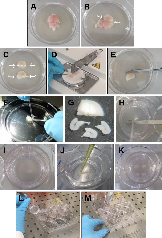
Figure 1: Detailed procedure for the preparation of rhinal cortex-hippocampus organotypic slices. (A) Remove the brain from the head and place it in ice-cold GBSS with the dorsal surface faced up. (B) Insert the forceps into the cerebellum. Open the brain through the midline and remove the excess tissue over the hippocampus. (C) With a spatula cut below the hippocampus, as indic.......
This article has been published
Video Coming Soon
Source: Carvalho, M., et al., A Model of Epileptogenesis in Rhinal Cortex-Hippocampus Organotypic Slice Cultures. J. Vis. Exp. (2021)
ABOUT JoVE
Copyright © 2024 MyJoVE Corporation. All rights reserved