A subscription to JoVE is required to view this content. Sign in or start your free trial.
Immunofluorescence-Based Visualization of DNA Repair Protein Interactions
In This Article
Overview
In this video, we describe the immunofluorescence method to visualize the localization of the DNA repair proteins γH2AX and 53BP1 at the sites of double-strand DNA breaks in irradiated cell nuclei.
Protocol
1. Nuclear extraction and cell fixation
- Prepare stock solutions: 0.2 M PIPES (pH 6.8), 5 M NaCl, 2 M sucrose, 1 M MgCl2, 0.1 M EGTA (pH 8.0).
- Prepare nuclear extraction buffer (NEB): dissolve 10 mM PIPES (pH 6.8), 100 mM NaCl, 300 mM sucrose, 3 mM MgCl2, 1 mM EGTA (pH 8.0) and 0.5% (v/v) Triton X-100 in ddH2O. Mix until dissolved completely.
- Prepare 4% (v/v) paraformaldehyde (PFA): dissolve 10 mL of 16% PFA aqueous solution in 30 mL PBS. Mix until dissolved completely.
- At appropriate time point (t=0, 1, 2, 4, 16 h), wash cells twice with 1 mL of PBS. Remove PBS completely. ....
Representative Results

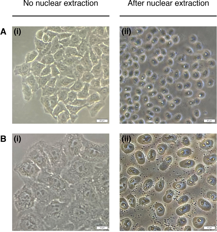
Figure 1. Nuclear extraction.
Representative images of cells prior to (left) and post (right) nuclear extraction. Cytoplasm should be digested but the nuclear structure should remain intact post-extraction (right). (A) 20x magnification; scale bar = 20 μm and (B) 40x magnification; scale bar = 10 μm.
....This article has been published
Video Coming Soon
Source: de la Peña Avalos, B., et al., Visualization of DNA Repair Proteins Interaction by Immunofluorescence. J. Vis. Exp. (2020).
Copyright © 2025 MyJoVE Corporation. All rights reserved