A subscription to JoVE is required to view this content. Sign in or start your free trial.
Lattice Light Sheet Microscopy to Visualize Receptor-Ligand Interactions in Live Cells
Overview
In this video, we describe the lattice light-sheet microscopy technique to visualize interactions between live T-cells and antigen-presenting cells. The method involves focusing an ultrathin lattice light sheet on an interacting cell pair to obtain a series of two-dimensional images, which are combined to obtain high-resolution four-dimensional images that reveal the spatiotemporal dynamics of the cell-cell interaction.
Protocol
1. Conducting LLSM Daily Alignment
NOTE: (Important) This alignment protocol is based on the LLSM instrument used (see the Table of Materials). Each LLSM may be different and require different alignment strategies, especially those that are home-built. Carry out the appropriate routine alignment and continue to section 2.
- Add 10 mL of water plus 30 µL fluorescein (1 mg/mL stock) to the LLSM bath (~10 mL volume), press Image (H.......
Representative Results

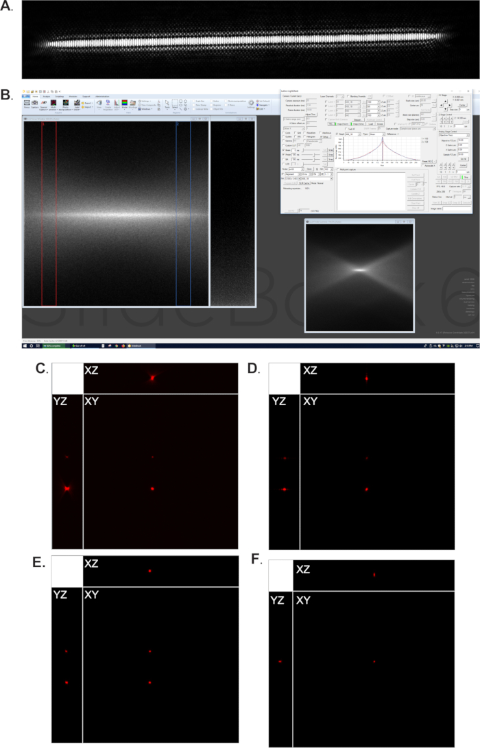
Figure 1: LLSM alignment. (A) Desired beam pattern for LLSM imaging experiment. (B) Screenshot of the beam alignment process; on the left is the focus window showing the narrowed, focused beam; at the top right is a graph showing that the beam is centered within the window; at the bottom right is the finder camera, which should also be a thin, focused beam. (C) .......
Reprints and Permissions
Request permission to reuse the text or figures of this JoVE article
Request PermissionThis article has been published
Video Coming Soon
Source: Rosenberg, J., et al. Visualizing Surface T-Cell Receptor Dynamics Four-Dimensionally Using Lattice Light-Sheet Microscopy. J. Vis. Exp. (2020).
ABOUT JoVE
Copyright © 2025 MyJoVE Corporation. All rights reserved