Imaging Protein-protein Interactions in vivo
In This Article
Summary
This protocol describes how to image protein-protein interactions using a FRET-based proximity assay.
Abstract
Protein-protein interactions are a hallmark of all essential cellular processes. However, many of these interactions are transient, or energetically weak, preventing their identification and analysis through traditional biochemical methods such as co-immunoprecipitation. In this regard, the genetically encodable fluorescent proteins (GFP, RFP, etc.) and their associated overlapping fluorescence spectrum have revolutionized our ability to monitor weak interactions in vivo using Förster resonance energy transfer (FRET)1-3. Here, we detail our use of a FRET-based proximity assay for monitoring receptor-receptor interactions on the endothelial cell surface.
Protocol
The protocol consists of three major steps. The first, step cloning of your gene of interest into a mammalian expression vector amino-terminally to the monomeric versions of CFP and/or YFP will only be discussed briefly. The second and third steps; transfection of vector DNA into EA.hy926 endothelial cells, and confocal imaging with FRET, are outlined in more depth below.
1. Construction of CFP and YFP Chimeric Receptors
Receptors of interest should be cloned amino-terminally to monomeric enhanced versions of CFP and YFP. Determination of FRET is empirical and is critically dependent upon the length of linker between the transmembrane spanning region and start of the fluorophore1. Therefore, several different lengths of linker must be explored for each new receptor pair under investigation.
- Into the pcDNA3.1 hygroR or neoR vector containing monomeric C- or YFP (this vector can be obtained from our lab), clone your receptor of interest into the NheI/EcoRI sites. *Note-If necessary, NheI is compatible several other enzymes including SpeI and XbaI.
- Transform your ligation reaction into an appropriate endA1 recA competent cell line (we routinely use Mach1). Screen colonies for the presence of insert followed by DNA sequencing to verify the absence of mutations.
- Upon obtaining conformation of successful molecular cloning, prepare transfection-grade vector DNA in sterilized water or TE using Qiagen, or similar, DNA kits. Evaluate concentration and purity using UV spectroscopy. Typical yields of DNA should be in the range of 100-200 μg, which is routinely diluted to a working stock of 1 μg/μL.
2. Transfection of EA.hy 926 Cells
- Split EA.hy 926 cells4, grown in complete-DMEM (DMEM+10% Fetal bovine serum+Pen./Strep.) into MatTek coverslip (No. 0) 35 mm dishes with 2 mL medium. You can conservatively obtain ~15 dishes (~90% confluent the following day) from a 15 cm confluent dish. Prepare one dish to evaluate expression of each individual receptor, as well as one dish for each receptor pair.
- Allow the cells to incubate overnight in 5%CO2 atmosphere at 37°C. The following day, the cultures should be ~ 70-90% confluent.
- On the day of transfection, prepare the nucleolipid complexes by mixing 2 μg of the chimeric receptor vector DNA (2 μL) with 8 μL of Fugene6 in 200 μL of pre-warmed OptiMEM. For receptor pairs, transfect with 1 μg of each receptor, for a total of 2 μg (See also discussion below).
- Mix gently by inversion.
- Continue to incubate at room temperature for 30 minutes.
- During the incubation period, prepare the cells for transfection. Aspirate off fresh media and wash each plate gently and briefly with warm PBS before the addition of 2 mL pre-warmed complete-DMEM without antibiotics (DMEM+FBS). It is important to avoid the presence of Penicillin and Streptomycin, as they are considerably more toxic to cells during treatment with Fugene6.
- Add the complex drop wise to cells and incubate for an additional 20-24 hours at 37°C in a 5% CO2 atmosphere.
- After 24 hours, carefully aspirate the media from the cells and pipette fresh complete-DMEM with antibiotics. Following transfection, cell viability should remain relatively high, i.e. little to no floating cells should be observed.
- Gene expression is typically maximal after an additional 48-60 hours post-transfection. However, imaging is typically feasible in as short as 24 hours. Time course experiments for optimal gene expression should be performed.
3. Confocal Imaging and FRET Analysis
Live cell imaging is performed on a Leica TCS-SP2 AOBS confocal laser scanning microscope equipped with blue diode (405 nm), Argon (458, 476, 488, 514 nm). green HeNe (543 nm), orange HeNe (594 nm), and red HeNe (633 nm) lasers, an HCX PI Apo 63x/1.3 n.a. glycerin-immersion objective lens, a motorized XY stage (Märzhäuser), and an environmentally controlled (temperature, humidity, and CO2) stage incubator (PeCon). Experimental controls are critical to eliminate fluorophore cross-talk between the emission channels as well as to evaluate expression issues and non-specific fluorophore multimerization. As such, single fluorophore transfections are utilized for setting up every image session. Negative FRET controls (using known noninteracting receptors) will need to be performed, in order to determine the background FRET that occurs due to over expression.
- To eliminate crosstalk between the emission channels for CFP and YFP, place the CFP expression control into the stage incubator. Using white light, adjust the Z-plate to focus on to the cells.
- Set SP window settings for emission detection of CFP to 465-505 nm and YFP to 525-600 nm.
- While monitoring both emission channels, excite the CFP at 458 nm. Using the AOTF, set the laser power to eliminate appreciable bleed over of CFP into the YFP emission channel. Save this setting.
- Perform the same control for YFP crosstalk (using the YFP expression control) in the CFP channel by adjusting the excitation power at 514 nm for cells expressing only YFP. Save this setting.
- Next place cells co-expressing CFP and YFP into the stage incubator. Using the sequence setting for the Leica confocal software, locate cells expressing similar levels of CFP and YFP with proper membrane localization.
- Using the Leica software acceptor photo-bleaching program, set the donor and acceptor settings to your saved CFP and YFP settings, respectably.
- Zooming onto the cell, highlight an ROI (region of interest) in which the photo-destruction of YFP is to occur on the cellular membrane and begin the program.
- For photo-destruction of the acceptor, adjust the AOTF to 100% at 514 nm and allow the laser bleaching to continue until 70% of the YFP emission has been depleted. To accelerate bleaching, ROIs are typically chosen in the raster axis of the laser (X-axis).
- During photo-bleaching, monitor the cellular movement and reject measurements obtained with appreciable movement in the XY-plane, as these measurements can report false FRET efficiencies.
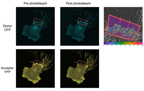
- Pre- and post-bleach images are recorded for both donor (CFP) and acceptor (YFP) and FRET efficiency is calculated as: FRETEff=(Dpost-Dpre)/Dpost for all Dpost > Dpre where Dpre and Dpost is the donor intensity before and after photobleaching respectively.
- JMP Software (SAS) is used to determine the extent of experimental variability.
4. Representative Results
Transfection efficiencies are usually between 30 to 40%. Despite previous findings by others, we have not seen that transfection and expression of one vector favors that of the other. Indeed, we frequently observe exclusive expression of one fluorophore chimera or the other. Typical FRET efficiencies vary among receptor systems. For the Tie receptor system, typical values are 20-28% for epithelial cells and 19-23% in endothelial cells. For negative controls, typical efficiencies are below 2-3%. The extent of variability will decrease considerably with experience.

Figure 1. Representative images of transfected EA.hy 926 cells. A) DIC image of EA.hy 926 cell monolayer. B) Fluorescence image of cells displayed in (A). C) Overlay of (A) and (B) demonstrating ~20-30% transfection efficiency.

Figure 2. Representative acceptor photo-bleaching analysis of FRET occurring between CFP and YFP in an EA.hy926 cell. The CFP emission channel (top panels) and YFP emission channel (bottom two panels) were monitored separately prior to and post acceptor photobleaching. Photobleaching experiments were restricted to, and FRET values calculated from, the region within the green box. FRET efficiency is displayed as an absolute range from high (red-1.0) to low (purple-0.0) on a magnified overlay of a CFP/YFP merged image for orientation purposes only.
Discussion
There are several steps that are critical to success. The most prominent among them is the relative levels of expression between the two chimeric receptors. To circumvent this issue, one may make stable cell lines expressing both proteins of interest, or identify optimal ratios of vector DNA to permit equivalent expression. Similarly, due to non-uniform transfection across a dish, protein levels will seldom be equivalent among cells. Therefore, attention must be paid to distinguish 'high' expressors from 'low'. Those that display 'average' levels of protein typically yield reliable and reproducible FRET efficiencies. One method our lab has pursued to relieve this issue is the use of adeno and lenti-viruses to 'even' gene expression. Furthermore, an alternative method to acceptor photo-bleaching for determining FRET efficiencies is sensitized emission. Although, despite the potential of sensitized emission to monitor single cells in real-time, we have found acceptor photo-bleaching to be more sensitive and more reliable.
Finally, using FRET to monitor protein interactions can be challenging, and requires careful selection of linker lengths for membrane bound receptors. Furthermore, addition of C/YFP often greatly influences protein expression levels and results in aggregation in the endoplasmic reticulum and golgi apparatus. However, for transient, or unstable, interactions, FRET is the ideal methodology to utilize.
Acknowledgements
We wish to acknowledge Dr Scott Henderson for help with confocal microscopy. This research was supported by grants from the National Institutes of Health 1RO1CA127501 to W.A.B as well as pilot project funding from the Massey Cancer Center and School of Medicine (VCU) to W.A.B. Microscopy was performed at the VCU-Dept. of Neurobiology & Anatomy Microscopy Facility, supported, in part, with funding from NIHNINDS Center core grant 5P30NS047463.
Materials
| Material Name | Type | Company | Catalogue Number | Comment |
|---|---|---|---|---|
| Name | Company | Catalog Number | Comments | |
| DMEM | Invitrogen | 11960-069 | ||
| Penicillin- Streptomycin | Invitrogen | 15070-063 | ||
| Fetal Bovine Serum | Hyclone | N/A | ||
| Opti-MEM | Invitrogen | 11058-021 | ||
| FUGENE 6 | Roche | 11814443001 | ||
| Coverslip Dishes | MatTek Corp. | P35G014C | ||
| pcDNA3.1(+) | Invitrogen | V790‐20 | ||
| Mach 1 Competent Cells | Invitrogen | C862003 |
References
- Kim, M., Carman, C. V., Springer, T. A. Bidirectional transmembrane signaling by cytoplasmic domain separation in integrins. Science. 301, 1720-1725 (2003).
- Zacharias, D. A., Violin, J. D., Newton, A. C., Tsien, R. Y. Partitioning of lipidmodified monomeric GFPs into membrane microdomains of live cells. Science. 296, 913-916 (2002).
- Seegar, T. C. M., Eller, B., Tzvetkova-Robev, D., Kolev, M., Henderson, S. C., Nikolov, D. B., Barton, W. A. Tie1-Tie2 interactions mediate functional differences between angiopoietin ligands. Molecular Cell. 37 (5), 643-655 (2010).
- Edgell, C. J., McDonald, C. C., Graham, J. B. Permanent cell line expressing human factor VIII-related antigen established by hybridization. Proc Natl Acad Sci U S A. 80 (12), 3734-3737 (1983).
This article has been published
Video Coming Soon
ABOUT JoVE
Copyright © 2025 MyJoVE Corporation. All rights reserved