A subscription to JoVE is required to view this content. Sign in or start your free trial.
A Technique to Establish a Bacterial Infection Model in an Ex Vivo Organ Culture
Overview
This video demonstrates a method to establish an infection model in placental tissues to study infection propagation. An ex vivo placental organ culture is infected with Listeria monocytogenes, a facultative intracellular parasite. The bacteria invade extravillous trophoblasts (EVTs) to enter the placental tissue and then propagate to other cells via cell-to-cell spread.
Protocol
All procedures involving human participants have been performed in compliance with the institutional, national, and international guidelines for human welfare and have been reviewed by the local institutional review board.
1. Preparation of Organ Culture Plates
NOTE: The following steps should be performed using sterile technique in a tissue culture hood. It is ideal for the dissecting microscope to be located in a sterile field. Howe.......
Representative Results

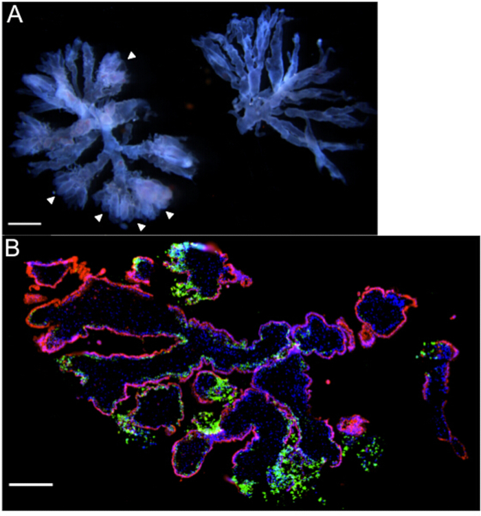
Figure 1: Villous organ cultures – Representative gross and microscopic images.
(A) Two terminal villous trees with a gestational age of 6 weeks, as viewed under a dissecting microscope. Note the "fluffy" ends (arrowheads) and prominent fetal vasculature coursing through the branches of the tree on the left that make this.......
This article has been published
Video Coming Soon
Source: Rizzuto, G. A., et al. Human Placental and Decidual Organ Cultures to Study Infections at the Maternal-fetal Interface. J. Vis. Exp. (2016)
ABOUT JoVE
Copyright © 2025 MyJoVE Corporation. All rights reserved