A subscription to JoVE is required to view this content. Sign in or start your free trial.
Long-Term Continuous Electroencephalography Monitoring in a Rodent Pup
In This Article
Overview
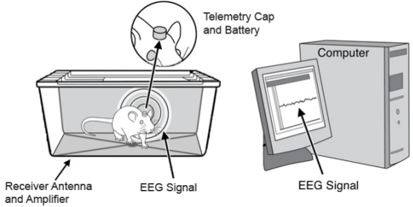
The video demonstrates long-term continuous electroencephalography (EEG) monitoring in rodent pups using a wireless transmitter system. The transmitter contains two recording electrodes and one reference electrode extending to the brain's surface. Upon placing the pup in a recording chamber, EEG can be continuously monitored throughout the pup's development into adulthood.
Protocol
All procedures involving animal models have been reviewed by the local institutional animal care committee and the JoVE veterinary review board.
1. Surgical Preparation
- Clean and prepare the transmitter to ensure safe and sterile surgery. Remove the transmitter from its anti-static packaging and either spray or soak in 70% ethanol. Rinse transmitter with sterile saline and place between sterile cotton sponges soaked in sterile saline or keep submerged in sterile saline.
- Collect and sterilize the tools required for surgery, steam autoclave for sterilization. See table of materials and reagents for the list of surgical tools.
2. Surgical Implantation
- Anesthetize animals and maintain anesthesia according to IACUC-approved protocol. At initiation and during surgery check the toe-pinch reflex every 15 min. The lack of response indicates sufficient level of anesthesia.
- For pups, use anesthesia by isoflurane (4%) with O2 (100%). For adults, use ketamine (100 mg/kg) with xylazine (10 mg/kg).
- Fix position in stereotaxic frame. Place the ear bar tips in the auditory meatus. Do not excessively tighten ear bars as the skull is very soft in young rat pups. Secure the anesthesia nose cone.
- Keep the animal warm during surgery by placing it on the heating pad set to 37 °C. In adult animals, apply lubricating ointment to the eyes of the animal.
- Sterilize incision site and maintain sterile surgical field.
- Swab the scalp with alternating applications of 70% ethanol and betadine. Start in the center of the scalp and make increasingly wider concentric circles.
- Cover the animal with drape and conduct the surgery over draped animal. Maintain the sterile surgical field by lining the surgical set-up with sterile drapes, spray equipment with 70% ethanol.
- Wear sterile surgical gloves and a gown (or as required by the institution). To help maintain a sterile field, use a surgical assistant.
- Make an incision on the scalp of the animal slightly behind the eyes along the midline, approximately 2 cm. Use caution when inserting the scalpel as the skull is still very soft in young rat pups. Make a single cut so the incision bleeds less and heals faster.
- Expose the skull. Prepare a clean and dry area to maximize the bond between the transmitter and the bones of the skull. Use aneurism clips to grasp scalp.
- Gently pull scalp away from midline at four corners. Look for anatomical landmarks such as bregma and lambda in the skull. Remember skull bones are not fused in animals at this age. Use the Paxinos atlas of stereotaxic coordinates to find the correct location for the burr hole.
- Use a Dremel-type tool with a burr-type drill bit. Create two burr holes in desired recording positions with the holes being bigger than 300 µm in diameter. Place the burr hole for the reference electrode over the cerebellum behind the lambda of the skull.
- Ensure that the wires on the transmitter are aligned with the burr holes. If the electrode wires are not aligned, glue contamination of the electrodes is likely, and will result in poor signal. To align the wires, check the fit of the transmitter and gently bend electrodes to line up over the intended sites for burr holes.
- Trim electrode leads. Use surgical scissors to trim the electrodes to the desired length. The electrode depth is important for the type of recording required for the experiment (i.e., place the electrodes above dura for EEG recordings, or use stereotaxic coordinates for defined brain structures).
- Liberally apply cyanoacrylate on the base of the transmitter to cover the area making sure to avoid coating the electrodes. Cyanoacrylate glue is an electrical insulator, contaminating electrodes with glue will result in no signal.
- If recording from deep brain structures, mount the transmitter on the cannula holder and place it in the stereotaxic arm for z-axis control. Lower the transmitter using the stereotaxic arm to appropriate depth and place cyanoacrylate gel around the transmitter.
- Thoroughly dry skull before placing transmitter to ensure strong adhesive bond. Apply transmitter coated with cyanoacrylate to the skull. Take care to align electrodes with corresponding burr holes.
- Try to avoid damaging major vascular structures. Hold the transmitter in place with slight pressure for one minute. Use slight pressure to form a strong bond between the transmitter and the skull.
- Apply additional cyanoacrylate, enough to completely seal transmitter/skull interface. To ensure a good fit and strong bond, maximize the surface area of the glue that contacts the skull. Apply the cyanoacrylate adhesive in a circle around the transmitter, making sure both skull and the wall of the transmitter are covered.
- Apply chemical accelerant (0.1 ml) through a syringe around the cyanoacrylate at the base of the implanted transmitter. Use accelerant sparingly, taking care not to apply to adjacent tissue.
Note: Chemical acceleration of the cyanoacrylate curing ensures that the strong bond between the transmitter and the skull is formed quickly. Cyanoacrylate accelerant is useful to speed curing of adhesive but is not necessary. - Remove the accelerant by washing the area thoroughly with sterile saline. Cyanoacrylate accelerator may cause tissue irritation if not washed from the area of the incision. To wash the area, fill a 1.0 ml syringe with sterile saline and irrigate the area through a syringe needle. Generally, 0.5 ml of saline is enough to wash out the accelerator.
- Suture the skin around the base of the transmitter, but do not cover the transmitter. The top of transmitter must be above skin to efficiently transmit neural signals. Skin should be reasonably tight around the transmitter and the glue around the unit. Use Vicryl or silk suture (soft thread); skin in immature animals is soft and is easily damaged if soft sutures are not used. For adult animals, use any suturing material.
- Remove animal from stereotaxic frame and place on heated blanket for recovery.
- Ensure animals are warm (37 °C) and ambulatory (i.e., completely recovered) before returning to the dam. Ensure that the animal is hydrated by pinching the skin on the animal's back (if the animal is dehydrated, the skin will remain deformed). If animal is dehydrated, administer sub-cutaneous injection of lactated Ringer's buffer. Do not leave the animal unattended until it has regained sufficient consciousness to maintain sternal recumbency.
- Administer buprenorphine (0.05 mg/kg) to animals for post-surgical pain management and a sub-cutaneous injection of 0.1 ml bupivacaine around the injection site.
Note: From start to finish the entire procedure should be completed in 5-10 min for animals of this age (postnatal day 6). Surgical time may take longer for older animals.
- Administer buprenorphine (0.05 mg/kg) to animals for post-surgical pain management and a sub-cutaneous injection of 0.1 ml bupivacaine around the injection site.
3. Care and Housing
Note: Some dams may not tolerate pups implanted with the device. Dams may need to be selected who are tolerant. It is acceptable for the dam to move pups around the cage by picking them up by the transmitter.
- Once animals are weaned, singly-house them to avoid the removal of the devices from their cage mate.
- Euthanize animals by lethal dose of pentobarbital (25 mg/kg) or isoflurane (in a bell jar) when signs of distress are present.
- Note: some animal housing cages with wire inserts may interfere with the implanted transmitters. Be sure to check the height of the wire insert to make sure that animals cannot get the transmitter caught between the 'bars' of the wire insert.
4. Recording EEG
- Place the animal in a cage by itself or co-housed with littermates and the dam. However, place only one implanted animal in a single cage. Do not leave pups alone in the recording chamber for more than 2 hr. Monitor the animals for signs of distress and dehydration.
- Connect the provided power supply to the receiver base and verify the power light is illuminated. Connect the receiver base to a data acquisition system using Bayonet Neil-Concelman (BNC) cables.
- Place the animal cage on top of the receiver base (Figure 1). The "signal" light should illuminate indicating a transmitter has been detected. Data may now be recorded.
- To record data, connect the receiver base to an analogue-to-digital converter and connect the converter to a computer (Figure 2).
- Set the sampling rate of the recording. Ensure the data is sampled properly. Select at least 250 Hz sampling rate (500 Hz recommended) for recording (bandwidth of the transmitter is 0.1-100 Hz).
- Save digitized data and analyze using signal-processing software packages such as Matlab.
Results

Figure 1: The transmitter and receiver. This particular wireless transmitter (A) weighs 4 g and displaces <1.4 cm3 of volume and with a footprint of 7 x 12 mm is easily mounted to the skull of rats and mice. The transmitter can amplify 2 channels of biopotentials for up to 6 months after which the battery is drained. Larger batteries can be used for longer recording time. Ani...
Disclosures
Materials
| Name | Company | Catalog Number | Comments |
| Sterile Surgical Gloves | Protective Industrial Products | 100-3201 PF | Powder Free Sterile Latex Surgical Glove |
| Scalpel Handle | FST | 10003-12 | |
| Scalpel Blade #15 | FST | 10015-00 | |
| Fine Scissors | FST | 14090-09 | |
| Burr tool | Ram Products, Inc. | Microtorque II | |
| Fine burr | FST | 19007-07 | |
| Aneurism clip | ROBOZ | RS-5422 | |
| Toothed Forceps | FST | 11022-14 | |
| Cotton-Tipped applicators | McKesson | 24-103 | |
| Needle Driver | WPI | 521725 | Olsen-Hegar Needle Holder |
| Cyanoacrylate gel | Henkel | Loctite 4541 | |
| Cyanoacrylate accelerant | Henkel | Loctite 7452 | |
| Suture | Ethicon | Vicryl RB-1 J304 | |
| Elecrocautery disposable | Bovie | AA01 | Fine Tip |
| Surgical Tray | FST | 20311-21 | |
| Epitel Receiver Base | Epitel Inc | N/A | |
| Epitel wireless transmitter | Epitel Inc | N/A | |
| Biopac digitizer | Biopac | MP-150 | |
| PC-compatible computer |
This article has been published
Video Coming Soon
Source: Zayachkivsky, A. et al. Long-term Continuous EEG Monitoring in Small Rodent Models of Human Disease Using the Epoch Wireless Transmitter System. J. Vis. Exp. (2015)
Copyright © 2025 MyJoVE Corporation. All rights reserved