Dissection of Human Vitreous Body Elements for Proteomic Analysis
In This Article
Summary
This video shows an effective technique for differentiating and dissecting the various semi-transparent structures of the human vitreous body in post mortem eyes.
Abstract
The vitreous is an optically clear, collagenous extracellular matrix that fills the inside of the eye and overlies the retina. 1,2 Abnormal interactions between vitreous substructures and the retina underlie several vitreoretinal diseases, including retinal tear and detachment, macular pucker, macular hole, age-related macular degeneration, vitreomacular traction, proliferative vitreoretinopathy, proliferative diabetic retinopathy, and inherited vitreoretinopathies. 1,2 The molecular composition of the vitreous substructures is not known. Since the vitreous body is transparent with limited surgical access, it has been difficult to study its substructures at the molecular level. We developed a method to separate and preserve these tissues for proteomic and biochemical analysis. The dissection technique in this experimental video shows how to isolate vitreous base, anterior hyaloid, vitreous core, and vitreous cortex from postmortem human eyes. One-dimensional SDS-PAGE analyses of each vitreous component showed that our dissection technique resulted in four unique protein profiles corresponding to each substructure of the human vitreous body. Identification of differentially compartmentalized proteins will reveal candidate molecules underlying various vitreoretinal diseases.
Protocol
1. Anterior Segment Dissection.
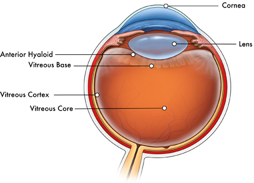
- The cornea is removed by first making an incision at the limbus into the anterior chamber using a 15° blade (Figure 1). Then one blade of curved cornea-scleral scissors is inserted into the anterior chamber. Circumferential cuts are made in the cornea, just anterior to the limbus.
- 0.12 Colibri forceps and Westcott scissors are used to cut the iris circumferentially anterior to the ciliary body.
- The lens nucleus is removed with 0.12 Colibri forceps or a 15° blade (supersharp) and the cortex and capsule with forceps.
2. Vitreous Core Aspiration.
- A 23-gauge needle on a 5-cc syringe is inserted into the mid-vitreous (Figure 1).
- Approximately 1 mL or more of vitreous core is gently aspirated.
- The sample is then placed in a microfuge tube and then into liquid nitrogen.
3. Anterior Hyaloid Dissection.
- The anterior hyaloid is seen as a semi-transparent ring by adjusting the incident light from a goose neck microscope light. Previous aspiration of the vitreous core separates the anterior hyaloid from other structures for easier identification.
- The sheet of elastic tissue is carefully pulled away from the ciliary body with Colibri or dressing forceps and cut with Vannas scissors. This maneuver is repeated.
- The sample is then placed in a microfuge tube and then into liquid nitrogen.
4. Vitreous Base Dissection.
- Using 0.12 forceps to stabilize the eye cup, Westcott scissors are used to "flower" the eye by making four equally spaced stress-relieving cuts from the ciliary body towards the optic nerve head.
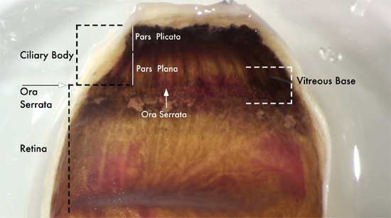
- The pars plicata of the ciliary body (Figure 2) is removed from each of the quadrants using Westcott scissors.
- The vitreous base is then grasped on either side of the ora serrata with forceps over the pars plana and the retina (Figure 2).
- Continuous traction on the vitreous base is applied with the forceps. Sequential cutting with Westcott scissors extracts a semi-transparent tissue with a "string of pearls" appearance as it is excised.
- The sample is then placed in a microfuge tube and then into liquid nitrogen.
5. Vitreous Cortex Removal.
- The pars plana is excised from each of the quadrants with Westcott scissors by cutting the leaflet approximately 3-mm posterior to the ora serrata (Figure 2).
- Between the tissue leaflets, the vitreous cortex is visualized as a semi transparent film.
- The elastic film is either grasped with 0.12 forceps or held by adherence to a Weck-Cel surgical sponge. The vitreous cortex is then pulled away from the retina and excised with Westcott scissors (Figure 1).
- The sample is placed in a microfuge tube and then into liquid nitrogen. All samples are stored at -80° C until utilized for experimentation.
6. Representative Results
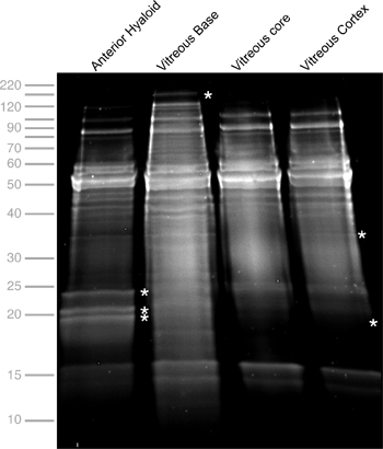
Tissue samples can be processed by a variety of methods for specific experiments. In our case, samples were submitted for protein analysis by SDS-PAGE (Figure 3).

Figure 1. Cross-sectional view of the human eye depicting different substructures of the vitreous body. The most anterior vitreous is a thin collagenous layer called the anterior hyaloid. The vitreous core comprises the entire central region of the vitreous body. This portion of the vitreous is more aqueous in contrast to the vitreous base, which is viscous enough to be grasped by forceps and is firmly attached to the underlying ciliary body and retina. Encompassing the vitreous core is a very thin collagenous shell called the vitreous cortex.

Figure 2. Vitreous base anatomy. The vitreous base is a semi-transparent substructure of the vitreous body located along the ora serrata (white arrows), which is the dividing line separating the ciliary body and retina. The anterior border of the vitreous base extends over the pars plana (white line) of the ciliary body. The posterior border of the vitreous base extends 2-3-mm posterior to the ora serrata (white dash). To excise the vitreous base, forceps are used to grasp the tissue and pull it away from the underlying ciliary body and retina. Once elevated, Westcott scissors are used to cut along the base.

Figure 3. One-dimensional SDS-PAGE of Vitreous Body Elements. Total protein concentrations for the anterior hyaloid, vitreous base, vitreous core, and vitreous cortex were 11.24, 20.1, 16.61, 14.24 mg/mL, respectively. Gel electrophoresis was performed at 200 kV for 45 minutes, stained with Flamingo (Bio-Rad), and visualized using a VersaDoc Imaging system (Bio-Rad). The profiles for the different tissues show several similar bands, indicating either conserved or cross-contaminating proteins, as well as unique bands (asterisk), indicating differentially localized proteins.
Discussion
The vitreous body is a semi-transparent gel whose molecular composition is poorly understood, especially at the level of its substructures: the vitreous base, core, cortex, and anterior hyaloid. The vitreous core contains collagens II, V, IX, and XI, along with chondroitin sulphate proteoglycans, heparan sulphate proteoglycans, and hyaluronan.1,2 Protein biomarkers in the vitreous core have been associated with diseases such as diabetic retinopathy.3-5 How these proteins are differentially expressed in each of the substructures, and in many cases the specific protein identities, are not known. These details may give insight to the origin of proteins associated with specific vitreoretinal diseases and help target future therapies. Although the optimum post mortem interval for tissue dissection has not been determined, protein degradation may affect downstream experiments. For example, immunohistochemistry is affected in 12-hour postmortem eyes and some specific enzyme activities may be reduced within a few hours (unpublished observation). All tissues in this study were collected between 2 and 8 hours of death without significant changes in protein expression or suitability for proteomics analsyis. The liquid nitrogen freezing method of preservation is chosen over fixation in order to prevent small changes in protein structure caused by fixative cross linking, which has been demonstrated in other tissues by LC-MS/MS.6 Proteomic studies will depend on the ability to accurately dissect the different compartments of the vitreous, as demonstrated in this video experiment. We have validated the dissection technique using 1-dimensional SDS-PAGE. As our results suggest, there are differentially expressed proteins in the various vitreous body substructures. Identifying these proteins will provide a more detailed understanding of vitreous compartmentalization.
Acknowledgements
Funding was provided by Fight for Sight. Tissues were obtained from the Iowa Lions Eye Bank.
Materials
| Name | Company | Catalog Number | Comments |
| Name | Company | Catalog Number | |
| 0.12 forceps | Storz Ophthalmics | E1502 | |
| 5-cc syringe | Becton-Dickinson | 309603 | |
| Straight Dressing Forceps With Serrations | Storz Ophthalmics | E1400 | |
| 23 gauge needle | Becton-Dickinson | 305145 | |
| Colibri forceps | Storz Ophthalmics | 2/132 | |
| Castroviejo angled corneal scissors | Storz Ophthalmics | E3223 | |
| Vannas Curved Capsulotomy Scissors | Storz Ophthalmics | E3387 | |
| Weck-Cel surgical spears | Medtronic | 0008680 | |
| Westcott Curved Tenotomy Scissors | Storz Ophthalmics | E3320 |
References
- Bishop, P. N. Structural macromolecules and supramolecular organisation of the vitreous gel. Prog Retin Eye Res. 19, 323-344 (2000).
- Sebag, J. Molecular biology of pharmacologic vitreolysis. Trans Am Ophthalmol Soc. 103, 473-494 (2005).
- Yoshimura, T. Comprehensive analysis of inflammatory immune mediators in vitreoretinal diseases. PLoS One. 4, e8158-e8158 (2009).
- Gao, B. B., Chen, X., Timothy, N., Aiello, L. P., Feener, E. P. Characterization of the vitreous proteome in diabetes without diabetic retinopathy and diabetes with proliferative diabetic retinopathy. J Proteome Res. 7, 2516-2525 (2008).
- Izuta, H. Extracellular SOD and VEGF are increased in vitreous bodies from proliferative diabetic retinopathy patients. Mol Vis. 15, 2663-2672 (2009).
- Azimzadeh, O. Formalin-fixed paraffin-embedded (FFPE) proteome analysis using gel-free and gel-based proteomics. J Proteome Res. 9, 4710-4720 (2010).
Explore More Articles
This article has been published
Video Coming Soon
ABOUT JoVE
Copyright © 2025 MyJoVE Corporation. All rights reserved