Intracellular Recording, Sensory Field Mapping, and Culturing Identified Neurons in the Leech, Hirudo medicinalis
In This Article
Summary
This article describes three nervous system preparations using leeches: intracellular recording from neurons in ventral ganglia, culturing neurons from ventral ganglia, and recording from a patch of innervated skin to map sensory fields.
Abstract
The freshwater leech, Hirudo medicinalis, is a versatile model organism that has been used to address scientific questions in the fields of neurophysiology, neuroethology, and developmental biology. The goal of this report is to consolidate experimental techniques from the leech system into a single article that will be of use to physiologists with expertise in other nervous system preparations, or to biology students with little or no electrophysiology experience. We demonstrate how to dissect the leech for recording intracellularly from identified neural circuits in the ganglion. Next we show how individual cells of known function can be removed from the ganglion to be cultured in a Petri dish, and how to record from those neurons in culture. Then we demonstrate how to prepare a patch of innervated skin to be used for mapping sensory or motor fields. These leech preparations are still widely used to address basic electrical properties of neural networks, behavior, synaptogenesis, and development. They are also an appropriate training module for neuroscience or physiology teaching laboratories.
Introduction
The leech preparation is useful for investigating the function of identifiable (sensory, motor and inter-neurons within the animal's central nervous system (CNS). The useful property of leech neurons is that the shape of their action potentials and biophysical properties are characteristic for a given cell type (i.e. P, N, T, Rz). Moreover neurons can be removed from the animal and cultured in a suitable medium in which the cells maintain their electrical characteristics for as long as 45 days1,2.
A distinct advantage of the leech ganglion for learning how to make electrophysiological recordings is that cells are characteristically and reliably arranged in the ganglion in a pattern that allows identification of distinct cell types from ganglion to ganglion and from animal to animal. The unique location and morphology of neurons in leech ganglia were noted by Retzius as early as 18913. Knowledge of the identity and function of a neuron is advantageous for investigating neural circuits and for making experiments with a given cell type. Due to the relatively large somata of the neurons within a ganglion they are visible with a dissecting microscope, and the somata can be selectively impaled with microelectrodes for recording their electrical properties.
Dyes and large molecules can be injected into individual cells and channel properties studied by patch clamp. Ionic currents that give rise to the various shapes of the characteristic action potentials in various neurons have been identified4-7. From investigations in the pattern of innervation and branching of individual neurons within the CNS, the synaptic connections have been reproduced in culture with identified neurons2,8. Studies in the leech ganglia have provided insight into the development of functional circuits that can be measured with electrophysiology as well as with whole animal behavior studies. The leech CNS has also served as a valuable preparation for investigating mechanisms involved with neuronal repair and regeneration in the whole animal and in culture4-6,9-12.
In mammalian brains it is a daunting task to explain behavior in terms of the properties and connections of individual identified neurons. In principle one can hope that the study of less complex systems such as the leech could help to define basic mechanisms used in higher animals. The connections that are used which underlie a swim pattern13,14 and rhythmicity of the heart15 have been well characterized in the leech. The ability of some leech neurons to form both electrical and chemical communication is intriguing. Some connections are bidirectional while others are unidirectional chemically but bidirectional electrically7. Understanding the synaptic connections within the animal as well as recreating the same type of connections in a culture dish are powerful tools for investigating synaptogenesis16-18 as well as pharmacological profiling of synapses19. The intracellular recording and stimulation techniques described here provide a foundation for pharmacological assays and research in neuroethology and development.
In addition, the segmental ventral nerve cord can be dissected with a patch of innervated skin. With the ability to keep a patch of skin and the neurons alive, sensory receptive fields can be probed by recording electrical signals from the cell body (i.e. soma) of sensory neurons within respective abdominal ganglion in the CNS. Thereby one can assess the sensitivity of the sensory endings by applying varying degrees of force on the skin and map receptive fields: light touch, pressure, and noxious (painful) stimuli20,21.
An advantage of this leech ganglion-skin preparation is that one can draw anatomically the receptive field map and trace the neuronal processes to the identified neuron once the electrical responses are being recorded. The three leech experiments described below can all be completed multiple times with a single specimen, which makes this a cost effective teaching module for academic laboratories.
Protocol
1. Preparation of Electrodes and Saline Solution
- Prepare physiological saline and electrodes before the animal is dissected. Leech Ringer’s solution consists of the following: 115.3 mM NaCl, 1.8 mM CaCl2, 4.0 mM KCl, 10 mM Tris/maleic acid or HEPES. Adjust the pH to 7.4.
- Prepare a Ag-Cl wire by dipping a cleaned silver wire into a small amount of bleach for 20 min or until the wire has a smooth dark gray finish. Wash the wire with distilled water before using. Then place the wire into a micropipette holder that plugs into the amplifier.
- Prepare a sharp glass electrode of borosilicate glass using a pipette puller. The electrode resistance should be on the order of 20 MΩ.
- Backfill the pipette with 3 M potassium acetate solution and insert in into the micropipette holder.
- For an extracellular electrode prepare a sharp glass pipette as described above, break the tip back by rubbing another sharp pipette near the sharp edge. Then fire polish the tip so that it does not tear the connectives. Plastic extracellular electrodes will also work as long as there is good contact between the electrode and the nerve.
- Insert the extracellular recording electrode into a micropipette holder (with Ag-Cl wire) that is fitted with a rubber tube and syringe for suction. Use the syringe to draw saline into the electrode at least to the level of the silver wire.
- Set up the hardware (amplifier and stimulus isolator) and software recording parameters by following the manual for your specific signal recording unit. See Leksrisawat et al.22 to view details on equipment used in the experiments described below.
2. Ventral Ganglion Dissection
- Dissect the leech in a large silicone elastomer-lined dish with leech Ringer’s solution. If the animal is stretched longitudinally it will be easier to remove the ventral nerve cord (see Figure 1A).
- With #10 or #11 size scalpel, make a longitudinal incision with very shallow cuts in the ventral midline. About the middle 2/3rd of the ventral cut is shown in Figure 1B. Try not to cut through the black stocking (the ventral blood sinus) until the animal is fully opened and the blood sinus is stretched without kinks. This blood sinus runs the length of the animal along the ventral midline. The VNC is contained inside of this blood vessel.
- After the skin has been cut the full length of the animal use the fine iris scissors to nick the blood sinus (Figure 2). Slip one blade of the fine iris scissors under the sinus and slightly raise the blade to separate it from the nerve tissue. Cut the sinus the length of the VNC while avoiding the nerve roots, segmental nerves, or the connectives between ganglia.
- Next, remove part of the ventral nerve cord by cutting the roots distal to where the blood vessel joins the roots. Repeat this process for each ganglion along the length of the ventral nerve cord.
- Transfer a segment of one or two ganglia to a smaller Petri dish. The ganglia or the single ganglion needs to be pinned taut with some slack.
- Pin the connectives and the roots so the ganglion glial sheath is slightly stretched to prevent the neuron cell bodies from moving around when impaling through the glial sheath and into the neuron of interest. The pins should be placed through the black sinus for the roots and in the middle of the two connectives to minimize damage of the axons (Figure 3).
- Place the Petri dish with preparation under the microscope and secure it with wax at the bottom of the dish to prevent movement.
- Carefully focus the light beam using a mirror and light condenser to see the neurons as in Figure 4A (schematically shown in Figure 4B). Notice the two large Retzius cells in the center of the ganglion.
- To make an intracellular recording of the Rz, T, P, and N neurons, carefully place the tip of the electrode over the cell of interest and gently lower the tip until a dimple is seen in the glial sheath.
- Tap the electrode holder or manipulator (very carefully) so that the electrode tip “pops” into the cell of interest.
- Inject short pulses (30-40 msec) of positive current (1-10 nA) into the cell to evoke action potentials.
3. Culturing Neurons from the Ventral Ganglion Preparation
- Pin the ganglia ventral side up in a clean dish containing L-15 with fetal calf serum (FCS) (Figure 3). Pin the roots so that the ganglia are slightly stretched as done above for intracellular recordings. With fine scissors, nick the glial capsule at one end, slip one scissors blade under the capsule, then cut across the glial capsule as shown in Figure 5A.
- Add 100 µl aliquot of Collagenase/Dispase to the dish and place on a shaker for 15 min. Examine cells by moving the culture media around the exposed cell bodies. Return the cells to the shaker for another 15 min or longer until the cells come out with minimal suction.
- Use a pipette attached to a syringe to gently extract cells from the ganglion. Fire polish the pipette tip so that it is slightly larger than the cell body diameter. For Rz cells in an adult leech, the diameter should be approximately 50 µm.
- Prepare a variety of pipettes with different diameters without a filament as this decreases the chances of the cells sticking inside the pipette. Store the pipettes in a bottle containing 80% ethanol (EtOH) using a cotton ball at the bottom to protect the tips from chipping. Remember to wash the EtOH out of the pipette with medium before aspirating cells.
- After the enzyme treatment, wash out the enzyme-containing medium with L-15 (with FCS) 2x. Identify the cells of interest and gently draw them into the pipette by pulling the syringe (Figure 6A). Discharge the aspirated cells into another dish which contains L-15 (with FCS).
- Until now, all procedures were non-sterile. Perform the following steps in a sterile hood. Place 3-4 large drops of sterile L-15 (with FCS) in different locations inside a sterile Petri dish.
- Pipette the cells into one of the before-mentioned drops, blowing them around in the drop to wash them. Repeat this in series in 3 or 4 drops of medium.
- Place cells in a clean Petri dish filled with L-15 (with FCS).
- Incubate the cells for 1-24 hr at room temperature. Afterwards the clean cells can be plated. By leaving the cells for a few hours or overnight the extracellular matrix comes off easily. Plate the cells immediately to generate longer processes.
- Use sterile substrate to coat a microwell dish and allow it to dry completely (overnight) before culturing experiments.
- After the dish has been coated, wash the dish gently with sterile water and then with a small amount of the L-15 medium. Remember to use the L-15 solution without the FCS added to allow the cells to adhere to the dish. The medium can be gently changed to L-15 with FCS after the cells have adhered to the substrate. If the cells are to be used a few hours after plating, then switching the medium to L-15 with FCS is not necessary.
- Position the cells next to each other at the time of plating to induce rapid synaptogenesis (Figure 6B). After 24 hr measure physiological activity in pairings of neurons.
- Use intracellular electrophysiological techniques to record RP or synaptic responses in cultured pairs of cells (stimulate 1 neuron through an intracellular electrode and record responses from the paired cell using a 2nd intracellular electrode).
4. Ganglion-body Wall Preparation
- Pin the leech dorsal side down in an elastomer-lined dish as described above.
- Pin the skin to one side with roots on the other side of the severed ganglion (Figure 7A, magnified in Figure 7B) or make a window on the ventral aspect of the leech over a ganglion (Figure 7C) with all the roots to the body wall intact.
- After obtaining a stable intracellular recording in one of the sensory cells (T, P, N) use a small glass rod that has been fire polished to lightly touch the skin in the same segment. If the neuron being recorded from is an N cell then more pressure will need to be applied to the skin. In most cases, the N cell will only respond if the skin is pinched with tweezers. For this reason these cells should be tried last as one might damage the skin or displace the intracellular recoding by disturbing the preparation in such a manner.
- One should be able to quickly sketch the skin and ganglion with details sufficient enough that a map of where one touched can be used to determine the receptive field for a particular neuron. After recording from as many T and P cells as possible on both sides of a ganglion try the N cells.
- In order to examine if the sensory neurons have processes that travel through the connectives in a rostral or caudal manner and then out the roots in the adjoining segment to the skin, the connectives between ganglia can be cut and the receptive fields reexamined in regards to the spread of detection. Basically one is examining if there has been any loss in the receptive field.
5. Injecting Fluorescent Dyes
- Prepare a leech ganglion as described above.
- Fill a microelectrode with a solution of Lucifer yellow-CH (3.0%) and lithium chloride (300 mM).
- Impale one of the sensory cells or Rz cells on the ventral side of the ganglion as described above.
- Inject dye into the cell by passing negative current (3nA) through the electrode for 15 min. Set the pulse duration to 100 msec and the frequency to 2 Hz.
- Visualize the dye-filled neurons by exciting the dye with a mercury lamp and a FITC/GFP filter set.
Representative Results
Intracellular recordings from sensory neurons in the leech ganglion are characterized by their distinct voltage-time traces shown in Figure 4C. The resting cell membrane potential in healthy leech ganglion cells is -45 to -60 mV. If potentials are smaller (less negative) one should change the glass electrode, or if the potential is still low, move on to another segment of the nerve cord. Spontaneous action potentials with small amplitude may indicate that the electrode is in a motor neuron. If this happens in the ganglion-body wall a current injection can be used to determine if the neuron innervates any of the muscles still attached to the skin. The annulus erector motor neuron (AE) is on the ventral side of the ganglion (Figure 4B) and it will cause the annuli (ridges or segments) to rise by activating the annulus erector muscle.
Injecting current into the sensory cells should evoke action potentials with the characteristic waveforms shown in Figure 4C. If the waveforms look different it could be due to an unbalanced bridge or offset capacitance compensation. One should consult the manual that accompanies the signal acquisition unit for details on how to balance the bridge inside a cell. Notice the stimulus artifacts at the beginning and end of the current pulses in Figure 4C. This is how the artifacts look when the bridge is balanced.
Primary cultures of neurons from the leech ganglion form electrical and chemical synapses in the dish. Initially the electrical properties of the cultured cells are not exactly the same as they are in vivo. However after a few days the impulses are nearly indistinguishable from those recorded in the ganglion. This is demonstrated in Figure 6C with a pair of Rz cells. After 7 hr in culture the amplitude of synaptic potentials was 1-2 mV. After 56 hr in culture synaptic potentials were larger than 10 mV (top traces in Figure 6C). The shape of the action potential also changes after 56hr in culture. Under the conditions described these cells are viable for several days.
Intercellular communication can be demonstrated by stimulating the nerve roots or connectives and recording from neurons in the ganglion. Stimulating these nerves generates robust synaptic potentials and action potentials in different ganglion cells. Traces in Figure 8B were recorded from a Retzius cell in a ganglion that had been removed from the animal and pinned out in a dish. The black trace was evoked by a subthreshold stimulus applied with an extracellular electrode to the connective. Increasing the stimulus voltage caused the cell to fire an action potential (red trace). Fluorescent dyes or horse radish peroxidase can be injected into these cells to study their morphology. Lucifer yellow was injected into an Rz neuron by passing a negative current through the electrode. An alternative is to inject the dye using puffs of air pressure. Figure 9A shows the dye shortly after the injection began. Thirty minutes later the dye already had diffused into the nerve roots (Figure 9B). The type of dye depends on the application, e.g. small molecular weight dyes that can pass through gap junctions are used to identify electrical synapses.

Figure 1. Ventral leech dissection. (A) The animal is thoroughly pinned ventral side up in an elastomer-lined dish filled with leech saline. (B) A shallow incision spanning the anterior: posterior axis is made just lateral to the midline to avoid damaging the nerve cord. The black blood sinus (which contains the ventral nerve cord) can easily be seen. Note the one exposed ganglion that is white compared to the sheath. Click here to view larger image.
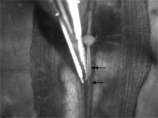
Figure 2. Dissection of the black sinus. The blood vessel (arrow) should be carefully cut on one side to expose the ventral nerve cord (double arrow). Leave segments of the blood vessel intact around the segmental roots for use as a handle to manipulate the ventral nerve cord when transferring between dissection and recording dishes. Click here to view larger image.
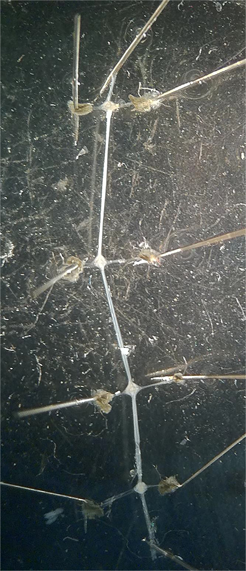
Figure 3. Isolated section of the ventral nerve cord. Pinning the ventral nerve cord by the blood vessel fragments remaining on the segmental roots and at the ends of the segmental connectives exposes the ganglia for electrophysiological recordings. A taut stretching of the ganglia allows one to see the identifiable cell bodies and will allow an easier penetration of the glial sheath with a sharp glass electrode. Click here to view larger image.

Figure 4. Cellular anatomy of the leech ganglion. (A) Ideally the Retzius cells (R) and other large cell bodies (P- pressure, T- touch, N-nociceptive, AE- annulus erector) will be clearly visible if the microscope and light condenser are set up properly. (B) Schematic view of cells in the ganglion. Slight differences in the location of the somas will occur in each ganglion due to how the ganglion is stretched and pinned taut. (C) Action potentials recorded from the soma of these neurons have characteristic waveforms. These spikes were evoked by injecting positive current into the cell (square pulse overlaying the voltage-time traces). Click here to view larger image.
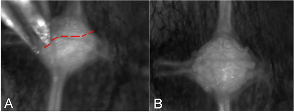
Figure 5. Desheathing the ganglion to remove individual cell bodies. (A) The glial sheath should be carefully cut across the ganglion to avoid damaging cells of interest. The red line indicates a cut to avoid hitting the soma of the Rz neurons. Opening the sheath gives the digestive enzymes access to the connective tissue matrix. (B) Healthy cell bodies bulging out of the ganglion after the glial sheath has been opened. Click here to view larger image.

Figure 6. Culturing neurons from the leech ganglion. (A) Cells are aspirated from the ganglion using a micropipette and transferred to a dish containing culture media. (B) Placing multiple cells near one another will increase the rate of synaptogenesis between identifiable neurons. Here the stumps of two neurons have formed a synapse. (C) Neurons in culture exhibit slightly different electrical properties. The top traces show synaptic potentials evoked by stimulating the adjacent cell 7 hr and 56 hr after the cells were plated. The bottom traces were evoked by injecting current directly into one of the Rz cells. Note the increase in synaptic potential amplitude and change in the action potential waveform after 56 hr in culture. Click here to view larger image.

Figure 7. The leech ganglion-body wall preparation for mapping receptive fields. (A) One approach is to remove an entire section of the body wall along with 1-3 ganglia. Here the roots on one side have been cut and the ganglion has been pinned out to the side. (B) A pinned out ganglion at higher magnification. (C) Another approach is to create a small window in the body wall and record from the intact ganglia while mapping a receptive field. (D-G) Each of the sensory neurons exhibits a characteristic response to a mechanical stimulus. (D) A light touch evokes activity in T cells but not in P or N cells. (E) The T cell response shuts off in response to stronger stimuli, giving way to activation of the P cell. As the stimulus gets stronger the N cells becomes activated (F-G). Click here to view larger image.
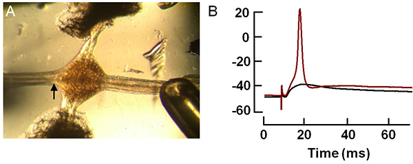
Figure 8. Evoked activity in the leech ganglion. Extracellular stimulation of the connectives can be used to drive synaptic transmission in the nerve cord. (A) This is done by attaching a suction electrode to one of the connectives and applying a small voltage. The sharp glass electrode (arrow) can be seen impaling a Retzius cell in the ganglion. (B) Voltage-time traces showing the stimulus artifact followed by a synaptic potential (black) and an action potential (red). Click here to view larger image.
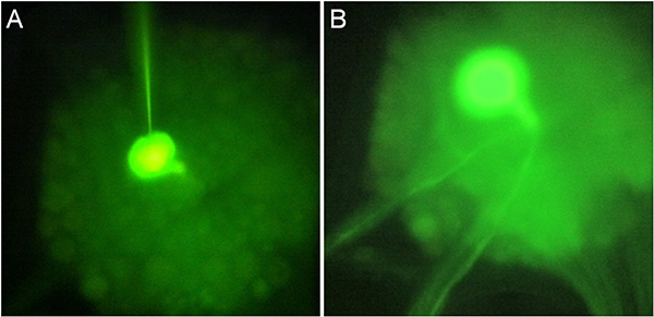
Figure 9. Injecting dye into leech neurons. (A) Lucifer yellow can be injected into the soma by passing current through the recording microelectrode. A mercury lamp and FITC filter set can then be used to visualize the fluorescence. (B) Dye has diffused into the Rz axons about 30 min after the injection. Click here to view larger image.
Discussion
The purpose of this set of experiments was 1) to teach and exhibit the fundamental principles of intracellular recordings from identifiable neurons and 2) to recognize the characteristic shapes of electrical signals that can arise from these particular neuronal types using adult leeches. There is a rich history of investigations using the leech for neurophysiological investigations and research is ongoing in addressing questions of neural circuitry and gene regulation during development as well as during synaptic repair and regeneration10. Neuromodulation, particularly through biogenic amine pathways, has also been a successful research direction in the leech. By adding dopamine and other pharmacological agents to the nervous system through the saline solution, it has been shown that dopamine modulates neural networks involved in decision-making23 and motor patterns for crawling behavior24,25. This experimental approach is a simple (and inexpensive) way to incorporate modulation into the teaching laboratory, and adjusting the ionic makeup of the saline solution is an effective way to teach the Nernst and Goldman-Hodgkin-Katz equations.
To be successful with the ganglion preparations it is important to have the ganglion taut prior to attempting to poke a soma with an intracellular electrode. Otherwise the soma will move when pushing down on the glial packet. In addition, when desheathing the ganglion for removing neurons for culture, avoid the region of interest when cutting across the ganglion so somas are not damaged as they are removed. Also, excessive incubation in the enzymes may result in the neurons being too fragile to be removed. It requires some trial and error to achieve an ideal incubation time because each batch of enzymes is slightly different in their actions.
Further investigation to optimize culture media could improve this technique. Various factors, such as the presence of insulin, sugars, or growth factors could be examined for longer maintenance of cultures. To determine if cells maintain their capacity to synthesize transmitters, such as serotonin synthesis in Rz cells, neutral red staining can be applied to label serotonergic processes. Also microglial invasion to injured and regenerating tissue can be quantified by nuclear stains days after an injury.
Although this preparation has many benefits to offer in examining regeneration and repair as well as neural circuitry, the pharmacological and molecular identification of the various receptor subtypes at synapses have not been fully characterized. An expressed sequence tag library is available26, and the medicinal leech genome is currently being assembled27, but the main limitation in leech compared to some model preparations is the ability to modify the genome for selective genes in specific cell types. In embryos this limitation has been addressed by injecting DNA or dsRNA to regulate gene expression28. The gene gun technique has recently been used to express invertebrate connexin (innexin) proteins in leech neurons. Expression of an innexin from Hirudo verbana (Hve-inx6) is sufficient for the formation of ectopic gap junctions in the medicinal leech29. The medicinal leech—or H. verbena as it were30 is sure to advance our understanding of the physiology and evolution of electrical synapses over the next few years.
Materials
| Name | Company | Catalog Number | Comments |
| Sylgard | Dow Corning | 182 silicone kit | |
| NaCl | Sigma-Aldrich | S7653 | |
| KCl | Sigma-Aldrich | P9333 | |
| CaCl2 | Sigma-Aldrich | C5670 | |
| Tris/maleic acid | Sigma-Aldrich | ||
| Bleach | Sigma-Aldrich | To chloride silver wire | |
| NaOH | Sigma-Aldrich | 221465 | To adjust pH |
| HCl | Sigma-Aldrich | H1758 | To adjust pH |
| CULTURING MATERIALS | |||
| Name of the Reagent | Company | Catalogue Number | Comments |
| Collagenase/Dispase | Boehringer Mannheim | 269-638 | |
| Leibovitz L-15 with L-Glutamine | Gibco | 041-01415H | |
| Fetal calf serum | Ready Systems Ag | ||
| 04-001 | |||
| D-Glucose-Monohydrate | Merck | 8342 | |
| Gentamycin sulfate | any supplier | 10 mg/ml | |
| Glutamine | Gibco | 043-0503H | |
| HEPES | Sigma | 3375 | Free acid, crystalline |
| Concanavalin A Type IV | Sigma | C 2010 | |
| Poly-L-Lysine Hydrobromide | Sigma | P 7890 | MW 15-30,000 |
| Microwell plate, 60 x 10 μl | Falcon | 3034 | Flat bottom, 10 in a bag |
| Material Name | Company | Catalogue Number | Comments |
| Dissecting tools | World Precision Instruments | assortment | |
| Intracellular electrode probe | |||
| Faraday cage | |||
| Insect Pins | Fine Science Tools, Inc | 26001-60 | |
| Dissecting microscope (100X) | |||
| Fiber optic lamp | |||
| Small adjustable mirror | To direct light beam toward the preparation. | ||
| Glass electrodes | Sigma-Aldrich | CLS7095B5X | Box of 200, Suction electrodes |
| Micromanipulator | World Precision Instruments | MD4-M3-R | Can fix for base or on a metal rod |
| Raised preparation stand | |||
| Silver wire (10/1,000 inch) | A-M Systems | 782500 | |
| Computer | any company | ||
| AC/DC differential amplifier | A-M Systems | Model 3000 | |
| PowerLab 26T | AD Instruments | 27T | |
| Head stage | AD Instruments | Comes with AC/DC amplifier | |
| LabChart7 | AD Instruments | ||
| Electrical leads | any company | ||
| Glass tools | make yourself | For manipulating nerves | |
| Cable and connectors | any company | ||
| Pipettes with bulbs | Fisher Scientific | 13-711-7 | Box of 500 |
| Beakers | any company | ||
| Wax or modeling clay | any company or local stores | ||
| Stimulator | Grass Instruments | SD9 or S88 | |
| Plastic tip for suction electrode | local hardware store (Watt’s brand) | ¼ inch OD x 0.170 inch ID | Cut in small pieces. Pull out over a flame and cut back the tip to the correct size. |
References
- Fuchs, P. A., Nicholls, J. G., Ready, D. F. Membrane properties and selective connexions of identified leech neurones in culture. J Physiol. 316, 203-223 (1981).
- Ready, D. F., Nicholls, J. Identified neurones isolated from leech CNS make selective connections in culture. Nature. 281, 67-69 (1979).
- Blackshaw, S. E., Muller, K. J., Nicholls, J. G., Stent, G. S. . Neurobiology of the Leech. , 51-78 (1981).
- Drapeau, P., Sanchez-Armass, S. Parallel processing and selection of the responses to serotonin during reinnervation of an identified leech neuron. J Neurobiol. 20, 312-325 (1989).
- Garcia, U., Grumbacher-Reinert, S., Bookman, R., Reuter, H. Distribution of Na+ and K+ currents in soma, axons, and growth cones of leech Retzius neurones in culture. J. Exp. Biol. , 1-17 (1990).
- Cooper, R. L., Miguel Fernandez, d. e., Adams, F., B, W., Nicholls, J. G. Synapse formation inhibits expression of calcium currents in purely postsynaptic cells. Proc. R. Soc. B-Biol. Sci. , 217-222 (1992).
- Liu, Y., Nicholls, J. Steps in the development of chemical and electrical synapses by pairs of identified leech neurons in culture. Proc. R. Soc. B-Biol. Sci. , 236-253 (1989).
- Fernandez de Miguel, F., Cooper, R. L., Adams, W. B. Synaptogenesis and calcium current distribution in cultured leech neurons. Proc. R. Soc. B-Biol. Sci. 247, 215-221 (1992).
- Briggman, K. L., Kristan, W. B. Multifunctional pattern-generating circuits. Annu. Rev. Neurosci. 31, 271-294 (2008).
- Macagno, E. R., Muller, K. J., Deriemer, S. A. Regeneration of axons and synaptic connections by touch sensory neurons in the leech central nercoys system. J. Neurosci. 5, 2510-2521 (1985).
- Becker, T., Macagno, E. R. CNS control of a critical period for peripheral induction of central neurons in the leech. Development. 116, 427-434 (1992).
- Marin-Burgin, A., Kristan, W. B., French, K. A. From Synapses to behavior: Development of a sensory-motor circuit in the leech. Dev. Neurobiol. 68, 779-787 (2008).
- Kristan, W. B. The Neurobiology of Swimming in the leech. Trends Neurosci. 6, 84-88 (1983).
- Brodfuehrer, P. D., Friesen, W. O. From stimulation to undulation: a neuronal pathway for the control of swimming in the leech. Science. 234, 1002-1004 (1986).
- Arbas, E. A., Calabrese, R. L. Slow oscillations of membrane potential in interneurons that control heartbeat in the medicinal leech. J. Neurosci. 7, 3953-3960 (1987).
- Ching, S. . Synaptogenesis between identified neurons. , (1995).
- Kristan, W. B., Eisenhart, F. J., Johnson, L. A., French, K. A. Development of neuronal circuits and behaviors in the medicinal leech. Brain Res. Bull. 53, 561-570 (2000).
- French, K. A. Gap junction expression is required for normal chemical synapse formation. J. Neurosci. 30, 15277-15285 (2010).
- Li, Q., Burrell, B. D. Two forms of long-term depression in a polysynaptic pathway in the leech CNS: one NMDA receptor-dependent and the other cannabinoid-dependent. J. Comp. Physiol. A Neuroethol. Sens. Neural Behav. Physiol. 195, 831-841 (2009).
- Nicholls, J. G., Baylor, D. A. Specific modalities and receptive fields of sensory neurons in CNS of the leech. J. Neurophysiol. 31, 740-756 (1968).
- Yau, K. W. Receptive fields, geometry and conduction block of sensory neurones in the central nervous system of the leech. J. Physiol. 263, 513-538 (1976).
- Leksrisawat, B., Cooper, A. S., Gilberts, A. B., Cooper, R. L. Muscle receptor organs in the crayfish abdomen: a student laboratory exercise in proprioception. J. Vis. Exp. (45), e2323 (2010).
- Crisp, K. M., Mesce, K. A. Beyond the central pattern generator: amine modulation of decision-making neural pathways descending from the brain of the medicinal leech. J. Exp. Biol. 209, (2006).
- Puhl, J. G., Mesce, K. A. Dopamine activates the motor pattern for crawling in the medicinal leech. J. Neurosci. 28, 4192-4200 (2008).
- Crisp, K. M., Gallagher, B. R., Mesce, K. A. Mechanisms contributing to the dopamine induction of crawl-like bursting in leech motoneurons. J. Exp. Biol. 215, 3028-3036 (2012).
- Macagno, E. R., et al. Construction of a medicinal leech transcriptome database and its application to the identification of leech homologs of neural and innate immune genes. BMC Genomics. 11, 407 (2010).
- Kandarian, B., et al. The medicinal leech genome encodes 21 innexin genes: different combinations are expressed by identified central neurons. Dev. Genes Evol. 222, 29-44 (2012).
- Shefi, O., Simonnet, C., Groisman, A., Macagno, E. R. Localized RNAi and ectopic gene expression in the medicinal leech. J. Vis. Exp. (14), e697 (2008).
- Firme, C. P., Natan, R. G., Yazdani, N., Macagno, E. R., Baker, M. W. Ectopic expression of select innexins in individual central neurons couples them to pre-existing neuronal or glial networks that express the same innexin. J. Neurosci. 32, 14265-14270 (2012).
- Siddall, M. E., Trontelj, P., Utevsky, S. Y., Nkamany, M., Macdonald, K. S. Diverse molecular data demonstrate that commercially available medicinal leeches are not Hirudo medicinalis. Proc. Biol. Sci. 274, 1481-1487 (2007).
Explore More Articles
This article has been published
Video Coming Soon
ABOUT JoVE
Copyright © 2025 MyJoVE Corporation. All rights reserved