Rodent Brain Microinjection to Study Molecular Substrates of Motivated Behavior
In This Article
Summary
Rodents are an appropriate model to investigate the molecular substrates of behavior and complex psychiatric disorders. Brain microinjection in awake rodents can be used to elucidate disease substrates. An efficient and customizable brain microinjection method as well as the execution of an operant paradigm that quantifies motivation is presented.
Abstract
Brain microinjection can aid elucidation of the molecular substrates of complex behaviors, such as motivation. For this purpose rodents can serve as appropriate models, partly because the response to behaviorally relevant stimuli and the circuitry parsing stimulus-action outcomes is astonishingly similar between humans and rodents. In studying molecular substrates of complex behaviors, the microinjection of reagents that modify, augment, or silence specific systems is an invaluable technique. However, it is crucial that the microinjection site is precisely targeted in order to aid interpretation of the results. We present a method for the manufacture of surgical implements and microinjection needles that enables accurate microinjection and unlimited customizability with minimal cost. Importantly, this technique can be successfully completed in awake rodents if conducted in conjunction with other JoVE articles that covered requisite surgical procedures. Additionally, there are many behavioral paradigms that are well suited for measuring motivation. The progressive ratio is a commonly used method that quantifies the efficacy of a reinforcer to maintain responding despite an (often exponentially) increasing work requirement. This assay is sensitive to reinforcer magnitude and pharmacological manipulations, which allows reinforcing efficacy and/ or motivation to be determined. We also present a straightforward approach to program operant software to accommodate a progressive ratio reinforcement schedule.
Introduction
Rodents and humans respond in remarkably similar ways to behaviorally relevant stimuli1-3. This suggests that rodents are appropriate subjects for elucidating the molecular substrates of behavior and complex psychiatric conditions4. Understanding the molecular substrates of complex behavioral processes, such as motivation, frequently requires brain microinjection. Both the brain microinjection technique and a primary motivation assay will be presented here. Rats will be used as subjects, but these procedures can readily be adapted to well-handled mice. Included herein are procedures for the manufacture of the required cannulae, obturators (dummy cannulae or stylets), and microinjectors. The method presented is significantly more flexible and more cost-efficient than prefabricated implements. This flexibility will prove valuable when optimizing conditions. Importantly, because the microinjection procedure can be used to test a myriad of hypotheses; the techniques presented here should be broadly applicable. For example, receptor ligands can be microinjected to understand neurochemistry3,5,6; cell-permeable peptides and small-molecules can be microinjected to understand intracellular signaling pathways7-10; toxins, ion channel blockers, or antagonist cocktails can be microinjected to understand circuitry1,11,12.
While the generic protocol presented here can be readily adapted by the user for their particular needs, the procedure is particularly well suited for behavioral assays since microinjection occurs in awake rodents that are only under mild hand restraint. No anesthesia or special restraints are required. This is possible because the brain itself lacks pain sensation. However, if anesthesia is not used, microinjection must occur through cannulae that were previously stereotaxically implanted. This is because nociceptors are present on the scalp, meninges,13 which are the membranes surrounding the brain, and the periosteum,14 which is the membrane covering the skull. It should be noted that microinjection under anesthesia is sometimes desirable. One example is when the virus is being injected, and one may wish to inject virus directly through either stainless steal needles15 or glass pipettes because this can reduce tissue damage and improve transduction efficiency.16,17 The microinjectors described below can be modified for this purpose and suggestions on how to do this can be found in the Discussion. Because other JoVE articles have demonstrated stereotaxic brain cannula implantation,18-20 these procedures will not be covered here.
We present these microinjection procedures together with an assay that quantifies motivation. Several rodent models of motivated behavior are currently in use, such as the runway box and barrier scaling. Here, we describe how to use an operant progressive ratio schedule of reinforcement to quantify motivation where operant responding is being maintained by a reinforcer. Responding on the progressive ratio is responsive to reinforcer magnitude.21,22 Accordingly, this assay is routinely used as a proxy for motivation and/or reinforcing efficacy. 21,23-30 Because several excellent reviews have covered this topic in detail,21,24 we will focus mainly on practical concerns.
Protocol
Procedures involving animal subjects have been approved by the Institutional Animal Care and Use Committee (IACUC) at Virginia Commonwealth University and adhere to the NIH Guide for the Care and Use of Laboratory Animals.
1. Preparation of Implements Prior to Surgery and Microinjection
- Set up for material preparation:
- Obtain 26 G cannula tubing, 33 G obturator wire, 33 G microinjection needle tubing, microinjector plastic tubing (PE20 or equivalent), two medium weight straight hemostats, lab tape, ruler, permanent marker, 70% ethanol (v/v), storage vials (e.g., 20 ml glass scintillation), super glue, small weigh boats, and rotary tool equipped with a non-heavy duty cut-off disc.
Note: Securing the rotary tool to a pipette tip box using straps or tape is helpful, as it elevates the tool. Wearing eye protection is important for avoiding injury when using a rotary tool. - Attach a piece of lab or autoclave tape to the bench.
- Obtain 26 G cannula tubing, 33 G obturator wire, 33 G microinjection needle tubing, microinjector plastic tubing (PE20 or equivalent), two medium weight straight hemostats, lab tape, ruler, permanent marker, 70% ethanol (v/v), storage vials (e.g., 20 ml glass scintillation), super glue, small weigh boats, and rotary tool equipped with a non-heavy duty cut-off disc.
- Preparation of cannulae:
Note: At least two cannulae are required per rat for a bilateral injection. One additional cannula will be used in a later step.- Acquire a section of cannula tubing of sufficient length to avoid bending during the process of marking and cutting.
- Draw a 15 mm reference line on the tape from step 1.1.2 using a fine-tipped pen. This will serve as a guide for the marking of sequential 15 mm sections on the tubing using a sharp permanent marker.
- Touch the cannula tubing, at marked points, to the slow spinning cut-off disc of the rotary tool in order to notch the tubing. Rotate the cannula 180˚ and notch the opposite side of the tubing. Do not cut through the tubing completely, because it may lead to occlusion.
- Sharply bend the tubing in order to break it into ~15 mm sections.
- Hold each cut end of the cannula perpendicular to the cut-off disc between the thumb and forefinger. Rotate the cannula tubing while touching the tip to the large forward surface of the cut-off disc (i.e., not the edge). This will generate a blunted tip. Note: Do not bend the cannula, for that could yield an off-site injection.
- Ream both ends of the cannula with a 26 G beveled needle (brown hub) to ensure that all burrs have been removed and that the cannula is unobstructed.
- Pass a sample of obturator wire cut to ~35 mm through the cut cannula to verify that there are no internal obstructions.
- Pick up each cannula individually using unclamped hemostats and measure each completed cannula with a ruler to ensure that it is exactly 14 mm in length.
- Preparation of obturators:
- Clamp one cannula at approximately half-length with medium weight, straight hemostats. Lay the locked hemostat on the bench. The cannula should be perpendicular to the bench.
Note: This cannula should not be used in surgery after being held by locked hemostats, as it is likely bent. - Feed one end of obturator wire into the cannula until it has reached the bench. Bounce the wire in the cannula to ensure that a burr is not preventing it from reaching the bench; i.e., reaching the bottom of the cannula. Proper obturator length will prevent clogging and additional tissue scarring.
- Bend the wire by pinching with fingers until a ~30˚ angle is achieved while maintaining contact between the obturator and the bench. Ensure that the angle of the bend is such that it lays flush with the head cap, and the excess faces forward, making it harder for the rat to remove.
- Remove the wire from the cannulae and cut off the excess portion at ~2.5 mm from the bend with small diagonal cutters (wire cutters).
Note: At least two obturators will be required per rat for a bilateral injection, as each implanted cannula requires an obturator. Creating an additional ~35˚ bend midway through the obturator will also aid in preventing removal by the animal. Store both the cannulae and obturators in separate vials (e.g., 20 ml glass scintillation) containing 70% ethanol (v/v) until use, but at least O/N.
- Clamp one cannula at approximately half-length with medium weight, straight hemostats. Lay the locked hemostat on the bench. The cannula should be perpendicular to the bench.
- Preparation and testing of microinjectors:
- Obtain a piece of microinjector needle tubing that is ~1 m long.
- Hold one end of the microinjector tubing perpendicularly with locked straight hemostats.
- Twist the hemostat while keeping tension on the tubing to coil it tightly around the hemostat at least one time.
- Measure 32 mm from the opposite, loose end of the microinjector tubing and to grasp this point perpendicularly with a second straight, medium weight hemostat and lock them. Grasp the microinjector tubing with hemostats on the side of the 32 mm mark closest to the loose end rather than on the side closest to the hemostat with wound wire.
- Bend the tubing back and forth while keeping tension on the tubing until it breaks.
Note: A single, sharp upward and downward bend will produce a blunt break, which is desirable to ensure that the microinjection occurs in desired brain region. - Inspect microinjector tubing to ensure that both ends are straight and that the inside diameter is unobstructed.
- Acquire cannula tubing that will be used to manufacture a microinjector collar.
- Measure and mark sequential 5 mm sections of cannula tubing using a permanent marker and a 5 mm reference line drawn on tape (step 1.1.2).
Note: Each microinjector requires one collar. Collars are simply short cannulae adhered to the microinjector wire. - Touch cannula tubing, at marked points, to the slow spinning cut-off disc of rotary tool in order to notch the tubing. Rotate the cannula 180˚ and notch the opposite side of the tubing. Cutting completely may occlude the tubing.
- Sharply bend microinjector collar tubing in order to break it into ~5 mm sections.
- Ream the inside diameter of both ends of the collar with a 26 G beveled needle (brown hub) to ensure that most of the burrs have been removed. Some small burrs will grab the microinjector wire and thus aid in gluing.
- Slide one collar onto each microinjector tube ~2 cm from the end.
- Create a small pool of super glue in corner of a small weigh boat.
- Use scrap cannula tubing cut to ~25 mm to apply a small amount of super glue to the microinjector tube ~5 mm from one end. Next, slide the collar over this super glue bead so that it is ~1 mm from the end of the microinjection tube. Cover both ends of the collar with glue.
Note: Avoid a large accumulation of super glue on the collar, as it will prevent the plastic tubing from sealing around the microinjector. While it is important to avoid getting super glue on the open end of microinjector tubing, as this will make the microinjector unusable, it is also important to slide the collar as close to the end as possible to minimize void (non-injected) volume. - Lean the completed microinjector on the outside of another small weigh boat with the collar side up and allow to dry for 24 hr.
- Obtain an ~8 cm piece of microinjector plastic tubing (PE20 or equivalent), 1 ml syringe filled with sterile water, and 26 G beveled needle (brown hub).
- Attach a 26 G (brown hub) needle to syringe and slide the cut tubing onto the needle, ensuring not to puncture the tube.
- Slide the plastic tubing over the collar end of the dried, complete microinjector.
- Squeeze the water-filled syringe plunger to test for patency.
Note: Water should spray very far and straight out of the microinjector tip. If the stream is not straight, the injection location will not be correct. The source of a sideways or diffuse spray is a poor break (step 1.4.5). A microinjector should not be used if the spray is sideways or diffuse. Saline should not be used, as it could clog the microinjector or needle. - Pull the syringe back in order to remove water from microinjector. Store in a dry, sealed vial; e.g., a 20 ml glass scintillation vial.
Note: Microinjectors can be autoclaved after manufacture with most super glues,31 but this must be empirically determined. Alternative sterilization techniques include ethylene oxide and gamma irradiation.
2. Preparing for and Performing Microinjections
- Obtain completed microinjectors, microinjector plastic tubing (PE20 or equivalent), microinjection pump, two small weigh boats, sterile water, 70% ethanol (v/v), acetone, two gastight 1 μl glass syringes with a blunt needle (25 G), cotton tipped wooden applicators, spare obturators, 1 ml syringe, 26 G (brown hub) needle, small curved forceps (style #7 is recommended), and lab wipes.
- Preparing microinjectors for injection:
- Bend completed microinjector 15 mm from the end opposite to the collar so that the angle between the two is ~95˚.
Note: A precise bend location is imperative for accurate injection location. Using #7 forceps (or similar) to grab the microinjector and bending upwards is useful. Always re-measure the length before performing microinjections. - Cut the plastic tubing (PE20) to ~70 cm and slide over microinjector collar.
Note: Adding tape tabs to one plastic tube near both the microinjector and loose end is useful when completing injections, especially when injecting two different solutions. Tabs are not recommended for both tubes as they become cumbersome during the injection.
- Bend completed microinjector 15 mm from the end opposite to the collar so that the angle between the two is ~95˚.
- Preparing pump for microinjections:
- Power on pump.
- Set diameter of microinjection needle, desired infusion rate, and target injection volume.
Note: The infusion rate and injection volume is dependent upon experimental demands. This is elaborated on in the Discussion. Some pumps have syringe tables pre-loaded. If not, a syringe table can be found on the manufacture website(s). - Set pump mode to ‘Volume’ so that a precise volume is delivered automatically. If ‘Pump’ mode is selected instead, the experimenter must manually stop the pump.
- Adjust the pump backstop to allow the syringe to fill to the injection volume plus an additional 0.2 μl.
- Preparing microinjection syringe and microinjector for injection:
- Lock each gastight microinjection syringe into the rack of the microinjection pump.
- Place the tip of the gastight syringe into the sterile water contained in a small weigh boat and gently flutter the plunger to lubricate the internal wire. Evenly pull the plunger to the backstop on the pump to fill with water.
- Slide the PE20 tubing that is connected to the microinjector (in step 2.2) onto a 26 G (brown hub) needle attached to a 1 ml syringe that is filled with sterile water. Spray sterile water through the microinjector, and inspect spray pattern and distance.
Note: Water should spray very far and straight out of the microinjector tip. If the stream is not straight, the injection location will not be correct. - Place the sterilized microinjector onto a sterile field.
- Slide the tubing off of the needle, keeping the microinjector tubing upright to prevent water from dripping out while also cutting the tubing below the area damaged by the needle.
- Push the microinjection syringe plunger slightly forward to create a droplet on the end of the needle and slide the water-filled PE20 tubing (that is connected to the microinjector) onto the needle.
Note: This will create a liquid-to-liquid connection that will allow appropriate backpressure that could dislodge a clog that could form at the tip of the microinjector. - Push the plunger completely forward to prepare for sample loading and ensure that no trace ethanol remains in the microinjector.
- Check the setup for leaks by touching the water droplet that has formed at the tip of microinjector to a clean lab wipe multiple times.
- Note: When touching to the lab wipe, only one wet spot should form. If there are more drops, there is a leak in the system.
- Avoid leaks by controlling super glue application (Step 1.4.14) and by trimming the PE20 tubing with sharp scissors or a razor blade any time that the PE20 tubing is removed from any connection. Additionally, repeat steps 2.4.3 through 2.4.8 to remedy the leak.
- Pull syringe plunger back 0.2 μl; creating a bubble between the sterile water in the PE20 tubing/ microinjector and the solution to be injected.
- Note: This bubble prevents dilution of the injectate, contamination of the water, and allows for monitoring of the injection process since it will move during the injection. A single, solid bubble should be produced. If it is broken, a leak is present or the microinjector tip is occluded; indicating that step 2.4.8 (and the accompanying note) should be repeated.
- Place the microinjector into the reagent to be injected and slowly pull the syringe plunger to the backstop.
- Preparing animal for injection:
- Hold the rat’s chest against the experimenter’s chest. Clean the area around cannulae with a cotton applicator soaked in 70% ethanol (v/v).
- Note: With proper handling, mice can be gently restrained in a cupped hand. Otherwise, scruffing may be required.
- Remove obturators using small forceps and place into weigh boat with 70% ethanol (v/v).
- Performing injections:
- Place filled microinjector (step 2.4.10) into cannula. Press start on microinjection pump and monitor bubble movement.
- Note: It may be necessary to apply light pressure to ensure that microinjectors do not lift. The bubble (formed in step 2.4.9) should uniformly advance and fully enter into the microinjector.
- Remove the microinjectors from the cannula and replace obturators, once the injection is complete and the post-injection diffusion period has passed.
- Note: Typical post-injection diffusion times are 2 – 4 min for pharmacological reagents, and 2 – 10 min for viruses. Leaving the microinjector in during the post infusion period prevents the injectate from refluxing back up the cannulae rather than diffusing into the tissue. Place obturators on the side of the ethanol-filled weigh boat to allow the ethanol to drain before reinserting into cannula.
3. Post-Injection Clean Up
- Obtain one small vial of each: sterile water, 70% EtOH, and acetone.
- Cleaning microinjector and microinjection syringe:
- Remove glass syringes from pump syringe rack and microinjection tubing from glass syringe.
- Disconnect microinjector tubing from glass syringe and attach a 1 ml syringe filled with sterile water to the microinjector tubing via a 26 G needle (brown hub). Push the 1 ml syringe plunger forward to expel ~0.3 ml. Next, touch the tip of the microinjector to a lab-wipe and pull air back through the microinjector.
Note: The microinjector and tubing can be re-used for similar experiments if deemed prudent. - Place glass syringe needle into sterile water and draw plunger fully back. Next, push the plunger fully forward to flush. Touch the glass syringe tip to a lab wipe to ensure that no liquid remains.
- Repeat step 3.2.3 with sterile water, air, 70% EtOH, and acetone in the following order: sterile water, sterile water, air, air, 70% EtOH, 70% EtOH, air, air, acetone, acetone, air, air. Performing all of these cleaning steps will help preserve the microinjection syringe.
- Place the microinjector syringe(s) into packaging and pull plunger back to 0.05 μl.
Note: This is important, as it allows the plunger to be pushed or pulled in order to dislodge a stuck plunger that may occur should any salt deposits form after storage.
4. Programming of Motivation Assay
- Log into Graphic State with administrator credentials.
- Select or create relevant database.
- Creating progressive ratio reinforcement schedule:
- Select Lists > Database Level Lists > Event Transition Parameter Lists.
- Select ‘Add’. Enter appropriate list number (L#) and description.
- Select ‘Add Events’.
- Enter the first reinforcement schedule value and select ‘Add’. Repeat.
Note: Values can be determined by using a formula where the progressive ratio schedule = 5e (j*(reinforcer earned+1))-5.7 In order to tailor this equation to properly test the hypothesis, the value j must be empirically determined or obtained from the literature. - Select ‘In Order’ within the ‘Selection Order’ box.
- Select ‘Hold Value = to: ____’ within the ‘When Finished’ box. Enter the reinforcement schedule value into the text box that far exceeds expected responding. This will be the required effort for all subsequent entries once the list has been exhausted.
- Creating an experimental protocol:
Note: Graphic State moves the subject through a series of states that are exited based upon either time or performance criteria. Set all discrete and contextual cues and parameters marked below as ‘$’ to appropriately test the hypothesis under examination.- Select File > Create Experiment Protocol.
- Enter the desired protocol number and description into appropriate text box.
- Select ‘State Creation’.
- Set all stimuli for both the ‘RDY – Ready State’ and ‘FIN – Finished State’.
- Select ‘New State’ and name it ‘S2 – PR Responding.
- Select ‘New State’ and name it ‘S3 – PR Reinforcer and Cue.’
- Select ‘New State’ and name it ‘S4 – Timeout’.
- Highlight ‘S1’ and name it ‘S1 – Habituation.’
- Select Add ‘Time Go To.’
- Enter appropriate time ($) and units ($), e.g., minutes and select ‘S2’ from TO S drop down box. The time unit ‘units’ was determined when the database was initially created.
Note: The created time transition should read similar to [After $ minutes GO P = 100%, TO S2] Figure 1 is an example of this event once created.

Figure 1. Representative programming example #1. In this instance a time delimited state is illustrated in which a user-defined value of $, shown here in minutes, indicates when the current state should exit to state 2 (S2).
- Highlight ‘S2 – PR Responding.
- Select Add ‘Event Go To.’
- Enter list name containing the progressive ratio schedule that was created in step 4.3 into the first text box as L$ (e.g., L1), select the appropriate operandum (e.g., lever or nose poke) from first drop down box, and select ‘S3’ from TO S drop down box.
Note: The created event may read similar to [IF L$1 – Active GO P = 100%, TO S3]. The identifier 1 – Active was designated during database creation. Here, 1 – Active is referring to the operandum plugged into switch one. - Select ‘Add Time Go To.’
- Check the ‘R’ box, enter appropriate time and units ($), and select ‘FIN’ from TO S drop down box.
Note: The created time event may read similar to [ R (checked) IF $ minutes GO P = 100%, TO FIN]. This will ensure that the session will end if there is no activity on that operandum after the set threshold; i.e., the session will timeout after the animal withholds responding on the reinforcer-paired operandum for the set period of time. - Select Add ‘Event Go To.’
- Check the ‘R’ box, enter 1 into the first text box, select the release of appropriate operandum from first drop down box, and select ‘S2’ from TO S drop down box.
Note: The created event may read similar to [ R (checked) After $ [1] Active GO P = 100%, TO S2]. This step ensures that upon release of the operandum (indicated by brackets above), that the state is reentered, thus resetting the Session timeout threshold (4.4.9.4). At this point, the state will exit when the progressive ratio contained in the list is completed (Step 4.4.9.2) or the session times out (Step 4.4.9.4). Thus, every time that the operandum is activated, it counts towards the progressive ratio. And, every time that the operandum is inactivated; e.g., the lever is released or the nose poke is exited, the Session timeout threshold (Step 4.4.9.4) is reset. Figure 2 represents all transitions programmed for ‘S2 – PR Responding.’

Figure 2. Representative programming example #2. Here, the state is exited upon either of two criteria. L$ is the progressive ratio schedule created in Step 4.3. Upon schedule completion, the state exits to State 3 (S3). Note that this criterion does not have an R, which allows the schedule to advance rather than beginning again upon each reentry into this state. The state can also exit after ‘$’ minutes to state FIN. This is the predetermined timeout threshold after which the session will terminate if no responding occurs within this window. This is explained further in the Discussion. Lastly, the Session timeout threshold is reset upon every operandum release. Thus, ‘[1]’ indicates release of the operandum ‘1’.
- Highlight ‘S3 – PR Reinforcer and Cue.’
- Select ‘Add Time Go To’
- Enter appropriate time and units to deliver the reinforcer ($), and select ‘S4’ from TO S dropdown box.
Note: The created time event may read similar to [After $ seconds GO P = 100%, TO S4].
- Highlight ‘S4 – Timeout.’
- Select Add ‘Time Go To’
- Enter appropriate time and units to remain in an inactive state ($) following reinforcer delivery, and select ‘S2’ from TO S dropdown box.
Note: The created time event may read similar to [After $ seconds GO P = 100%, TO S2]. State ‘S4 – Timeout’ may not be required for all designs.
- Select resolve states.
Note: This protocol is now ready to run.
Representative Results
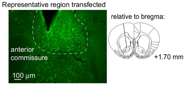
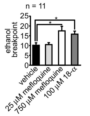
Here we illustrate examples of results that can be obtained with the above procedure. Shown first is a typical area that is transfected by viral vectors (Figure 3). In general, a transfected volume of ~1 mm3 is obtained in striatal tissue. The volume of the transfected region can be quantified across serial sections using Cavalieri’s method.32 Importantly, the transfected volume depends on many factors such as the type of tissue being transfected; viral serotype; promoter; rate and volume injected; number of particles injected; injectate pH; and whether hyperosmolar reagents, such as mannitol, were used.33,34 Typically, we microinject 1012 particles/ml, 1 µl/side over 10 min, and allow 7 additional min prior to replacing obturators. Additionally, the injectate is generally around pH 8. Next, we show that motivation can be manipulated by either transgene expression (Figure 4) or pharmacological reagent microinjection (Figure 5).
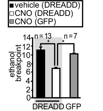
In Figure 4, we expressed a Designer Receptor Exclusively Activated by Designer Drugs (DREADDs). The DREADD receptor-coding region was followed by an Internal Ribosome Entry Site and a mCitrine cassette. The mCitrine allows convenient visualization of transfected cells. The DREADD was coupled to the heterotrimeric G-protein Gαq. Activation of the Gαq-coupled DREADD can stimulate astrocytes,28,35 and the DREADD itself can be activated by systemic administration of clozapine-N-oxide (CNO, 3 mg/kg, i.p).14,22,36 Rats were trained to lever respond for ethanol reinforcement, where 3 lever presses yielded 1 chance to drink during daily 1 hr sessions over 60 contiguous days. Next, rats were forced into abstinence, and Gαq-coupled DREADDs were expressed in nucleus accumbens core astrocytes. After 3 week abstinence, the motivation to self-administer ethanol was measured by breakpoint.21,27-30 Activation of nucleus accumbens core astrocytes, via systemic CNO administration, decreased the motivation of rats to resume ethanol self-administration after abstinence compared to vehicle. Importantly, CNO had no effect in an equally trained cohort that was expressing Green Fluorescent Protein instead of the DREADD.

Figure 3. Representative ventral striatal region transfected by microinjected virus. This image illustrates the region of nucleus accumbens astrocytes that were transfected by following the above method. Data are reprinted from Bull et al.13 with permission of the copyright holder. Details can be found in that publication and the accompanying supplementary material. Please click here to view a larger version of this figure.

Figure 4. Motivation of rats to self-administer ethanol was reduced by activating a transgene that was over-expressed in nucleus accumbens core astrocytes. Virus was microinjected and motivation measured via breakpoint after allowing one week for the virus to express. The transgene expressed was a Designer Receptor Exclusively Activated by Designer Drugs (DREADDs). A one-way ANOVA (F(2,30)=3.29, p=0.04) followed by a Scheffé post-hoc revealed that activation of the DREADD by systemic administration of clozapine-N-oxide (CNO, 3 mg/kg, ip) significantly reduced the motivation of rats to self-administer ethanol after abstinence compared to vehicle CNO had no effect in an equally trained cohort that was expressing Green Fluorescent Protein (GFP) instead of the DREADD. Data are reprinted from Bull et al.13 with permission of the copyright holder. Details can be found in that publication and the accompanying supplementary material.

Figure 5. Microinjection of gap junction blockers increased the motivation of rats to self-administer ethanol after abstinence. Two gap junction blockers were evaluated for their effect on the motivation (measured via breakpoint) of rats to self-administer ethanol: mefloquine and 18-α-glycyrrhetinic acid (18-α). A two-way ANOVA revealed that microinjection of gap-junction blockers into the nucleus accumbens core increased the motivation of rats to self-administer ethanol after abstinence (F(3,40)=5.56, p=0.003). Data are reprinted from Bull et al.13 with permission of the copyright holder. Details can be found in that publication and the accompanying supplementary material.
Discussion
The procedure presented here is an efficient means to manufacture microinjection cannulae and microinjectors that will aid in elucidating the molecular substrates of motivated behavior. This method offers several advantages. First, by manufacturing one’s own implants and microinjectors, novel experimental parameters can be rapidly optimized; i.e., one does not need to wait for custom made components to arrive. Second, due to the small diameter of the cannula, more cannulae can be simultaneously implanted. This shortens the required surgical time, which can improve survivability, and also allows multiple implants per animal. Third, the software used to control the operant chambers readily accommodates progressive ratio schedules since a fixed ratio paradigm can be rapidly converted to a progressive ratio paradigm by simply applying an event transition parameter list that contains the desired reinforcement schedule.
To be broadly useful, a generic microinjection procedure was presented that should be broadly applicable for the microinjection of nearly any reagent currently available. Consequently, we anticipate that this technique will continue to be of similar high utility in the future with minor modification. By changing only a few variables, this approach can be applied to a wide number of reagents. Parameters that would most commonly be manipulated include the length that the microinjector protrudes from the cannula, volume of injection, and rate of injection. For example, one may want the injector to protrude further from the cannula tip to avoid the glial scar that typically forms around chronic implants. Additionally, one may wish to inject a larger volume. For striatal virus microinjections, a volume of 1 μL is typically used and this volume is typically injected over a longer period of time (frequently 7 – 10 min plus 3 – 10 min additional diffusion time) compared to that used for pharmacological reagents (typically 0.3 – 0.5 μL over 2 – 3 min plus 1 – 3 min additional diffusion time). The user should consult the literature and/ or empirically determine the parameters best suited for their needs. Regardless, the success of this procedure is critically dependent upon 4 variables: 1) cannula length, 2) microinjector length, 3) quality of microinjector spray pattern, and 4) the system integrity prior to injection. Because microinjection location is dependent on the depth that the microinjector protrudes from the cannula, it is imperative that both cannulae (Step 1.2.8) and microinjector length (post-bending, Step 2.2.1) are both precisely known and uniform between all subjects. This can easily be controlled by readily rejecting any implement that is not the required length at the final re-measuring. Moreover, the injection location can only be predicted if it occurs immediately beneath the guide cannula. Thus, any microinjector that does not spray a long, fine stream upon testing (Steps 2.4.6) should be rejected. A quality injection is also related to the integrity of the system prior to injection. If after dispensing all water from the injector (prior to filling with reagent) multiple spots are observed on the lab-wipe, then a leak needs to be remedied (Note on Step 2.4.8). Further, if the bubble (Step 2.4.9) that separates the drug from the water in the PE20 tubing is not one, single bubble (after filling the microinjector with reagent), then the injector is partially clogged. This clog could either prevent or divert the injection. This too can be readily remedied (Note on step 2.4.8).
Should one wish to microinject while the animal is in the stereotaxic frame there are three alternatives. First, one could increase the length of the microinjector collar such that it can be held firmly by the stereotaxic manipulator and also extend far enough to allow connection to PE20 tubing. Second, one could temporally implant a cannula and use the standard microinjector presented here. Third, one could use drawn and polished glass pipettes. 16,17
A significant limitation of the procedure presented here is that it is best conducted in well-handled rats that are familiar with the procedure. Rats used for the data described in the results section required no special handling procedures because the same investigator handled the rats on a daily basis for over 2 months. This included daily observation and manipulation of the surgical implant for at least 2 weeks. However, rats can be rapidly habituated by a number of techniques that are used to prior to the pre-pulse inhibition assay, which can be affected by stress. These special habituation techniques have been nicely detailed previously.43 In addition to these procedures, it is advisable that rats be habituated to the microinjection procedure where shortened microinjectors are used during ‘sham’ injections. During these sham injections, it is critical that the microinjector not protrude into the tissue in order to limit tissue damage. In other words, the microinjector should be bent no longer than 14 mm. Thus, the thorough habituation required for optimal implementation of this technique could be viewed as a limitation.
While several behavioral paradigms exist to measure motivation, the progressive ratio is commonly used to quantify the effort that the subject is willing to exert to obtain a reinforcer. The progressive ratio paradigm produces a measure known as breakpoint, which is often defined as the maximal number of lever presses in the last completed ratio; i.e., maximal responding that generated a reinforcer.21 The progressive ratio is sensitive to reinforcer magnitude. For example, higher cocaine (or sucrose) doses produce a higher breakpoint and lower cocaine (or sucrose) doses yield a lower breakpoint.21,22 Accordingly, breakpoint is a routinely used proxy for motivation and/or reinforcing efficacy.21,23-26 Because the intention of the breakpoint is to determine when the animal stops responding, an important parameter of the progressive ratio paradigm is session length. Finite session lengths can put a false cap on breakpoint values and this can be exacerbated by pre-treatments that abnormally decrease the rate of self-administration or that increase post-reinforcement pausing. This confound can be overcome by any number of approaches; e.g., sessions that terminate when the animal has withheld responding by some multiple of the average inter-infusion interval.44 A more commonly applied variant of this approach is to terminate sessions once responding has been withheld by some empirically determined value that is held constant across subjects. We have provided the method to apply this approach in Step 4.4.9.11.
Acknowledgements
MSB is supported by the Alcohol Beverage Medical Research Foundation, a Center for Translational Research Award (UL1 TR000058), the National Institutes for Alcohol Abuse and Alcoholism (P50 AA022537), and startup funds provided by the Virginia Higher Education Equipment Trust Fund and the VCU School of Medicine.
Materials
| Name | Company | Catalog Number | Comments |
| Cannula Tubing | Amazon Supply/ Small Parts | HTXX-26T-60 | 26 gauge, Hypotube S/S 316-TW 26GA |
| Obturator | Amazon Supply/ Small Parts | GWXX-0080-30-05 | 33 gauge, Wire S/S 316LVM 0.008 IN |
| Microinjector Wire | MicroGroup | 33RW 304 | 33 gauge |
| Super Glue | Loctite | 3924AC | Liquid, Non-gel, can be autoclaved |
| Microinjector Plastic Tubing | Becton Dickson | 427406 | PE20 |
| Medium Weight Hemostats | World Precision Instruments | 501241-G | |
| Ruler | Fisher | 09-016 | 150 mm |
| #7 Forceps | Stoelting | 52100-77 | Dumont, Dumostar |
| Rotary Tool | Dremmel | 285 | Two-speeds |
| Cut-off Disc | McMaster Carr | 3602 | 15/16" x 0.025" |
| Microinjection Pump | Harvard Apparatus | PhD 2000 | |
| 1 ul Glass Syringe | Hamilton | 7001KH | Needle Style: 25s/2.75"/3 |
| Cotton Tipped Applicator | Fisher | 23-400-101 | |
| Lab Wipes | Kimwipes | 34133 | |
| Operant Software | Coulbourn | Graphic State | |
| Operant Chambers | Coulborun | Habitest |
References
- Koob, G. F., Volkow, N. D. Neurocircuitry of addiction. Neuropsychopharmacology. 35 (1), 217-238 (2010).
- Kalivas, P. W., Peters, J., Knackstedt, L. Animal models and brain circuits in drug addiction. Mol Interv. 6 (6), 339-344 (2006).
- Bossert, J. M., Marchant, N. J., Calu, D. J., Shaham, Y. The reinstatement model of drug relapse: recent neurobiological findings, emerging research topics, and translational research. Psychopharmacology (Berl). 229 (3), 453-476 (2013).
- Connor, E. C., Chapman, K., Butler, P., Mead, A. N. The predictive validity of the rat self-administration model for abuse liability). Neurosci Biobehav Rev. 35 (3), 912-938 (2011).
- Watson, D. J., et al. Selective blockade of dopamine D3 receptors enhances while D2 receptor antagonism impairs social novelty discrimination and novel object recognition in rats: a key role for the prefrontal cortex. Neuropsychopharmacology. 37 (3), 770-786 (2012).
- Takahashi, A., Shimamoto, A., Boyson, C. O., DeBold, J. F., Miczek, K. A. GABA(B) receptor modulation of serotonin neurons in the dorsal raphe nucleus and escalation of aggression in mice. J Neurosci. 30 (35), 11771-11780 (2010).
- Spencer, R. C., Klein, R. M., Berridge, C. W. Psychostimulants act within the prefrontal cortex to improve cognitive function. Biol Psychiatry. 72 (3), 221-227 (2012).
- Bowers, M. S., et al. Activator of G protein signaling 3: a gatekeeper of cocaine sensitization and drug seeking. Neuron. 42 (2), 269-281 (2004).
- Abudara, V., et al. The connexin43 mimetic peptide Gap19 inhibits hemichannels without altering gap junctional communication in astrocytes. Front Cell Neurosci. 8, 306 (2014).
- Cahill, E., et al. D1R/GluN1 complexes in the striatum integrate dopamine and glutamate signalling to control synaptic plasticity and cocaine-induced responses. Mol Psychiatry. 19 (12), 1295-1304 (2014).
- McFarland, K., Kalivas, P. W. The circuitry mediating cocaine-induced reinstatement of drug-seeking behavior. J Neurosci. 21 (21), 8655-8663 (2001).
- Fuchs, R. A., Branham, R. K., See, R. E. Different neural substrates mediate cocaine seeking after abstinence versus extinction training: a critical role for the dorsolateral caudate-putamen. J Neurosci. 26 (13), 3584-3588 (2006).
- Ebersberger, A. Physiology of meningeal innervation: aspects and consequences of chemosensitivity of meningeal nociceptors. Microsc Res Tech. 53 (2), 138-146 (2001).
- Netter, F. H. . Musculoskeletal System Part I: Anatomy; Physiology, and Metabolic Disorders. 8, (1987).
- Ferguson, S. M., Eskenazi, D., Ishikawa, M., Wanat, M. J., Phillips, P. E., Dong, Y., Roth, B. L., Neumaier, J. F. Transient neuronal inhibition reveals opposing roles of indirect and direct pathways in sensitization. Nat Neurosci. 14 (1), 22-24 (2011).
- Gonzalez-Perez, O., Guerrero-Cazares, H., Quinones-Hinojosa, A. Targeting of deep brain structures with microinjections for delivery of drugs, viral vectors, or cell transplants. J Vis Exp. (46), (2010).
- Inquimbert, P., Moll, M., Kohno, T., Scholz, J. Stereotaxic injection of a viral vector for conditional gene manipulation in the mouse spinal cord. J Vis Exp. (73), (2013).
- Schier, C. J., Mangieri, R. A., Dilly, G. A., Gonzales, R. A. Microdialysis of ethanol during operant ethanol self-administration and ethanol determination by gas chromatography. J Vis Exp. (67), (2012).
- Fornari, R. V., et al. Rodent stereotaxic surgery and animal welfare outcome improvements for behavioral neuroscience. J Vis Exp. (59), e3528 (2012).
- Geiger, B. M., Frank, L. E., Caldera-Siu, A. D., Pothos, E. N. Survivable stereotaxic surgery in rodents. J Vis Exp. (20), e880 (2008).
- Richardson, N. R., Roberts, D. C. Progressive ratio schedules in drug self-administration studies in rats: a method to evaluate reinforcing efficacy. J Neurosci Methods. 66 (1), 1-11 (1996).
- Cheeta, S., Brooks, S., Willner, P. Effects of reinforcer sweetness and the D2/D3 antagonist raclopride on progressive ratio operant performance. Behav Pharmacol. 6 (2), 127-132 (1995).
- Roberts, D. C., Morgan, D., Liu, Y. How to make a rat addicted to cocaine. Prog Neuropsychopharmacol Biol Psychiatry. 31 (8), 1614-1624 (2007).
- Arnold, J. M., Roberts, D. C. A critique of fixed and progressive ratio schedules used to examine the neural substrates of drug reinforcement. Pharmacol Biochem Behav. 57, 441-447 (1997).
- Hodos, W. Progressive ratio as a measure of reward strength. Science. 134 (3483), 943-944 (1961).
- Depoortere, R. Y., Li, D. H., Lane, J. D., Emmett-Oglesby, M. W. Parameters of self-administration of cocaine in rats under a progressive-ratio schedule. Pharmacol Biochem Behav. 45 (3), 539-548 (1993).
- Bowers, M. S., et al. Nucleus accumbens AGS3 expression drives ethanol seeking through G betagamma. Proc Natl Acad Sci U S A. 105 (34), 12533-12538 (2008).
- Bull, C., et al. Rat Nucleus Accumbens Core Astrocytes Modulate Reward and the Motivation to Self-Administer Ethanol after Abstinence. Neuropsychopharmacology. 39 (12), 2835-2845 (2014).
- Hopf, F. W., Chang, S. J., Sparta, D. R., Bowers, M. S., Bonci, A. Motivation for alcohol becomes resistant to quinine adulteration after 3 to 4 months of intermittent alcohol self-administration. Alcohol Clin Exp Res. 34 (9), 1565-1573 (2010).
- Hopf, F. W., et al. Reduced nucleus accumbens SK channel activity enhances alcohol seeking during abstinence. Neuron. 65 (5), 682-694 (2010).
- Salerni, C. M. . Engineering Adhesives for Repeated Sterilization. , (2015).
- Gundersen, H. J., Jensen, E. B. The efficiency of systematic sampling in stereology and its prediction. J Microsc. 147 (Pt 3), 229-263 (1987).
- Mastakov, M. Y., Baer, K., Xu, R., Fitzsimons, H., During, M. J. Combined injection of rAAV with mannitol enhances gene expression in the rat brain. Mol Ther. 3 (2), 225-232 (2001).
- Burger, C., Nguyen, F. N., Deng, J., Mandel, R. J. Systemic mannitol-induced hyperosmolality amplifies rAAV2-mediated striatal transduction to a greater extent than local co-infusion. Mol Ther. 11 (2), 327-331 (2005).
- Agulhon, C., et al. Modulation of the autonomic nervous system by acute glial cell Gq-GPCR activation in vivo. J Physiol. 591 (22), 5599-5609 (2013).
- Armbruster, B. N., Li, X., Pausch, M. H., Herlitze, S., Roth, B. L. Evolving the lock to fit the key to create a family of G protein-coupled receptors potently activated by an inert ligand. PNAS. 104 (12), 5163-5168 (2007).
- Cruikshank, S. J., et al. Potent block of Cx36 and Cx50 gap junction channels by mefloquine. PNAS. 101 (33), 12364-12369 (2004).
- Miguel-Hidalgo, J., Shoyama, Y., Wanzo, V. Infusion of gliotoxins or a gap junction blocker in the prelimbic cortex increases alcohol preference in Wistar rats. J Psychopharmacol. 23 (5), 550-557 (2008).
- Bowers, M. S., Lake, R. W., Rubinchik, S., Dong, J. Y., Kalivas, P. W. Elucidation of Homer 1a function in the nucleus accumbens using adenovirus gene transfer technology. Ann N Y Acad Sci. 1003, 419-421 (2003).
- Mackler, S. A., et al. NAC-1 is a brain POZ/BTB protein that can prevent cocaine-induced sensitization in the rat. J Neurosci. 20 (16), 6210-6217 (2000).
- Geyer, M. A., Swerdlow, N. R. Measurement of startle response, prepulse inhibition, and habituation. Curr Protoc Neurosci. 8, Unit 8.7 (2001).
- Bedford, J. A., Bailey, L. P., Wilson, M. C. Cocaine reinforced progressive ratio performance in the rhesus monkey. Pharmacol Biochem Behav. 9 (5), 631-638 (1978).
This article has been published
Video Coming Soon
ABOUT JoVE
Copyright © 2025 MyJoVE Corporation. All rights reserved