Fabrication of Amyloid-β-Secreting Alginate Microbeads for Use in Modelling Alzheimer's Disease
In This Article
Summary
This protocol illustrates a cell encapsulation method by rapid physical gelation of alginate to immobilize cells. Obtained microbeads allow controlled and sustained secretion of amyloid-β over time and can be used to study the effects of secreted amyloid-β in in vitro and in vivo models.
Abstract
According to the amyloid cascade hypothesis, the earliest trigger in the development of Alzheimer’s disease (AD) is the accumulation of toxic amyloid-β (Aβ) fragments, eventually leading to the classical features of the disease: amyloid plaques, neurofibrillary tangles and synaptic and neuronal loss. The lack of relevant non-transgenic preclinical models reflective of disease progression is one of the main factors hindering the discovery of effective drug treatments. To this end, we have developed a protocol for the fabrication of alginate microbeads containing amyloid-secreting cells useful for the study of the effects of chronic Aβ production.
Chinese hamster ovary cells previously transfected with a human APP gene, secreting Aβ (i.e., 7PA2 cells), were used in this study. A three-dimensional (3D) in vitro model for the sustained release of Aβ was fabricated by encapsulation of 7PA2 cells in alginate. The process was optimized to target a bead diameter of 500-600 μm for further in vivo studies. Optimization of 7PA2 cell encapsulation in alginate was performed altering fabrication parameters, e.g., alginate concentration, gel flow rate, electrostatic potential, head vibration frequency, gelling solution. Levels of secreted Aβ were analyzed over time and compared between alginate beads and standard cell culture methods (up to 96 h).
A concentration of 1.5 x 10⁶ 7PA2 cells/mL and an alginate concentration of 2% (w/v) buffered with HEPES and subsequent gelation in 0.5 M calcium chloride for 5 min were found to fabricate the most stable microbeads. Fabricated microbeads were 1) of uniform size, 2) with an average diameter of 550 μm, 3) containing about 100-150 cells per microbead and 4) able to secrete Aβ.
In conclusion, our optimized method for the production of stable alginate microbeads containing amyloid-producing 7PA2 cells might enable the modeling of important aspects of AD both in vitro and in vivo.
Introduction
Modeling neurodegenerative disease is challenging due to the complex and intricate nature of the brain. In Alzheimer’s disease (AD), the progressive loss of synaptic function and death of neurons is believed to be a downstream effect of sustained overproduction and accumulation of amyloid beta (Aβ) peptides following abnormal processing of the amyloid precursor protein (APP) according to the amyloid cascade hypothesis1.
In order to understand the mechanisms of this amyloid-induced pathology and to aid in the identification of novel treatment targets, scientists have developed various in vivo preclinical models. One category of models utilizes a bolus injection of a synthetic Aβ peptide into the rat brain2,3,4. The main limitation of such models is that they rely on a single-point or repeated treatments with Aβ peptides in high concentrations deposited all in one go. This is inconsistent with the chronic, sustained nature of the release of Aβ in disease5. Another category of in vivo models is transgenic animal models expressing one or more genetic mutations linked with the familial variations of the disease6,7,8,9,10. However, since familial AD only accounts for fewer than 5% of all Alzheimer’s cases11, the relevance of these models in translating to sporadic AD in humans is questionable12. Another drawback of the transgenic approach is the accelerated Aβ formation from birth, which translates into deficits in cognitive function and pathological changes too quickly and aggressively to resemble disease progression in sporadic AD in patients12. For example, the 5x FAD model produces plaques in as little as 1.5 months13.
Interestingly, both these categories result in changes in cognitive function of relevance to AD research2,3,4,5,6, and sometimes they are accompanied by the appearance of pathological hallmarks of the disease such as amyloid plaques6,8, tau phosphorylation6,7 and/or synaptic and neuronal loss7,9,14. But while these types of models may give us an insight into the effects of high levels of amyloid in the brain, which are often associated with later stages of AD, they fail to reflect the earlier changes exhibited in response to the chronic and sustained exposure to the Aβ peptide12, such as altered expression of synaptic markers15 and components in the extracellular matrix16. Therefore, there still remains a need for creating a chronic model that more accurately illustrates the effects of sustained Aβ secretion on in vivo cognition and illustrates changes in pathology.
To this end, we have developed a system which allows the constant, sustained secretion of Aβ in a controlled manner by immobilizing amyloid-secreting cells within hydrogel microbeads, which can be subsequently implanted within the adult rat brain to model aspects of sporadic AD.
Alginate was the selected biomaterial as it is biocompatible and does not induce any adverse responses when implanted in vivo17. Cell encapsulation in alginate hydrogels has been well established over the last four decades. The first example of its translation to the clinic was reported for the treatment of type 1 diabetes mellitus17. The earliest report of successful encapsulation of islets of Langerhans dates back to 1980. Transplantation of microbeads containing insulin-secreting cells revolutionized treatment options for diabetic patients as it restored pancreatic function, eliminating the need for insulin injection therapy18. These works report on how cell encapsulation can protect them from external stresses, whether mechanical or chemical. In fact, alginate beads act as a barrier and isolate cells from the surrounding environment preserving their phenotype, whilst allowing sufficient access to the surrounding media for nutrients and clearance of cellular byproducts19. Moreover, the use of alginate allows matching of mechanical properties of soft tissue20. Alginate hydrogels can be tuned to have a stiffness of 1-30 kPa, by simply varying alginate concentration and cross-linking density20,21. This is an essential aspect, not only to maintain the phenotypic expression of encapsulated cells in vitro, but also to avoid any inflammatory effects after engraftment in vivo22.
In this protocol, 7PA2 cells - a Chinese hamster ovary cell line that is stably transfected with a human APP V717F mutated gene23 - are used. These cells continuously produce catalytic products of APP, including Aβ1-4224,25, and have been used to generate Aβ as an alternative to synthetic production in preclinical, acute in vivo studies26. We describe a fabrication method for immobilizing 7PA2 cells within 'soft' alginate microbeads, designed to allow the sustained secretion of biomolecules. As a proof of concept, we report on the release of the Aβ1-42 peptide over time. The alginate that is used is a low-viscosity alginate with a molecular weight of 120,000-190,000 g/mol and a mannuronic to guluronic ratio of 1.56 (M/G).
In further studies, these microbeads can be safely transplanted within regions of the rat brain of relevance to AD (e.g., the hippocampus) to study the effects of chronic Aβ secretion on behavior in vivo and pathology ex vivo. In addition, this system can be used to study the effects of chronic Aβ release in in vitro and ex vivo applications. For instance, 7PA2-containing alginate microbeads can be co-cultured in vitro with neuronal or astrocytic cultures to assess the effects of chronic Aβ exposure on cellular mechanisms associated with AD. Furthermore, this method can be used to examine the relationship between chronic Aβ production and long-term potentiation in ex vivo electrophysiology.
The highlight of this protocol is the modularity and flexibility of the fabrication method, which enables the fabrication of alginate beads with a target dimension by fine tuning fabrication parameters. Depending on the application, the protocol can be adjusted to obtain bespoke targets with regards to microbead size, density of the encapsulated cells, and microbead stiffness. This protocol can be used for the encapsulation of a variety of cell types, developing more relevant three-dimensional (3D) in vitro models to study different pathologies. We recently reported on how alginate-encapsulated cells can be used to model early stages of cancer progression20.
The concept of the encapsulation process is based on the extrusion of a laminar jet of cells suspended in alginate solution through a nozzle. A vibrating head disrupts the jet with a controlled frequency resulting in equally sized alginate-based droplets. An external electric field allows separation of the formed alginate-based droplets, which upon contact with a solution enriched in divalent ions, such as calcium ions, can quickly cross-link, preserving their spherical shape. Incubation in the gelation solution allows formation of spherical microbeads containing cells within a homogeneous physical hydrogel27. Target size of microbeads and alginate hydrogels allows nutrients and oxygen exchange with cell culture media for long periods of time (weeks). Figure 1A show a schematic representation of the encapsulation apparatus used (Figure 1B).
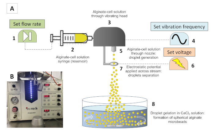
Figure 1: The encapsulation system. (A) Schematic representation of the encapsulation system. An alginate-cell suspension is loaded into a syringe (2) and fed through a reservoir at an extrusion speed set at syringe pump (1). In the reservoir, a vibration hat (3) vibrates at a frequency set by a waveform generator (4) to disrupt the stream at equal intervals, forming equally-sized droplets. As the solution is fed through a nozzle (5) and droplets are formed, an electrostatic potential is applied across an electrode (7) set by a voltage generator (6), which slightly charges the surface of the droplets, allowing the stream to spread as a result of repelling electrostatic forces. As droplets engage with the gelation bath (8), Ca2+-driven cross-linking of alginate results in the formation of spherical microbeads. (B) Photograph of the encapsulator before fabrication of alginate microbeads. Please click here to view a larger version of this figure.
Microbead size can be altered depending on the intended use. In order to control the size of the microbead, the various parameters outlined in Figure 1A and in the protocol are adjusted accordingly. The internal diameter of the nozzle used has a substantial impact on the size of the droplets; further adjusting encapsulation parameters, namely extrusion speed, vibration frequency and voltage, is key to achieving a consistent size distribution. Table 1 outlines how the different parameters can alter the size of microbeads achieved with this system.
| Parameter | Nozzle size | Vibration frequency | Flow rate | Electrode voltage |
 |  |  |  | |
| Bead size |   |  |  | – |
Table 1: Fabrication parameters and their influence on microbead size. The table illustrates how each parameter can influence the resultant size of fabricated microbeads, irrespective of the nozzle and the viscosity of the solution used.
Protocol
1. Preparations
- Prepare 100 mL of ~4% (w/v) alginate stock solution.
- Weigh 4.4 g of alginate sodium salt and add the dry powder to 100 mL of HEPES-buffered saline (HBS, 20 mM HEPES (0.477 g) with 150 mM NaCl (0.877 g) in deionized water allowing hydration.
- Use a magnetic stirrer at ~500 revolutions per minute (rpm) and heat to up to 50 °C to allow complete alginate hydration.
NOTE: The alginate may need one or two hours to be completely hydrated. To save on time, alginate solution can be made up a day in advance of the encapsulation protocol and stored at 4 °C until the next day. When alginate has been refrigerated, heat gently to 37 °C using a water bath or a hot plate and agitate with a magnetic stirrer immediately before use. - Sterile filter the alginate solution using a 0.22 µm pore polyethersulfone (PES) filter before use. Filtering is recommended at 37 °C to allow easy flow of alginate. The viscosity will increase if the temperature is allowed to drop.
- Prepare 1,000 mL of 0.5 M CaCl2.
- Weigh 55.49 g of anhydrous CaCl2 and dissolve in 1,000 mL of HBS (20 mM HEPES and 150 mM NaCl in 1,000 mL of deionized water).
- Sterile filter using a 0.22 µm pore PES filter.
- Prepare 100 mL of dissolution mix.
- Prepare a 100 mM HEPES (23.83 g) and 500 mM trisodium citrate dihydrate (147.05 g) solution in phosphate-buffered saline.
- Adjust to pH 7.4 using NaOH or HCl.
2. Setting up the encapsulation system
- Clean the laminar flow hood.
- Sterilize the laminar flow hood containing the encapsulator using UV exposure followed by spraying with 70% v/v ethanol solution.
- Flush the encapsulator system with 10 mL of 70% v/v ethanol solution followed by 10 mL of sterile deionized water.
- Attach the 300 µm nozzle and repeat step 2.1.2.
- Spray the bottle containing the filtered alginate solution, the bottle containing filtered calcium chloride solution and any tools, equipment and empty culture plates that will be used with 70% v/v ethanol solution and place them into the laminar flow hood. Prior to use, switch on a UV light again for 30 min to thoroughly sterilize.
- Prepare a waste beaker filled with a biological grade disinfectant solution and place the beaker into the hood.
- Fill a beaker with 100 mL of sterile CaCl2 (aqueous) solution and add in a magnetic stirrer. Set the stirring speed to 100 rpm. Place the beaker on a magnetic platform at a height of 18 cm from the tip of the nozzle. This beaker will be used to collect alginate beads and allow their gelation.
- Prepare cells for encapsulation.
- Remove cells from the incubator and detach from the near-confluent flask using 0.25% trypsin-EDTA solution and incubate at 37 °C for 5-10 min.
- Isolate a sample for estimation of cell density and then centrifuge the remaining cells at 1,000 rpm for 5 min to obtain a cell pellet.
- Resuspend the pellet in HBS (20 mM HEPES and 150 mM NaCl) to double the final desired cell concentration (e.g., for a final concentration of 1.5 x 10⁶ cells/mL, resuspend the cells in HBS to achieve a concentration of 3 x 10⁶ cells/mL).
- In a 50 mL centrifuge tube, mix the cell suspension in a 1:1 ratio with ~4% (w/v) alginate solution to obtain a final suspension containing the desired cell concentration (e.g., 1.5 x 10⁶ cells/mL) in a ~2% (w/v) alginate solution.
- Set the encapsulation parameters.
- Set the speed of the encapsulator machine to the maximum extrusion speed (8.9 mL/min), the voltage to 1.0 kV and the frequency to 5,500 Hz.
NOTE: These parameters were previously optimized to obtain microbeads of 550 µm in diameter.
- Set the speed of the encapsulator machine to the maximum extrusion speed (8.9 mL/min), the voltage to 1.0 kV and the frequency to 5,500 Hz.
3. Fabrication
- Fabricate the microbeads.
- In a 20 mL syringe, load 5 mL of the cell-alginate suspension and attach a syringe to the encapsulator.
- Start the encapsulator by activating the flow which will push the cell-alginate suspension through the feeder. A stream of droplets will be extruded through the nozzle.
- Collect the first 1 mL in the waste beaker to void the initial non-uniform stream.
- Continue to run the remaining 4 mL allowing the droplets to fall into the CaCl2 gelation bath. Each milliliter can be run separately (but successively) and collected in four different gelation baths if required.
NOTE: Upon contact with the gelation bath, the alginate in the droplets will instantly cross-link with the calcium ions in the gelation bath and form spherical microbeads. - After one minute, remove the gelation beaker from the magnetic platform and allow the microbeads to sit for a further 4 min without agitation (time necessary to allow complete gelation across the microbeads at room temperature).
- Retrieve microbeads.
- Remove any large alginate debris or artefacts using a pair of sterile tweezers, and then use a sterile plastic pipette to transfer to a 74 µm mesh filter to retrieve the microbeads from the gelation bath.
- Transfer the microbeads into a centrifuge tube using the appropriate culture medium and allow to equilibrate for 5 min in the cell culture medium.
- Transfer to a flask, plate or Petri dish for incubation and further experiments.
NOTE: Gelation bath should not be reused.
4. Testing and use of microbeads
- Test microbead quality.
- To assess the cell viability (or other biological readouts) after encapsulation and culture, gently disrupt the microbeads to release the encapsulated cells using the dissolution mix.
NOTE: The required volume of dissolution mix is dependent on the number of microbeads used per test. A suggested ration is 4 mL of dissolution mix to every 1 mL of encapsulated cells. This step only needs to be performed on a single sample fro each microbead population. - Incubate the cells in a cell culture incubator supplemented with 5% CO2 at 37 °C for 10 min.
- Estimate cell viability for cells now in solution as usual by staining cells with trypan blue and using a hemocytometer chamber.
NOTE: If using automated cell counters for estimations of cell viability, care should be taken to avoid alginate artefacts interfering with the counts. In those cases, naual cell counting is advised. - To assess microbead stability, measure the average diameter for a sample from each microbead population over a time course using a microscope and imaging software. Dramatic subsequent variations in diameter can be indicative of alginate degradation.
- To assess the cell viability (or other biological readouts) after encapsulation and culture, gently disrupt the microbeads to release the encapsulated cells using the dissolution mix.
- Use microbeads for detection of secreted Aβ.
- Sample cell culture media from 7PA2 cells cultured in standard conditions (2D) at 24 h intervals and store at -20 °C for further analysis.
- Sample cell culture media from encapsulated 7PA2 cells cultured in standard conditions (3D) at 24 h intervals and store at -20 °C for further analysis.
- Detect secretion of Aβ from 7PA2 cells into collected conditioned media by enzyme-linked immunosorbent assay (ELISA) (Figure 4).
- Use the microbeads for further study in relevant applications.
NOTE: Encapsulated 7PA2 cells can be used to assess the effects of sustained Aβ secretion in any or model.- For microbead engraftment within the rat hippocampus in brains, generate 1 mm thick coronal sections of the rat brain and use surgical tools to insert microbeads into the desired area (Figure 5).
- Encapsulate different cell types in alginate microbeads following the described protocols to assess the sustained release of the biomolecules of interest. Relevant in vitro and in vivo systems can be further modeled.
- Detect the expression of membrane-bound markers of encapsulated cells using flow cytometry and/or immunofluorescence.
NOTE: An example of the use of a similar method for the fabrication of microbeads modeling early stages of tumor mass growth and biomarker expression is described by Rios 20.
Representative Results
7PA2 cells are successfully encapsulated in alginate microbeads
After preparation, uniform and spherical alginate microbeads are successfully generated using this protocol. The images in Figure 2A below showcase an example of how altering one of the parameters (i.e., voltage) changes the flow and dispersion of the alginate droplets stream. Figure 2B shows an example of obtained alginate beads immediately after the gelation process.
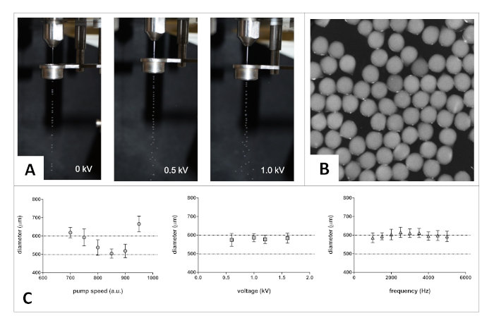
Figure 2: Fabrication method optimization. (A) Photographs showing changes in stream dispersion. (B) Bright-field image showing alginate spherical microbeads immediately after fabrication. (C) Example of optimization step: tuning selected fabrication parameters to achieve the target microbead size. Selected graphs to illustrate the relationship between each parameter and the size of the resultant microbeads (size distribution from at least n = 100 microbeads). Note that the nozzle internal diameter, viscosity of ejected solution and gelling conditions can also influence the size of fabricated beads. Error bars represent S.D. Please click here to view a larger version of this figure.
The described protocol is flexible and allows the fabrication of different sizes of alginate-based microbeads, varying in composition according to the application. In Figure 2C, we report the influence of alginate flow rate, voltage and frequency on microbead size using a 2% (w/v) alginate solution in HEPES (pH 7.2) extruded through a 300 μm nozzle.
As the scope of this study was to produce microbeads that can be embedded within the rat brain, we adjusted the fabrication parameters to obtain an average microbead diameter in the range of 500-600 μm. For quality control purposes, the method was optimized to have minimal variability across the population of fabricated beads (i.e., narrow size distribution). Please note that (1) produced beads can be injected in the rat brain using a Hamilton syringe equipped with a 20G needle; and (2) one hemisphere of rat brain can host up to two or three beads of this size.
After optimization, encapsulating 7PA2 cells at a concentration of 1.5 x 10⁶ cells/mL suspended in a 2% (w/v) alginate solution buffered with HEPES and dropped in a 0.5 M calcium chloride gelation bath yielded the desired microbeads. Figure 3A below shows encapsulated 7PA2 cells evenly distributed in microbeads after one day in standard cell culture conditions. 7PA2 cell proliferation was tested using an MTS assay as per manufacturer’s protocol. There was no significant difference between the behavior of 7PA2 cells grown with or without alginate over a seven-day period (Figure 3B). When incubating encapsulated cells in standard culture conditions, cells are expected to continue to proliferate over the duration of the experiment, and small degrees of cell escape can be expected as a result, as reported in other works28. The effects of this can be mitigated by encapsulating at a smaller cell density or reducing the serum concentration in which microbeads are incubated.
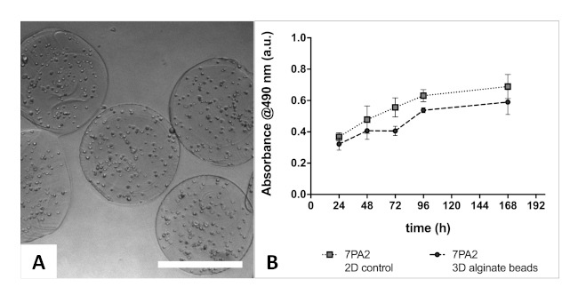
Figure 3: Encapsulated 7PA2 cells. (A) Bright-field image of encapsulated 7PA2 cells showing even cell distribution throughout the alginate microbead. Fabrication parameters were set to obtain ~150 7PA2 cells per bead. No significant variation in alginate beads was observed over 7 days of culture. (B) There was no observed difference between the overall proliferation of 7PA2 cells incubated with alginate versus cells incubated without alginate. 7PA2 cells grow and migrate across the volume of the alginate beads; reduction of serum concentration can be considered to slow down cell growth. Error bars represent S.D. Please click here to view a larger version of this figure.
Alginate microbeads encapsulating 7PA2 cells are stable over time
To investigate the size and shape of obtained alginate microbeads, microscope analysis was performed after fabrication and gelation. The average diameter for the microbeads obtained using the selected protocol is 550 ± 2 μm.
Theoretically, the number of cells expected in a 550 μm-diameter microbead can be calculated as follows:

where V = volume and r = radius. The volume of a single microbead is V = 8.8 x 10⁻⁵ mL; therefore, the number of cells per microbead at time of encapsulation = (1.5 × 106) x (8.8 x 10-5) ≈ 130 cells.
Experimentally, we counted the number of encapsulated cells immediately after fabrication. Alginate beads were gently disrupted using the dissolution mix and cells were stained with trypan blue solution. Viability estimation and counting of cells was performed using a hemocytometer; obtained results showed an average of 116 ± 17 live cells per microbead (n = 5, data not reported). As expected, a minor difference between theoretical and experimental cell count immediately after encapsulation was observed. For some applications, and in particular to determine the amount of released Aβ over time, it is important to predict the number of cells encapsulated in each alginate bead.
Interestingly, the encapsulation process does not have a significant impact on cell viability. Results are reported in a previous work, in which colorectal cancer cells (i.e., HCT-116) were encapsulated using a similar method, with no differences in cell viability in alginate microbeads compared to 2D controls20.
To measure the stability of the alginate used in the encapsulation process, we measured microbead diameters over a 14-day period (n = 100). There were no observed changes in average diameter 14 days after encapsulation as compared with immediately after encapsulation (data not reported).
Encapsulated 7PA2 cells release Aβ over time
Conditioned media analyzed from 2D and 3D cultures of 7PA2 cells reveal a constant increase in Aβ1-42 levels. Cell culture media was sampled every 24 h, and up to four days, and analyzed using ELISA. Our data shows that the rate of release of Aβ1-42 from microbeads (3D) is similar in profile to that released from the 2D culture (Figure 4).
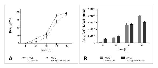
Figure 4: Rate of Aβ1-42 secretion from 7PA2 cells. (A) Aβ release (% normalized after 4 days) from 2D and 3D in vitro models has a similar profile. Both models show a constant increase in Aβ levels over the four-day period and, as expected, a steady concentration is not reached. (B) Concentration of Aβ1-42 normalized to initial cell number, showing similar secretion of Aβ1-42 over time, regardless of the culturing methods. Neither the presence of alginate nor the process of encapsulation (3D in vitro model) alter the secretion of Aβ1-42. Error bars represent S.D. Please click here to view a larger version of this figure.
Potential application of encapsulated 7PA2 cells
We report that 7PA2 cells encapsulated in alginate microbeads can be effectively used for the sustained release of Aβ1-42, and hence used to test the effect of chronic Aβ secretion in any preclinical model. In Figure 5 below, we show the advantages of using alginate microbeads that can be easily injected and located within the brain of a rat. Ex vivo sections are here used for illustration purposes, comparing a millimeter alginate bead versus fabricated alginate microbeads.

Figure 5: Using microbeads for relevant preclinical applications. Microbeads for engraftment in the rat brain must be small enough to be embedded without creating a lesion too large to avoid damage to the brain parenchyma and adversely affect normal brain function. Image (A) shows a size comparison between a millimeter-scaled bead and a micrometer-scaled bead side by side. (B) Implanting a millimeter-scaled bead within the brain for in vivo purposes would not work. Image (C) shows a microbead fabricated using this protocol. The size is suitable for insertion within the hippocampus of the rat without having a detrimental effect on normal physiology. Please click here to view a larger version of this figure.
The images above highlight the importance of controlling the size of the microbead for such studies. Here, we demonstrate the advantage of using microbeads of diameters under 600 µm. This allows the use of minimally invasive injection (e.g., Hamilton syringe) to better control the location of injected beads within the brain.
In summary, encapsulating 7PA2 cells gives control over the size of microbeads, number of encapsulated cells and the prediction of Aβ secreted from the microbeads (e.g., concentration, release profile). Controlling the size of the microbeads is essential for two reasons: 1) to permit fine-tuned control over the concentration of released Aβ, and 2) to allow implantation in a controlled region of the rat brain. The results obtained here describe the facile tuning of alginate microbeads and highlight potential applications for further studies.
Discussion
The method outlined in this article is useful for encapsulating cells achieving a narrow size distribution of alginate microbeads27. It also affords the advantage of growing cells in an immuno-isolated environment17,19, protecting them from external stress. Additionally, encapsulation of cells in alginate more closely mimics physiological conditions, specifically with regards to cell-to-cell interactions and matrix stiffness20. These factors are all particularly crucial for subsequent use in relevant applications, such as in vivo engraftment, precluding potential immune reactions in the surrounding tissue17. Furthermore, a major advantage of this protocol is the easy tuneability according to the application of interest: it is possible to modify the protocol and optimize the parameters for fabrication of larger or smaller, and softer or stiffer beads. The described method is used to fabricate beads small enough to be injected with minimally invasive methods, and sufficiently large enough to host a number of cells and provide a sufficient release of Aβ to result in observable behavioral and pathological effects upon injection in animal models.
The success of this protocol is dependent on a number of critical steps. Careful cell handling and optimal cell culturing techniques are important to maintain cell viability and function20,28. Using standard culture conditions ensures the conservation of normal function of 7PA2 cells, as observed. This allows a similar release profile for Aβ1-42 from encapsulated cells when compared with 2D cultures. In addition, for the protocol to work optimally, a low-viscosity alginate solution guarantees better results compared with a high viscosity alginate solution. This ensures that a homogenous laminar jet is extruded through the nozzle and an even distribution of cells within the matrix of the fabricated beads27. Materials used to encapsulate cells must have a very rapid gelation mechanism, allowing retention of shape.
Another critical step in this protocol is the handling of fabricated microbeads following gelation. Here, we show the retrieval of microbeads by using a large aperture plastic pipette to transfer the beads. Alternatively, pouring the calcium chloride solution containing the microbeads into a mesh filter can be used for bead retrieval. A large (5, 10 or 25 mL) serological pipette can be used to draw up the microbeads and then washed through the mesh filter instead of pouring. The advantage of this is a higher confidence in the sterility of the procedure compared to pouring. However, of the limitations is that some beads may be distorted if they are compressed by the pipette, in addition to risking a lower yield if a large proportion of microbeads are not rescued.
This approach has been used to encapsulate different cell lines to model and study different diseases (e.g., release of insulin from encapsulated pancreatic islet). The novelty of our approach is the engraftment of the microbeads generated using this protocol as a useful method in modeling important aspects of Alzheimer’s disease in vivo. When comparing the release profile of Aβ from encapsulated cells (Figure 4) to the levels of Aβ generated by a bolus injection (such as that reported in other studies3,26), a more chronic and sustained release of Aβ can be expected. Figure 6 illustrates the predicted tendency that may be achieved. Using this system for in vivo modeling is more relevant to the way the disease progresses and can be more useful in drug discovery and development.
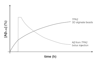
Figure 6: Predicted release profile of Aβ1-42 from encapsulated 7PA2 cells compared with a bolus injection. The profile of Aβ release from engrafted 7PA2-containing microbeads allows the testing of the effects of chronic and sustained Aβ in an animal model of relevance to AD. Conversely, a bolus injection would create a spike in Aβ levels over a short period of time, followed by a rapid clearance of Aβ from the brain. Please click here to view a larger version of this figure.
Acknowledgements
The authors would like to thank Mr Kajen Suresparan, Dr Jonathan Wubetu, Mr Dominik Grudzinski, Miss Chen Zhao, and Dr Thierry Pilot for their help in the optimization of alginate microbead fabrication, cell culture and Aβ detection, and useful scientific discussions.
Materials
| Name | Company | Catalog Number | Comments |
| µManager software | Vale Lab, UCSF, USA | v.1.46 | |
| 0.22 um PES filter | Merck, UK | SLGP033RS | |
| 15mm Netwell insert 74 um mesh filter | Constar, usa | #3477 | |
| Alginic acid sodium salt from brown algae | Sigma-Aldrich,uk | A0682 | |
| Calcium chloride | Sigma-Aldrich,uk | C1016 | |
| CellTiter96 AQueous One Solution cell proliferation assay | Promega, USA | G3580 | |
| Encapsulator | Inotech | IE-50 | serial no. 05.002.01-2005 |
| HEPES | Sigma-Aldrich,uk | H4024 | |
| Hu Aβ 1-42 ELISA | ThermoFisher, UK | KHB3441 | |
| ImageJ software | ImageJ | v1.49p | |
| Inverted light microscope | Olympus | CKX41 | |
| Leica microscope | Leica microsystems, UK | DMI6000B | |
| Neo sCMOS Camera | Ander, UK | 5.5 | |
| Phosphate buffered saline | Sigma-Aldrich,uk | D1408 | |
| Sodium chloride | Sigma-Aldrich,uk | 433209 | |
| Trispdium citrate dihydrate | Sigma-Aldrich,uk | W302600-K | |
| Trypsin-EDTA solution | Sigma-Aldrich,uk | T4049 |
References
- Hardy, J. A., Higgins, G. A. Alzheimer's disease: the amyloid cascade hypothesis. Science. 256, 184-185 (1992).
- Karthick, C., et al. Time-dependent effect of oligomeric amyloid-β (1-42)-induced hippocampal neurodegeneration in rat model of Alzheimer's disease. Neurological Research. 41 (2), 139-150 (2018).
- Watremez, W., et al. Stabilized Low-n Amyloid-β Oligomers Induce Robust Novel Object Recognition Deficits Associated with Inflammatory, Synaptic, and GABAergic Dysfunction in the Rat. Journal of Alzheimer's Disease. 62 (1), 213-226 (2018).
- Brouillette, J., et al. Neurotoxicity and memory deficits induced by soluble low-molecular-weight amyloid-β1-42 oligomers are revealed in vivo by using a novel animal model. Journal of Neurosciences. 32 (23), 7852-7861 (2012).
- Solana, C., Tarazona, R., Solana, R. Immunosenescence of Natural Killer Cells, Inflammation, and Alzheimer's Disease. International Journal of Alzheimer's Disease. , 3128758 (2018).
- Sturchler-Pierrat, C., et al. Two amyloid precursor protein transgenic mouse models with Alzheimer disease-like pathology. Proceedings of the National Academy of Sciences of the United States of America. 94 (24), 13287-13292 (1997).
- Tomiyama, T., et al. A mouse model of amyloid beta oligomers: their contribution to synaptic alteration, abnormal tau phosphorylation, glial activation, and neuronal loss in vivo. Journal of Neurosciences. 30 (14), 4845-4856 (2010).
- Saito, T., et al. Single App knock-in mouse models of Alzheimer's disease. Nature Neurosciences. 17 (5), 661-663 (2014).
- Oddo, S., et al. Triple-Transgenic Model of Alzheimer's Disease with Plaques and Tangles: Intracellular A and Synaptic Dysfunction. Neuron. 39 (3), 409-421 (2003).
- Leon, W. C., et al. A Novel Transgenic Rat Model with a Full Alzheimer's-Like Amyloid Pathology Displays Pre-Plaque Intracellular Amyloid-β-Associated Cognitive Impairment. Journal of Alzheimer's Disease. 20 (1), 113-126 (2010).
- Prince, M., et al. Dementia UK: Second Edition - Overview. Alzheimer's Society. , 61 (2007).
- Cavanaugh, S. E., Pippin, J. J., Barnard, N. D. Animal models of Alzheimer disease: historical pitfalls and a path forward. Alternatives to animal experimentation. 31 (3), 279-302 (2014).
- Oakley, H., et al. Intraneuronal β-Amyloid Aggregates, Neurodegeneration, and Neuron Loss in Transgenic Mice with Five Familial Alzheimer's Disease Mutations: Potential Factors in Amyloid Plaque Formation. The Journal of Neuroscience. 26 (40), 10129-10140 (2006).
- Forny-Germano, L., et al. Alzheimer's Disease-Like Pathology Induced by Amyloid-β Oligomers in Nonhuman Primates. Journal of Neuroscience. 34 (41), 13629-13643 (2014).
- Masliah, E., et al. Altered expression of synaptic proteins occurs early during progression of Alzheimer's disease. Neurology. 56 (1), 127-129 (2001).
- Lepelletier, F. X., Mann, D. M. A., Robinson, A. C., Pinteaux, E., Boutin, H. Early changes in extracellular matrix in Alzheimer's disease. Neuropathology and Applied Neurobiology. 43 (2), 167-182 (2017).
- Calafiore, R., Basta, G. Clinical application of microencapsulated islets: Actual prospectives on progress and challenges. Advanced Drug Delivery Reviews. 67-68, 84-92 (2014).
- Lim, F., Sun, A. M. Microencapsulated islets as bioartificial endocrine pancreas. Science. 210 (4472), 908-910 (1980).
- Tran, N. M., et al. Alginate hydrogel protects encapsulated hepatic HuH-7 cells against hepatitis C virus and other viral infections. PLoS One. 9 (10), 109969 (2014).
- Rios de la Rosa, J. M., Wubetu, J., Tirelli, N., Tirella, A. Colorectal tumor 3D in vitro models: advantages of biofabrication for the recapitulation of early stages of tumour development. Biomedical Physics & Engineering Express. 4 (4), 045010 (2018).
- Tirella, A., Orsini, A., Vozzi, G., Ahluwalia, A. A phase diagram for microfabrication of geometrically controlled hydrogel scaffolds. Biofabrication. 1 (4), 045002 (2009).
- Smalley, K. S. M., Lioni, M., Herlyn, M. Life ins't flat: Taking cancer biology to the next dimension. In Vitro Cellular & Developmental Biology - Animal. 42 (8-9), 242-247 (2006).
- Podlisny, M. B., et al. Aggregation of secreted amyloid beta-protein into sodium dodecyl sulfate-stable oligomers in cell culture. Journal of Biological Chemistry. 270 (16), 9564-9570 (1995).
- Portelius, E., et al. Mass spectrometric characterization of amyloid-β species in the 7PA2 cell model of Alzheimer's disease. Journal of Alzheimer's Disease. 33 (1), 85-93 (2013).
- Welzel, A. T., et al. Secreted amyloid β-proteins in a cell culture model include N-terminally extended peptides that impair synaptic plasticity. Biochemistry. 53 (24), 3908-3921 (2014).
- O'Hare, E., et al. Orally bioavailable small molecule drug protects memory in Alzheimer's disease models. Neurobiology of Aging. 34 (4), 1116-1125 (2013).
- Nedović, V., Willaert, R. Fundamentals of Cell Immobilisation Biotechnology. Methods and Technologies for Cell Immobilisation/Encapsulation. , 185-204 (2004).
- Omer, A., et al. Long-term Normoglycemia in Rats Receiving Transplants with Encapsulated Islets. Transplantation. 79 (1), 52-58 (2005).
This article has been published
Video Coming Soon
ABOUT JoVE
Copyright © 2025 MyJoVE Corporation. All rights reserved