Standardizing a Non-Lethal Method for Characterizing the Reproductive Status and Larval Development of Freshwater Mussels (Bivalvia: Unionida)
In This Article
Summary
Freshwater mussel conservation is dependent on monitoring reproductive patterns and processes of species. This study standardizes a non-lethal protocol for sampling gill contents, characterizing larval development, and providing a digital repository for data collected. This protocol-database package will be an important tool for mussel researchers in recovery of imperiled species.
Abstract
Actively monitoring the timing, development, and reproductive patterns of endangered species is critical when managing for population recovery. Freshwater mussels are among the most imperiled organisms in the world, but information about early larval (glochidial) development and brooding periods is still lacking for many species. Previous studies have focused on the complex life history stage when female mussels are ready to parasitize host fish, but few studies have focused on the brooding period and timing of larval development. The protocol described here allows researchers to non-lethally evaluate the state of gravidity for female mussels. The results of this study show that this method does not affect a female mussel’s ability to stay gravid or become gravid again after sampling has been performed. The advantage of this method may permit its use on federally threatened or endangered species or other populations of high conservation concern. This protocol can be adapted for use on both preserved or live individuals and was tested on a variety of mussel species. The database provided is a repository for a breadth of information on timing of reproductive habits and will facilitate future freshwater mussel research, conservation, and recovery efforts.
Introduction
The persistence of populations in freshwater systems depends on the success of reproduction and recruitment. For parasitic organisms, identifying the intricacies of the life cycle (e.g., stages of larval development and host attraction strategies) can give insight into an organism’s reproductive habits and critical processes that influence recruitment. Such information becomes important when species are imperiled, and successful recruitment is needed to sustain remaining populations, or if recovery necessitates the use of captive propagation for reestablishing extirpated populations.
Freshwater mussels (Bivalvia: Unionida) are considered one of the most imperiled groups of organisms worldwide and a compilation of species-specific reproductive habits could aid in research efforts1,2,3,4,5. With over 800 currently recognized species distributed across the globe, freshwater mussels have hotspots of diversity in North and South America, and southeastern Asia but essential life history information is unknown for many species2,5,6,7. Families within this order are characterized by having parasitic larval stages that complete metamorphosis into free-living juveniles during attachment to a host7,8. This unique life history stage contributes to the biodiversity in freshwater systems, which are currently in crisis9. High levels of imperilment can be attributed to many anthropogenic threats including pollution of waterways, habitat alteration and destruction, reductions in the abundance and diversity of host fishes, and introduction of invasive species1,10. As benthic filter feeders, mussels burrow into the substrate and are susceptible to contaminants and pollutants that drain into the watershed11. Recovery of mussel species is pertinent as they provide a wide variety of ecosystem services, including carbon sequestration, a food source, and water purification by filter feeding11. In addition, mussels have been found to indicate ecosystem health, promote biodiversity, and in turn, increase resiliency of an ecosystem12.
Many freshwater mussel studies have focused on investigating early life history requirements to better inform species status assessments and management strategies. The freshwater mussel families relevant to this study (e.g., Hyriidae, Margaritiferidae, Unionidae) have a unique life history strategy where females brood larvae (glochidia) in their marsupial gills8. Through a variety of strategies, the female mussel expels mature glochidia from marsupial gills to parasitize a vertebrate host with glochidia13. Research on glochidial development within the gills was modified from a technique utilizing hypodermic syringes to sample gonadal fluid from live mussels and evaluate gamete production14,15,16. As researchers validated this non-lethal methodology for gonad sampling, it was adapted for marsupial gill sampling to evaluate brood development15,16. Brood development can be used to decipher phylogenetic relationships as some mussel species can brood glochidia in only the outer two gills (ectobranchus), only the inner two gills (endobranchus), or in all four gills (tetrabranchus) but this characteristic is not known for every species17. Brooding patterns have previously been used to classify mussel species by whether female mussels brood glochidia over winter (bradytictic) or for a short period in the summer (tachytictic)18. The over wintering of mussel broods was supported when the reproductive cycle of Anodonta was studied19. However, basic reproductive biology was studied more thoroughly over the years and found this dichotomy was a gross generalization and brooding periods of some species are much more complex than originally presumed20,21. For example, species of the genus Hyridella (family Hyriidae), Glebula, and Elliptio (family Unionidae) have been observed with upwards of three broods per breeding season22,23,24. The complexity of species-specific, and sometimes even population-specific20, reproductive habits has led to a gap in knowledge about the timing and duration of brooding, and number of broods a female mussel may produce.
Although hypodermic syringes have been used to extract gill contents, reporting the results is complicated due to the lack of standardization to ensure comparable results across all studies. Previously, four developmental stages of glochidia (i.e., egg, embryo, immature, fully developed) have been identified in Unionidae but have not been adopted into standard procedure16,25,26. Other studies observing members of Margaritiferidae have substituted the classification of ‘immature glochidia’ with ‘developing glochidia’, leading to potential confusion27,28. The lack of consistency in characterizing the different larval development stages has left many researchers to generally describe brooding females as ‘gravid’, which does not encompass the intricacies of larval development. Life history studies conducting host-fish trials have prioritized the need for gravid females with fully-developed glochidia, but this information is scattered throughout published and unpublished literature29,30. Currently, data are lacking about the reproductive habits of many mussel species, including timing of the transition between egg, immature glochidia, and fully developed glochidia ready for attachment to hosts. For most species, it is unclear how long females brood glochidia and how quickly fertilized eggs fully develop. The knowledge gaps are often wider for species of conservation concern, which presents the need for a standardized method of extracting gill contents that has been tested for non-lethal effects and can be promoted to the scientific community to supplement mainstream data collection methodologies, without posing a threat to protected populations24,31,32.
This study had three objectives: 1) formalize a gill sampling technique and test it for lethal and non-lethal effects on female mussels in situ, 2) characterize different stages of glochidial development and describe a standardized method of identifying and reporting various larval stages, and 3) create a public repository for the data collected. Field surveys, long-term monitoring projects, and museum collections all represent opportunities for the protocol described here to be implemented and additional data to be collected for a wider body of interest. The formalized protocol includes visuals and character descriptions for differentiating each stage of larval development. By standardizing the categories, the results collected can be compared among all occurrences and species. Once data are collected, all can be submitted to the Freshwater Mussel Gravidity Almanac (FMGA), which is a database for gravidity information collected using this protocol. A final product to store and compile all the gravidity information collected will provide a research tool to facilitate future research, conservation, and recovery efforts. The incorporation of this methodology into various mussel projects and the submission of data to FMGA would expand upon the breadth of knowledge concerning gravidity status of mussel species throughout the year. As a highly imperiled group of organisms, this protocol and resulting database on the reproductive habits of freshwater mussels is essential to understanding population dynamics and facilitating the conservation of these species.
Protocol
1. Gravid female collection
NOTE: Reference the Fish and Wildlife Service’s Freshwater Mussel Survey Protocol33 for guidance on how to adequately survey a sampling site for threatened or endangered species. Proper federal permits must be obtained before field collection of protected species and state permits for all species present.
- Collect live mussels from the field using tactile-visual methods (step 1.2) or utilize preserved specimens from a museum (step 1.3).
NOTE: It is important to keep live mussels cool and wet after collection to prevent desiccation and reduce stress, and minimal handling of gravid mussels is important to avoid females prematurely releasing gill contents34. - Evaluate female gravidity either by visual inspection during collection (e.g., presence of mantle lure, conglutinates, etc.) or by visual inspection after collection (e.g., gently prying open valves enough to look inside and see whether gills are inflated, see Figure 1).
NOTE: Species vary in how glochidia are brooded within the marsupial gills as sometimes only the two outer gills (ectobranchus), only the two inner gills (endobranchus), or all four gills (tetragenous) are marsupial17. The protocol can be paused here, and gravid females can be transported back to the lab for gill sampling.
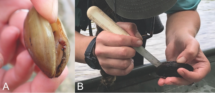
Figure 1: Gently prying individuals open. To check gravidity of a live mussel, gently pry open the valves with thumbs (A) or cautiously use a speculum or reverse pliers to pry open the valves (B). Review step 2.3 in the protocol for cautions associated with this method. Please click here to view a larger version of this figure.
- Perform a visual inspection on preserved specimens by opening the valves and inspecting the gills to determine whether the individual is a gravid female (Figure 2).
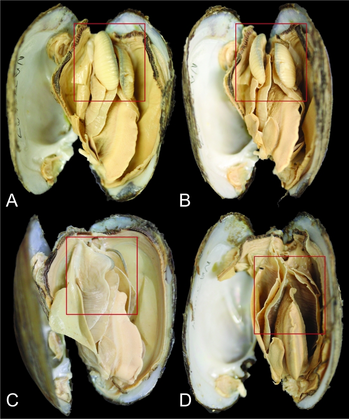
Figure 2: How to identify a gravid female. Female mussel marsupial gills appear inflated when the female is gravid and brooding. Photos A and C show gills from a lateral perspective while photos B and D provide a ventral view of the gills. Red boxes outline the gills to highlight the differences between a gravid (A/B) and not gravid (C/D) female Lampsilis straminea mussel. The total lengths of the individuals are 79 mm (A/B) and 88 mm (C/D). Please click here to view a larger version of this figure.
2. Gill content sampling
NOTE: This protocol can be adapted whether sampling occurs on live mussels in the field and laboratory, or on preserved specimens.
- Prepare a 1.5 mL plastic microcentrifuge collection tube with approximately 1 mL of either sterile water if gill contents will be evaluated within 24 h of extraction35 or ethanol (EtOH) if sample evaluation cannot occur within 24 h of collection or if gill contents are from a museum specimen preserved in EtOH. If glochidia are intended for scanning electron microscope (SEM) imaging, use 70% EtOH, and if glochidia will be used for genetic testing, use non-denatured 95% EtOH36.
- Remove the paper wrapping for one sterile 20 G bevel-tip needle on a 10 mL syringe. Unscrew the cap to expose the needle and prepare a 1.5 mL plastic tube for gill content collection. Push the handle of the syringe all the way down so the black stopper is at the 0 mL/cc line.
NOTE: A sterile syringe should be used each time gill contents are sampled. A used syringe can be sterilized in the field by dipping the tip in a 10% bleach solution, then rinsing the syringe by filling it with 1 mL of sterile water and depressing the plunger back to 0 mL/cc, and finally drying the syringe with a clean cloth. - Pick up the gravid female and gently pry open the two valves using the tips of the thumbs.
CAUTION: Be careful to not harm the animal. Opening the valves too wide or too fast can overextend adductor muscles and cause mortality. Thin-shelled specimens (e.g., species of Anodonta, Leptodea, Utterbackia, etc.) and young individuals are especially vulnerable in this step. Forcefully handling fragile-shelled species may crack the shells and cause mortality. In some cases, squeezing thin-shelled animals from the anterior and posterior shell margins, while looking at the ventral surface, will cause the shell to flex and gape slightly, allowing one to observe the gills or pry open the shells and avoid damaging the fragile shell margin.
NOTE: Tools can be used to assist with this step but can also cause mortality if not used with care and should be avoided whenever possible. For example, a speculum or modified set of reverse pliers may be used to help pry the individual open and a wedge can be used to help prop the valves open. These instruments may not be necessary if another person is available to assist (i.e., one person holds the animal open while another maneuvers the syringe for extraction). Damaging or separating the mantle tissue from the periostracum can cause growth deformities and mortality37; therefore, it is critical to avoid severing the connection between the mantle tissue and outside margin of the shell. - Use the needle tip of the syringe to gently penetrate a single water tube of the inflated marsupial gill. Next, gently scoop the gill contents out by utilizing the beveled tip of the needle.
NOTE: Gill contents usually have a milky-white consistency, which should be visible on the beveled tip of the needle.- Deposit the contents of the syringe directly into a Petri dish if a microscope is readily available. Otherwise, store the contents in a 1.5 mL plastic microcentrifuge tube with designated liquid (see step 2.1) for later evaluation.
NOTE: Minimize disturbance and handling of glochidia samples during transportation to avoid damage and reduced viability32,35.
- Deposit the contents of the syringe directly into a Petri dish if a microscope is readily available. Otherwise, store the contents in a 1.5 mL plastic microcentrifuge tube with designated liquid (see step 2.1) for later evaluation.
- Record information on the genus-species identification, gravidity status, length of female (mm), collector and contact information, state, county, drainage, specific collection location, latitude and longitude, a unique identifier for the gill sample, a unique identifier for the survey site, and the date of collection if gill contents were extracted (Figure 3). Record a unique identifier on each collection vessel to ensure accurate data records during transport.
- Photograph the outside right valve of the mussel for identity validation and include the tube labeled with the unique identifier legible in the picture. Optionally, collect other abiotic and biotic parameters to supplement the information on the environment and community the mussel was found in (see Figure 3 for suggestions).
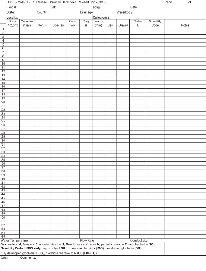
Figure 3: Example of a field gravidity datasheet. Accurate data reporting is necessary if a gill sample is taken to produce reliable information. This is an example of a field datasheet with the minimum fields and extra abiotic parameters to be collected along with each gill sample. For more comprehensive information, please see step 4.1 in the protocol. Please click here to view a larger version of this figure.
3. Laboratory evaluation of gill contents
- If gill contents are in a 1.5 mL tube, transfer them into a Petri dish and fill the bottom of the dish with water. Gently swirl the Petri dish in a circular motion to collect contents in the center of the dish for a more concentrated view of the sample.
NOTE: The 1.5 mL tube may need to be flushed out using a squirt bottle or transfer pipet filled with water if gill contents are sticking to tube walls. - Place the Petri dish under a dissecting microscope to evaluate the sample. If possible, take a photograph of the gill sample under the microscope and label it with the unique identifier for that sample.
- Record results of which developmental stages are present in each gill sample. Use Figure 4 as a guide to characterize each developmental stage. In some cases, females may be brooding larvae at multiple developmental stages; therefore, report every developmental stage observed within a given sample (e.g., ‘EGG/DG/IMG/FDG’). Once preserved glochidia have been evaluated, proceed to section 4. If fully developed glochidia are identified and EtOH was not used for preservation, proceed to step 3.3.
NOTE: EGG, egg masses; DG, developing glochidia; IMG, immature glochidia; FDG, fully developed glochidia.
- Record results of which developmental stages are present in each gill sample. Use Figure 4 as a guide to characterize each developmental stage. In some cases, females may be brooding larvae at multiple developmental stages; therefore, report every developmental stage observed within a given sample (e.g., ‘EGG/DG/IMG/FDG’). Once preserved glochidia have been evaluated, proceed to section 4. If fully developed glochidia are identified and EtOH was not used for preservation, proceed to step 3.3.
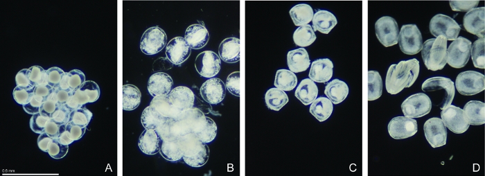
Figure 4: Representations for various stages of glochidia development in the marsupial gills. (A) Egg masses (EGG) have a membrane that makes eggs clump together. Within each egg membrane there is an opaque spherical mass of differentiating cells. The opaque spherical mass may split into multiple spherical masses during early cell division but should still be recorded as EGG until a distinct bivalve shape is observed. (B) Immature glochidia (IMG) have a distinct bivalve-shaped mass contained within the egg membrane. (C) Developing glochidia (DG) have a distinct bivalve shape, no egg membrane, and unorganized tissue inside, often fuzzy in appearance. Developing glochidia (DG) are not reactive when exposed to NaCl and classified as ‘DG(T)’ when data are recorded. (D) Fully developed glochidia (FDG) have the distinct bivalve shape and obvious adductor muscle tissue allowing glochidia to close. Fully developed glochidia (FDG) are often observed as two open valves after preservation. Two open valves will usually snap closed, or snap open and closed, when exposed to NaCl and are classified as ‘FDG(T)’. Please click here to view a larger version of this figure.
- Conduct a sodium chloride (NaCl) test to further evaluate viability of any fully developed glochidia by adding a crystal of NaCl to a subset droplet of the gill sample35. Viable glochidia will respond to NaCl by closing their valves from an open position. Report any salt-tested glochidia with ‘(T)’ at the end of the designation when data are recorded.
NOTE: Fully developed glochidia may also be observed actively snapping open and closed without exposure to NaCl.
4. Report to database
- Access the FMGA web (http://arcg.is/089uee), which was developed using online software programs38,39,40. The FMGA page provides a link to the desktop data entry form and an application download for mobile devices. The mobile app enables data entry in the field and automated georeferencing41.
NOTE: The gravidity calendar and other graphics associated with freshwater mussel species life history events can also be found on the FMGA Dashboard.- Use the mobile app or the desktop site to record results in the data entry form by utilizing drop-down menus and text entry fields. For large, pre-existing datasets, contact the authors for the template spreadsheet. Enter recorded data under the appropriate column headings, keeping in mind that each record, or row on the spreadsheet, represents observations of a gill sample from one gravid individual.
- Submit the results and they will be added to the FMGA database after being validated by an administrator, who may contact the collector to request further details or photos.
NOTE: Once data are validated and compiled in the FMGA database, all gravidity calendars and other interactive graphics displayed on the FMGA dashboard will be updated.
Representative Results
This protocol was applied during a capture-mark-recapture study that monitored the freshwater mussel community within a 750 m2 stretch of Bruce Creek (Walton County, Florida) from January 2015 to December 2015. Field sampling was scheduled to occur every four weeks; however, due to high-flow events, sampling was not conducted in April or September of 2015. State and federal agencies including U.S. Geological Survey, U.S. Fish and Wildlife Service, and Florida Fish and Wildlife Conservation Commission assisted in field surveys and gill sampling. Each gravid female encountered during the survey was subjected to in-field gill content sampling using the protocol described above, tagged (see Table of Materials), and placed back into the river substrate. The gill samples were stored in 95% EtOH and transported to the U.S. Geological Survey's Wetland and Aquatic Research Center’s laboratory for evaluation of gill contents.
By tagging females and recapturing them at monthly intervals throughout the year, we evaluated both lethal and non-lethal impacts of the gill sampling protocol on a total of 90 individuals. The following seven species were recaptured during this study: Elliptio pullata (n = 5), Fusconaia burkei (n = 1), Hamiota australis (n = 19), Obovaria choctawensis (n = 1), Strophitus williamsi (n = 1), Villosa lienosa (n = 60), and Villosa vibex (n = 3). Our sampling included individuals ranging from 24 mm to 80 mm in total length and two species (F. burkei and H. australis) protected by the U.S. Endangered Species Act. All data utilized in this study are publicly available We have provided access to our dataset on ScienceBase (https://doi.org/10.5066/P90VU8EN)42.
Survivorship was assessed by how many individuals were recaptured alive after gill sample collection. We observed high survivorship (97%) during the study with some mortality, possibly contributable to predation, indicated by on-site observations. Results showed around 51% of individuals (46 of 90) were found to stay gravid between consecutive sampling events. Another 10% of individuals (9 of 90) were found gravid, recaptured not gravid, and found gravid again. About 39% of individuals (35 of 90) in this study were found gravid, a gill sample was taken, but when recaptured again throughout the year, they were never found gravid a second time. The results indicate that the protocol described here is neither lethal nor sub-lethal and does not substantially disturb the current brooding period after the gill was sampled.
Although sample sizes in this study are unequal across species, results from this study highlight the beneficial and practical applications of this protocol. The gravidity calendar for V. lienosa illustrates gravid females brooding FDG were found in almost every month of the year except August, when only females brooding EGG were found (Figure 5A). Female H. australis were found not gravid (NG) in July, August, and December. A larger proportion of females were brooding FDG in January and February but were also found in October and November (Figure 5B). No individuals of E. pullata were found brooding FDG although females were brooding EGG from May to June, and one gravid female recorded (GFR) in June (Figure 5C). The only gravid F. burkei female was found GFR in June and recaptured NG in July. The same O. choctawensis individual was collected FDG in February and recaptured NG in July. Only one S. williamsi was found and was recaptured three times. This female was found FDG in March, NG in May, GFR in June, and EGG in August (Figure 5C). Gravid females of V. vibex brooding FDG were found between February and June (Figure 5C).
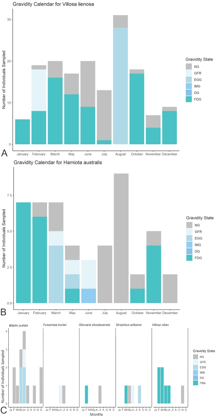
Figure 5: Results of the study in Bruce Creek, FL displayed in a gravidity calendar format. (A) Gravidity calendar for Villosa lienosa captures/recaptures. (B) Gravidity calendar for Hamiota australis captures/recaptures. (C) Gravidity calendars for all species with less than 10 individuals sampled. The y-axis includes abbreviations for the months January (Ja), February (F), March (Mr), May (My), June (Jn), July (Jl) August (A), October (O), November (N) and December (D). Please click here to view a larger version of this figure.
Discussion
Significance
Conservation of imperiled species depends on successful recruitment within extant populations. In some cases, artificial propagation may be necessary to augment recruitment of these at-risk populations. This requires researchers being informed on the timing of active reproduction for each species and possibly applying different methodologies or management practices to mitigate impact on recruitment. As an imperiled group of organisms, it is paramount to establish a standardized and non-lethal approach for studying reproductive habits, and to provide a platform on which to compile and visualize data to inform the scientific community with the most up-to-date information available. This study provides a step-by-step protocol to ensure precautions are taken, and gill contents can be adequately sampled and evaluated from female mussels. This protocol was tested for lethal and non-lethal effects, allowing researchers and managers to responsibly implement this methodology. We also developed a suite of database management tools and applications to facilitate compilation of gravidity information on a publicly available, user-friendly dashboard. Studies on epidemiology, glochidia morphology, life history, phylogenetics, propagation, and translocations can all benefit and utilize this repository of temporal gravidity information for all species of freshwater mussels.
This study alone supported previous studies’ findings of some species reproductive habits but also revealed novel information on others. Although V. vibex was collected in fewer numbers than V. lienosa, similarities can be found between the two based on the gravidity data. Both species of Villosa seem to brood fully developed glochidia during a large portion of the year, which characterizes them as an overwintering brooder. This is consistent with previous studies on other Villosa species43,44,45. The results of this study suggest H. australis can be found gravid from October and overwintering into June, except no captures were found gravid in December. A previously published study identified congener H. altilis with a gravidity period of four months, March through June46,47. This finding illustrates a longer gravidity period than previously thought and generally groups H. australis as an overwintering brooder. As federally protected species, varying brooding periods for H. altilis and H. australis could impact management decisions to better protect populations during reproductively active times. Elliptio pullata were only found gravid with EGG in May and June which corresponds to their characterization as a tachytictic species with a very short brooding period24,48,49,50. As data are compiled on Elliptio species using this protocol, detailed information can make field efforts more efficient when certain glochidial development stages are targeted, since glochidia are only found a few months out of the year. Inference from the other species with lower sample sizes is limited but as data are compiled into the database, higher sample sizes will give insight into reproductive habits of additional mussel species.
Procedural comments
Freshwater mussels and their glochidia are known to be susceptible to anthropogenic stressors10,35. During gravidity inspection, the mussel valves may not be easy to open, and carelessly forcing the valves open can cause unintentional harm and result in stress or mortality. Some fragile-shelled species (e.g., species of Anodonta, Leptodea, Utterbackia, etc.) and smaller sized individuals may have very fragile shells and weak adductor muscles that can break and tear easily. Gill sampling could be considered a stressor if handling is not done responsibly and with caution. A previous study found that handling and aerial exposure of mussels during reproductively active times may cause various physiological stress, including premature release of gill contents34. However, a study utilizing a similar methodology as described here, found handling gravid female mussels during gill sampling did not interrupt the present brood or cause premature release in both short- and long-term brooding species16. Furthermore, a sterile syringe needs to be used during this protocol to prevent any unintended infection or cross contamination when puncturing gills of multiple individuals. Additionally, glochidia are fragile and broods can be matured and stressed but not expelled. Mature glochidia in poor health can result in fewer individuals reacting to salt tests35. When making the distinction between DG(T) and FDG(T) it is important to salt test with a large sample size, make notes on observations to carefully identify distinctions between DG and FDG glochidia using the descriptions provided in this study. When proper care is taken, the minimal stress induced by this procedure can allow for female mussels to continue brooding glochidia naturally and reduce impacts on recruitment in the population.
Additional data can be recorded to supplement the database and provide broad context for reproductive habits of freshwater mussels. Some species (e.g., species of Fusconaia), have been observed to have gills of different colors based on the development stage of the glochidia51. During an initial gravidity check of the female, a description of the gill color may be included in the reported data to allow for future investigation. Also, at this point in the protocol, researchers can note whether the brooding female was found brooding glochidia in the two outer gills (ectobranchus), two inner gills (endobranchus), or all four gills (tetragenous)17. This information can be added to FMGA and help fill in data gaps regarding brooding for each species investigated. Environmental conditions, specifically water temperature, can be collected and recorded in the field for a more comprehensive observation of the gravidity status and timing of species at various latitudinal ranges. Research shows that environmental parameters, such as temperature, photoperiod, flow rate, and food availability, may induce reproductive events in freshwater mussels52,53,54,55,56. Additional fields may be added to the database as they are submitted to promote future research on abiotic factors influencing gravidity. A capture-mark-recapture modification modeled after our study can also be added to this protocol, which would allow researchers to monitor a specific mussel’s reproductive habits and reveal information on multiple broods per year.
The accuracy of information in the FMGA depends on the source. For example, misidentification of freshwater mussels is common due to many species having similar external characteristics that make it difficult to distinguish between species57. A gill sample from a misidentified individual could create confusion and false information for a species’ brooding period. If a gill sample is taken, photographs should be taken of inside both valves (if individual is not alive), outside of right valve, and the umbo (hinge where two valves connect) and submitted with gravidity data through the desktop site or mobile application. We also welcome photos of the gill contents. Within the submission forms there is a drop-down menu enabling the collector to indicate their level of confidence regarding species identification. Before the record is validated, this information will be taken into consideration when checking collector identification against plausible distribution, etc. Due to the high degree of intraspecific morphological variation in species of Unionidae, submission of tissue samples is encouraged and may be necessary to facilitate molecular identification.
Future implications
As a non-lethal method, this protocol can be applied to both common and imperiled species. The gravidity calendars for imperiled species can assist conservation managers involved with endangered species legislation and recovery planning by providing information on time periods when species are reproductively active. The state and federal agencies that manage at-risk species can better advise permit allocations for times when the species is not vulnerable and reproducing, and even limit the harvest of host fish during times mussels are brooding fully developed glochidia. Additionally, field surveys can target species during non-reproductive periods to minimize impact to recruitment processes. The publicly accessible database, FMGA, provides a tool for researchers and managers to obtain important reproductive information on any target freshwater mussel species. The database will also highlight data gaps, encouraging further research on species-specific brooding patterns. Since understanding a species reproductive pattern allows for adequate management decisions to be implemented, we hope that our protocol and database facilitate future freshwater mussel research, conservation, and recovery.
Acknowledgements
The authors would like to thank the funding sources: U.S. Fish and Wildlife Service and U.S. Geological Survey. A special thanks to Andrew Hartzog and Sandra Pursifull for organizing field crews and data collection, along with Lauren Patterson and Chris Anderson for their valuable contributions to database development. We would also like to thank everyone who helped in the field and the laboratory including Sherry Bostick, Mark Cantrell, Sahale Casebolt, Jordan Holcomb, Howard Jelks, Gary Mahon, John McLeod, Kyle Moon, Cayla Morningstar, Emma Pistole, Matt Rowe, Channing St. Aubin, and Jim Williams. Any use of trade, firm, or product names is for descriptive purposes only and does not imply endorsement by the U.S. Government.
Materials
| Name | Company | Catalog Number | Comments |
| 1.5 mL snap cap centrifuge tubes | USA Scientific | 1615-5510 | Snap cap tubes are important in the field so the loose screw cap is not lost. |
| 20 G needle on 10 mL disposable syringe | Exelint International | 26255 | sterile 10 mL disposable syringe with needle Model: 10ml Luer Lock Tip W/20G X 1 1/2" |
| Dissecting Microscope | any | any | |
| Marking Pen | Fisher Scientific | 13-379-4 | This is what we used but any marker that can write on small plastic tubes will do. This one is fairly ethanol and water proof. |
| Molecular grade ethanol | any | any | Needed if preserving gill contents. Non-denatured 95% is needed for genetic work, 70% is needed for SEM imaging work. |
| Paper | any | any | Needed to record information on samples collected. |
| Pen/pencil | any | any | If in the field, better to write on waterproof paper with pencil so it doesn't smear. If in the museum/lab, any writing utensil is fine. |
| Petri dish | DWK Life Sciences (Kimble) | 23000-9050 | This is what we used but any petri dish available is fine. It is nicer to have the taller walls in case too much water is used. |
| Sodium Chloride | any | any | Needed for NaCl test for reactive glochidia. Preserved samples do not need this. |
| Speculum | any | any | Only needed if you want help opening the valves of a live mussel. |
| Sterile water | any | any | Added to gill samples to be evaluated for reactivity within 24 hours of collection. |
| Super glue | Gorilla | Gorilla super glue gel | Used to apply tags and only needed if conducting a capture-mark-recapture study. |
| Tags | Hallprint | FPN 8x4 | Only needed if conducting a capture-mark-recapture study. |
| Transfer Pipet | Thermo Scientific Samco | 225 | This is what we use but any transfer pipet or squirt bottle is applicable. |
| Tweezers | any | any | Needed to move crystals of NaCl for salt test. Preserved samples do not need this. |
| Waterproof paper | RainWriter | any | Only needed if conducting work in the field. This allows you to record information on each individual gill contents are extracted from. |
| Wooden pick | any | any | Only needed if you want help opening the valves of a live mussel. |
References
- Williams, J. D., Warren, M. L., Cummings, K. S., Harris, J. L., Neves, R. J. Conservation status of freshwater mussels of the United States and Canada. Fisheries. 18 (9), 6-22 (1993).
- Lopes-Lima, M., et al. Conservation status of freshwater mussels in Europe: state of the art and future challenges. Biological Reviews. 92 (1), 572-607 (2017).
- Zieritz, A., et al. Diversity, biogeography and conservation of freshwater mussels (Bivalvia: Unionida) in East and Southeast Asia. Hydrobiologia. 810 (1), 29-44 (2018).
- Haag, W. R., Williams, J. D. Biodiversity on the brink: an assessment of conservation strategies for North American freshwater mussels. Hydrobiologia. 735 (1), 45-60 (2014).
- Ferreira-Rodríguez, N., et al. Research priorities for freshwater mussel conservation assessment. Biological Conservation. 231, 77-87 (2019).
- Graf, D. L., Cummings, K. S. Review of the systematics and global diversity of freshwater mussel species (Bivalvia: Unionoida). Journal of Molluscan Studies. 73 (4), 291-314 (2007).
- Lydeard, C., Cummings, K. . Freshwater mollusks of the world: a distribution atlas. , (2019).
- Wächtler, K., Dreher-Mansur, M. C., Richter, T., Bauer, G., Wächtler, K. Larval types and early postlarval biology in naiads (Unionoida). Ecology and evolution of the freshwater mussels Unionoida. , 93-125 (2001).
- Dudgeon, D., et al. Freshwater biodiversity: importance, threats, status and conservation challenges. Biological Reviews. 81 (2), 163-182 (2006).
- Downing, J. A., Van Meter, P., Woolnough, D. A. Suspects and evidence: a review of the causes of extirpation and decline in freshwater mussels. Animal biodiversity and Conservation. 33 (2), 151-185 (2010).
- Vaughn, C. C. Ecosystem services provided by freshwater mussels. Hydrobiologia. 810 (1), 15-27 (2018).
- Aldridge, D. C., Fayle, T. M., Jackson, N. Freshwater mussel abundance predicts biodiversity in UK lowland rivers. Aquatic Conservation: Marine and Freshwater Ecosystems. 17 (6), 554-564 (2007).
- Barnhart, M. C., Haag, W. R., Roston, W. N. Adaptations to host infection and larval parasitism in Unionoida. Journal of the North American Benthological Society. 27 (2), 370-394 (2008).
- Saha, S., Layzer, J. B. Evaluation of a nonlethal technique for determining sex of freshwater mussels. Journal of the North American Benthological Society. 27 (1), 84-89 (2008).
- Tsakiris, E. T., Randklev, C. R., Conway, K. W. Effectiveness of a nonlethal method to quantify gamete production in freshwater mussels. Freshwater Science. 35 (3), 958-973 (2016).
- Gascho, L., Andrew, M., Stoeckel, J. A. Multi-stage disruption of freshwater mussel reproduction by high suspended solids in short-and long-term brooders. Freshwater Biology. 61 (2), 229-238 (2016).
- Bauer, G., Wächtler, K. . Ecology and evolution of the freshwater mussels Unionoida. , (2012).
- Ortmann, A. E. Notes upon the families and genera of the najades. Annals of the Carnegie Museum. 8 (2), 222 (1912).
- Heard, W. H. Sexuality and other aspects of reproduction in Anodonta (Pelecypoda: Unionidae). Malacologia. 15, 81-103 (1975).
- Heard, W. H. Brooding patterns in freshwater mussels. Malacological Review. 7, 105-121 (1998).
- Watters, G. T., O'Dee, S. H. Glochidial release as a function of water temperature: beyond bradyticty and tachyticty. Proceedings of the Conservation, Captive Care, and Propagation of Freshwater Mussels Symposium. , 135-140 (1998).
- Parker, R. S., Hackney, C. T., Vidrine, M. F. Ecology and Reproductive Strategy of a South Louisiana Freshwater Mussel, Glebula Rotundata (Lamarck) (Unionidae: Lampsilini). Freshwater Invertebrate Biology. 3 (2), 53-58 (1984).
- Jones, H. A., Simpson, R. D., Humphrey, C. L. The reproductive cycles and glochidia of fresh-water mussels (Bivalvia: Hyriidae) of the Macleay River, Northern New South Wales, Australia. Malacologia. 27 (1), 185-202 (1986).
- Price, J. E., Eads, C. B. Brooding patterns in three freshwater mussels of the genus Elliptio in the Broad River in South Carolina. American Malacological Bulletin. 29 (1), 121-126 (2011).
- Haag, W. R., Staton, J. L. Variation in fecundity and other reproductive traits in freshwater mussels. Freshwater Biology. 48 (12), 2118-2130 (2003).
- Soler, J., Wantzen, K. M., Jugé, P., Araujo, R. Brooding and glochidia release in Margaritifera auricularia (Spengler, 1793) (Unionoida: Margaritiferidae). Journal of Molluscan Studies. 84 (2), 182-189 (2018).
- Smith, D. G. Notes on the biology of Margaritifera margaritifera margaritifera (Lin.) in Central Massachusetts. American Midland Naturalist. , 252-256 (1976).
- O'Brien, C., Nez, D., Wolf, D., Box, J. B. Reproductive biology of Anodonta californiensis, Gonidea angulata, and Margaritifera falcata (Bivalvia: Unionoida) in the Middle Fork John Day River, Oregon. Northwest Science. 87 (1), 59-73 (2013).
- Haag, W. R., Warren, M. L. Host fishes and reproductive biology of 6 freshwater mussel species from the Mobile Basin, USA. Journal of the North American Benthological Society. 16 (3), 576-585 (1997).
- O'Dee, S. H., Watters, G. T. New or confirmed host identifications for ten freshwater mussels. Proceedings of the Conservation, Captive Care, and Propagation of Freshwater Mussels Symposium. , 77-82 (1998).
- Johnson, N. A., McLeod, J. M., Holcomb, J., Rowe, M., Williams, J. D. Early life history and spatiotemporal changes in distribution of the rediscovered Suwannee moccasinshell Medionidus walkeri (Bivalvia: Unionidae). Endangered Species Research. 31, 163-175 (2016).
- McLeod, J. M., Jelks, H. L., Pursifull, S., Johnson, N. A. Characterizing the early life history of an imperiled freshwater mussel (Ptychobranchus jonesi) with host-fish determination and fecundity estimation. Freshwater Science. 36 (2), 338-350 (2017).
- Carlson, S., Lawrence, A., Blalock-Herod, H., McCafferty, K., Abbott, S. Freshwater mussel survey protocol for the Southeastern Atlantic Slope and Northeastern Gulf drainages in Florida and Georgia. US Fish and Wildlife Service, Ecological Services and Fisheries Resources Offices and Georgia Department of Transportation, Office of Environment and Location. , (2008).
- Waller, D. L., Rach, J. J., Cope, G. W., Miller, G. A. Effects of handling and aerial exposure on the survival of unionid mussels. Journal of Freshwater Ecology. 10 (3), 199-207 (1995).
- Fritts, A. K., et al. Assessment of toxicity test endpoints for freshwater mussel larvae (glochidia). Environmental Toxicology and Chemistry. 33 (1), 199-207 (2014).
- Christian, A. D., Monroe, E. M., Asher, A. M., Loutsch, J. M., Berg, D. J. Methods of DNA extraction and PCR amplification for individual freshwater mussel (Bivalvia: Unionidae) glochidia, with the first report of multiple paternity in these organisms. Molecular Ecology Notes. 7 (4), 570-573 (2007).
- Henley, W. F., Grobler, P. J., Neves, R. J. Non-invasive method to obtain DNA from freshwater mussels (Bivalvia: Unionidae). Journal of Shellfish Research. 25 (3), 975-978 (2006).
- ESRI. ArcGIS Desktop 10.6.1.9270. Environmental Systems Research Institute (ESRI). , (2017).
- ESRI. ArcGIS Online. ESRI Geospatial Cloud: Survey123 Field Application for ArcGIS 3.3.64. Environmental Systems Research Institute (ESRI). , (2019).
- ESRI. ArcGIS Online. ESRI Geospatial Cloud: Survey123 Connect for ArcGIS 3.3.51. Environmental Systems Research Institute (ESRI). , (2019).
- ESRI. ArcGIS Online. ESRI Geospatial Cloud: Operations Dashboard for ArcGIS. Environmental Systems Research Institute (ESRI). , (2019).
- Johnson, N. A., Beaver, C. E. Empirical data supporting a non-lethal method for characterizing the reproductive status and larval development of freshwater mussels (Bivalvia: Unionida). U.S. Geological Survey data release. , (2019).
- Ortmann, A. E. A monograph of the naides of Pennsylvania. Memoirs of the Carnegie Museum. 8, (1919).
- Posey, W. R. Life History and Population Biology of the State Special Concern Ouachita Creekshell, Villosa arkansasensis (I. Lea 1862). Arkansas Game and Fish Commission. , (2007).
- Asher, A. M., Christian, A. D. Population characteristics of the mussel Villosa iris (Lea) (rainbow shell) in the Spring River watershed, Arkansas. Southeastern Naturalist. 11 (2), 219-239 (2012).
- Haag, W. R., Warren, M. L., Shillingsford, M. Host fishes and host-attracting behavior of Lampsilis altilis and Villosa vibex (Bivalvia: Unionidae). The American Midland Naturalist. 141 (1), 149-158 (1999).
- Roe, K. J., Hartfield, P. D. Ham,iota a new genus of freshwater mussel (Bivalvia: Unionidae) from the Gulf of Mexico drainages of the southeastern United States. Nautilus-Sanibel. 119 (1), 1-10 (2005).
- Williams, J. D., Butler, R. S., Warren, G. L., Johnson, N. A. . Freshwater mussels of Florida. , (2014).
- Jirka, K. J., Neves, R. J. Reproductive biology of four species of freshwater mussels (Molluscs: Unionidae) in the New River, Virginia and West Virginia. Journal of Freshwater Ecology. 7 (1), 35-44 (1992).
- Watters, G. T., O'Dee, S. H., Chordas, S. Patterns of vertical migration in freshwater mussels (Bivalvia: Unionoida). Journal of Freshwater Ecology. 16 (4), 541-549 (2001).
- Richardson, F., Martínez, P. Anodonta propagation studies: determination of mussel sexual maturity and Glochidia release agents. Proceedings of the Gulf and Caribbean Fisheries Institute. 48, 535-538 (2004).
- Heinricher, J. R., Layzer, J. B. Reproduction by individuals of a nonreproducing population of Megalonaias nervosa (Mollusca: Unionidae) following translocation. The American Midland Naturalist. 141 (1), 140-148 (1999).
- Watters, G. T., O'Dee, S. H. Glochidia of the freshwater mussel Lampsilis overwintering on fish hosts. Journal of Molluscan Studies. 65 (4), 453-459 (1999).
- Hastie, L. C., Young, M. R. Timing of spawning and glochidial release in Scottish freshwater pearl mussel (Margaritifera margaritifera) populations. Freshwater Biology. 48 (12), 2107-2117 (2003).
- Galbraith, H. S., Vaughn, C. C. Temperature and food interact to influence gamete development in freshwater mussels. Hydrobiologia. 636 (1), 35-47 (2009).
- Kobayashi, O., Kondo, T. Reproductive ecology of the freshwater pearl mussel Margaritifera togakushiensis (Bivalvia: Margaritiferidae) in Japan. Venus (Japan). 67 (3-4), 189-197 (2009).
- Shea, C. P., Peterson, J. T., Wisniewski, J. M., Johnson, N. A. Misidentification of freshwater mussel species (Bivalvia: Unionidae): contributing factors, management implications, and potential solutions. Journal of the North American Benthological Society. 30 (2), 446-458 (2011).
This article has been published
Video Coming Soon
ABOUT JoVE
Copyright © 2024 MyJoVE Corporation. All rights reserved