
Abstract
Biology
Aislamiento de progenitores de megacariocitos de ratón
ERRATUM NOTICE
Important: There has been an erratum issued for this article. Read more …Los megacariocitos de la médula ósea son células poliploides grandes que aseguran la producción de plaquetas sanguíneas. Surgen de las células madre hematopoyéticas a través de la megacariopoyesis. Las etapas finales de este proceso son complejas y clásicamente involucran a los progenitores bipotentes de megacariocitos-eritrocitos (MEP) y los progenitores de megacariocitos unipotentes (MKp). Estas poblaciones preceden a la formación de megacariocitos de buena fe y, como tales, su aislamiento y caracterización podrían permitir el análisis robusto e imparcial de la formación de megacariocitos. Este protocolo presenta en detalle el procedimiento para recolectar células hematopoyéticas de la médula ósea de ratón, el enriquecimiento de progenitores hematopoyéticos a través del agotamiento magnético y, finalmente, una estrategia de clasificación celular que produce poblaciones MEP y MKp altamente purificadas. Primero, las células de la médula ósea se recolectan del fémur, la tibia y también la cresta ilíaca, un hueso que contiene un alto número de progenitores hematopoyéticos. El uso de huesos de la cresta ilíaca aumenta drásticamente el número total de células obtenidas por ratón y, por lo tanto, contribuye a un uso más ético de los animales. Se optimizó un agotamiento del linaje magnético utilizando perlas magnéticas de 450 nm que permiten una clasificación celular muy eficiente por citometría de flujo. Finalmente, el protocolo presenta la estrategia de etiquetado y gating para la clasificación de las dos poblaciones progenitoras de megacariocitos altamente purificadas: MEP (Lin-Sca-1-c-Kit+CD16/32-CD150+CD9dim)y MKp (Lin- Sca-1-c-Kit+CD16/32-CD150+CD9brillante) ). Esta técnica es fácil de implementar y proporciona suficiente material celular para realizar i) caracterización molecular para un conocimiento más profundo de su identidad y biología, ii) ensayos de diferenciación in vitro, que proporcionarán una mejor comprensión de los mecanismos de maduración de los megacariocitos, o iii) modelos in vitro de interacción con su microambiente.
Erratum
Erratum: Isolation of Mouse Megakaryocyte ProgenitorsAn erratum was issued for: Isolation of Mouse Megakaryocyte Progenitors. A figure was updated.
Figure 2 was updated from:
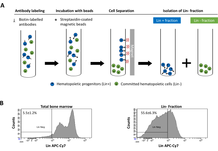
Figure 2: Magnetic depletion of lineage committed (Lin) cells. (A) Schematic representation of the magnetic depletion protocol. First, unsorted bone marrow cells are labeled with the biotin-conjugated rat anti-mouse antibody cocktail. Cells are then incubated with anti-rat Ig coated magnetic beads and subsequently subjected to the magnetic depletion using a strong magnet. The magnet will retain the labeled magnetic Lin+ fraction against the tube walls, while the unlabeled non-magnetic Lin- negative fraction will be collected in a new tube. (B) Lineage committed cells can be identified using fluorescent conjugated streptavidin. Typical analysis of the lineage expression in cells prior to magnetic depletion (total bone marrow) and after magnetic depletion (Lin- Fraction) N = 21. Please click here to view a larger version of this figure.
to:
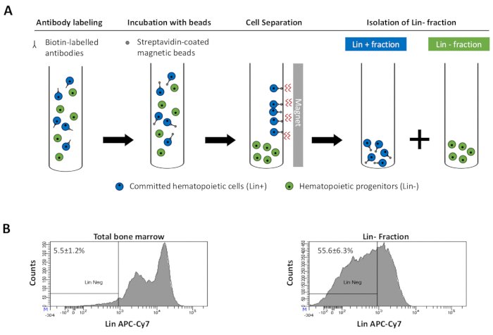
Figure 2: Magnetic depletion of lineage committed (Lin) cells. (A) Schematic representation of the magnetic depletion protocol. First, unsorted bone marrow cells are labeled with the biotin-conjugated rat anti-mouse antibody cocktail. Cells are then incubated with anti-rat Ig coated magnetic beads and subsequently subjected to the magnetic depletion using a strong magnet. The magnet will retain the labeled magnetic Lin+ fraction against the tube walls, while the unlabeled non-magnetic Lin- negative fraction will be collected in a new tube. (B) Lineage committed cells can be identified using fluorescent conjugated streptavidin. Typical analysis of the lineage expression in cells prior to magnetic depletion (total bone marrow) and after magnetic depletion (Lin- Fraction) N = 21. Please click here to view a larger version of this figure.
Explore More Videos
ABOUT JoVE
Copyright © 2024 MyJoVE Corporation. All rights reserved