A subscription to JoVE is required to view this content. Sign in or start your free trial.
Image-Based Methods to Study Membrane Trafficking Events in Stomatal Lineage Cells
In This Article
Summary
Several commonly used methods are introduced here to study the membrane trafficking events of a plasma membrane receptor kinase. This manuscript describes detailed protocols including the plant material preparation, pharmacological treatment, and confocal imaging setup.
Abstract
In eukaryotic cells, membrane components, including proteins and lipids, are spatiotemporally transported to their destination within the endomembrane system. This includes the secretory transport of newly synthesized proteins to the cell surface or the outside of the cell, the endocytic transport of extracellular cargoes or plasma membrane components into the cell, and the recycling or shuttling transport of cargoes between the subcellular organelles, etc. Membrane trafficking events are crucial to the development, growth, and environmental adaptation of all eukaryotic cells and, thus, are under stringent regulation. Cell-surface receptor kinases, which perceive ligand signals from the extracellular space, undergo both secretory and endocytic transport. Commonly used approaches to study the membrane trafficking events using a plasma membrane-localized leucine-rich-repeat receptor kinase, ERL1, are described here. The approaches include plant material preparation, pharmacological treatment, and confocal imaging setup. To monitor the spatiotemporal regulation of ERL1, this study describes the co-localization analysis between ERL1 and a multi-vesicular body marker protein, RFP-Ara7, the time series analysis of these two proteins, and the z-stack analysis of ERL1-YFP treated with the membrane trafficking inhibitors brefeldin A and wortmannin.
Introduction
Membrane traffic is a conserved cellular process that distributes membrane components (also known as cargoes), including proteins, lipids, and other biological products, between different organelles within a eukaryotic cell or across the plasma membrane to and from the extracellular space1. This process is facilitated by a collection of membranes and organelles named the endomembrane system, which consists of the nuclear membrane, the endoplasmic reticulum, the Golgi apparatus, the vacuole/lysosomes, the plasma membrane, and multiple endosomes1. The endomembrane system enables the modification, packaging, and transport of membrane components using dynamic vesicles that shuttle between these organelles. Membrane trafficking events are crucial to cell development, growth, and environmental adaptation and, thus, are under stringent and complex regulation2. Currently, multiple approaches in molecular biology, chemical biology, microscopy, and mass spectrometry have been developed and applied to the field of membrane trafficking and have greatly advanced the understanding of the spatiotemporal regulation of the endomembrane system3,4. Molecular biology is used for classical genetic manipulations of the putative players involved in membrane trafficking, such as altering the gene expression of the protein of interest or labeling the protein of interest with certain tags. Tools in chemical biology include the usage of molecules that specifically interfere with the traffic of certain routes4,5. Mass spectrometry is powerful for identifying the components in an organelle that has been mechanically isolated by biochemical approaches3,4. However, membrane traffic is a dynamic, diverse, and complex biological process1. To visualize the membrane trafficking process in live cells under various conditions, light microscopy is an essential tool. Continuous progress has been made in advanced microscope techniques to overcome the challenges in measuring the efficiency, kinetics, and diversity of the events4. Here, this study focuses on the widely adopted methodologies in chemical/pharmacological biology, molecular biology, and microscopy to study membrane trafficking events in a naturally simplified and experimentally accessible system, the stomatal developmental process.
Stomata are micropores on plant aerial surfaces that open and close to facilitate gas exchange between the inner cells and the environment6,7,8. Hence, stomata are essential for photosynthesis and transpiration, two events that are crucial for plant survival and growth. Stomatal development is dynamically adjusted by environmental cues to optimize the plant's adaptation to the surroundings9. Dating back to studies in 2002, the identification of the receptor protein Too Many Mouths (TMM) opened the door to a new era of investigating the molecular mechanisms of stomatal development in the model plant Arabidopsis thaliana10. After just a few decades, a classical signaling pathway has been identified. From upstream to downstream, this pathway includes a group of secretory peptide ligands in the epidermal patterning factors (EFP) family, several cell-surface leucine-rich-repeat (LRR) receptor kinases in the EREECTA (ER) family, the LRR receptor protein TMM, a MAPK cascade, and several bHLH transcription factors including SPEECHLESS (SPCH), MUTE, FAMA, and SCREAM (SCRM)11,12,13,14,15,16,17,18,19,20,21,22,23,24,25,26. Previous work indicates that one of the receptor kinases, ER-LIKE 1 (ERL1), demonstrates active subcellular behaviors upon EPF perception20. ERL2 also dynamically traffics between the plasma membrane and some intracellular organelles27. Blocking the membrane trafficking steps causes abnormal stomatal patterning, resulting in stomatal clusters on the leaf surface28. These results suggest that membrane traffic plays an essential role in stomatal development. This study describes a protocol to spatiotemporally investigate the ERL1 dynamics using protein-protein subcellular co-localization analysis combined with pharmacological treatment using some membrane trafficking inhibitors.
Protocol
1. Preparation of the solutions
- Prepare seed sterilization solution by mixing 15 mL of bleach with 35 mL of distilled water and 50 µL of Triton X-100.
- Prepare brefeldin A (BFA) solution by dissolving the BFA powder in ethanol to a final concentration of 10 mM (stock). Prepare wortmannin (Wm) solution by dissolving the Wm powder in DMSO to a final concentration of 10 mM (stock).
2. Sowing the seeds
- Aliquot 10-50 seeds from each of the required transgenic plants into 1.5 mL microcentrifuge tubes. Add 1 mL of seed sterilization solution into each tube, and mix thoroughly by inverting the tube gently for 10 min on a shaker.
- Discard the seed sterilization solution, and wash the seeds with 1 mL of autoclaved distilled water five times.
- Add 300 µL of autoclaved distilled water into each tube, and sow the seeds on half-strength MS media containing 1% (w/v) sucrose and 0.75% (w/v) agar. Supplement the medium with the corresponding antibiotics as needed.
- Keep the plate upside-down at 4 °C in the dark for 2 days to synchronize the germination.
- After 2 days, transfer the plate into a growth room with a 16 h light/8 h dark cycle (80 µmol/m2/s1) at 22 °C. This is considered the first day post germination (1 dpg).
- Transplant the seedlings into soil at 10 dpg for further growth.
3. Preparation of dual-colored F1 transgenic plants
- Grow homozygous transgenic plants bearing ERL1-YFP (plant A) and homozygous transgenic plants bearing an RFP-labeled marker protein RFP-Ara7 (plant B)20 side by side until flowering in a growth room with a 16 h light/8 h dark cycle (80 µmol/m2/s1) at 22 °C.
- Choose a young inflorescence from plant A. Keep one flower that is just about to open on the inflorescence for genetic crosses. Remove all the older flowers and siliques. Additionally, carefully remove the younger flowers and floral meristems to avoid future confusion.
- Gently dissect the unopened flower by removing the sepals, petals, and stamen using a pair of sharp tweezers. Only leave the pistil on the inflorescence.
- Take a mature stamen from an opened flower on plant B, and deposit the pollen grains onto the stigma of the dissected pistil on plant A.
- Label this manually crossed flower by indicating its father, mother, and the date of the genetic cross. Let the flower mature, and harvest the F1 seeds when the silique turns yellow/brown (~20 days after the cross). Check for a successful F1 plant that has both YFP and RFP signals.
4. Pharmacological treatment
- For the BFA treatment, remove the cotyledons of 7-day-old seedlings, immerse the rest of the seedlings into either a mock solution (0.3% ethanol) or 30 µM BFA solution, apply vacuum for 1 min, and keep the sample immersed for 30 min before imaging.
- For the wortmannin treatment, remove the cotyledons of 7-day-old seedlings, immerse the rest of the seedlings into either 0.25% DMSO solution or 25 mM wortmannin, apply vacuum for 1 min, and keep the sample immersed for 30 min before imaging.
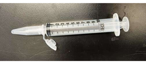
- For the vacuum treatment of samples, in a 1.5 mL microcentrifuge tube, add 500 µL of the corresponding drug solution, and immerse the dissected seedlings into the solution. Tightly attach a 10 mL syringe to the microcentrifuge tube, and apply vacuum for 1 min (Figure 1). Remove the syringe, and keep the sample in the solution for the required time. Gently take out the seedlings for imaging preparation.

Figure 1: Simple vacuum device. A 10 mL syringe is attached to a 1.5 mL microcentrifuge tube for the vacuum treatment. Please click here to view a larger version of this figure.
5. Sample preparation for imaging
- Dissect the true leaf from the treated sample using a sharp razor blade. Gently put the true leaf in a drop of water on a glass slide, and keep the abaxial side up. Slowly cover the true leaf with a coverslip while avoiding trapping any bubbles.
6. Confocal imaging
NOTE: A Leica SP8 inverted scanning confocal microscope was used to image the fluorescence signal of the samples in this work.
- Beam path setup
- Select a 514 nm laser for YFP excitation. Use a high laser power to increase the signal intensity and, thus, obtain high image quality. However, laser powers above 5% run the risk of photobleaching, and the health of the sample may be affected. If the sample signal is not too dim, start at low laser intensity.
- Turn on PMT/HyD for detection, and define the upper and lower emission band thresholds based on the spectrum for the YFP fluorophore. Set a detection window of 530-570 nm to collect the YFP signal.
- Sequential imaging for the second color: Click on the Seq button, and add a new channel. By default, the previously designed YFP beam path will be Seq 1. In Seq 2, select a 561 nm laser for the RFP excitation, and set a detection window of 570-630 nm to collect the RFP signal.
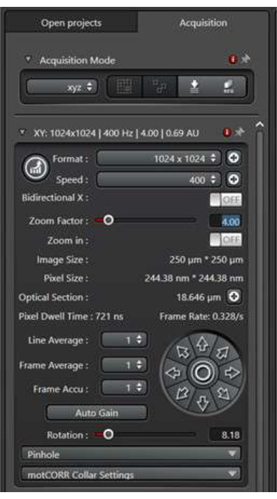
- Scan conditions setup (Figure 2)
- To image the subcellular membrane traffic activity within stomatal precursor cells, choose a 63x/1.2 W Corr lens on the system for imaging.
- The format refers to the image size in pixels. Start with 1,024 pixels x 1,024 pixels, and then optimize this based on the zoom factor and the objective settings for a good resolution of publication quality.
- The speed refers to the speed of the scanhead as the laser passes over each pixel. Although slow scan speeds often result in a better signal-to-noise ratio, they may not efficiently capture the rapidly changing membrane trafficking events. Start with a speed of 400 Hz, and optimize this based on the specific situation of the samples.
- The zoom factor is used to zoom in on a region of interest without changing the objective lens. Start with a zoom factor of 1, and optimize this based on the specific needs of the experiment.
- The line average refers to the number of times each X-line will be scanned to obtain an average result. A greater line average reduces the noise in the resulting image, but it also increases the scanning time and exposure time to the laser light. To image membrane trafficking events, do not use a high line average. Start with a 2x line average, and optimize as needed.
- Collecting a time series
NOTE: When studying membrane trafficking events, a time series is often needed to record the rapid movement of the subcellular endosomes.- In the Acquisition Mode, select the xyt scan mode to enable the time series utility.
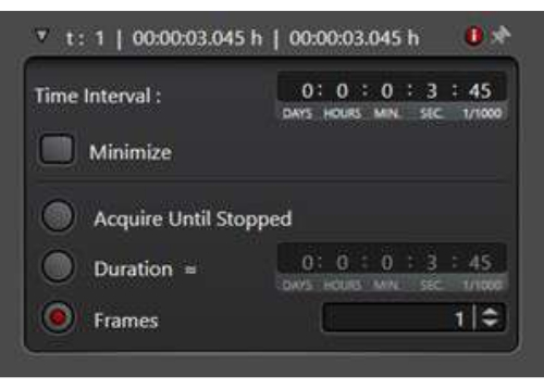
- Decide a waiting period between the time points under Time Interval. Alternatively, click on Minimize to take the images immediately one after another. A 7 s time interval was used in the following time series experiment.
- Select Duration, and enter the total time for the experiment to run. Alternatively, define the number of frames to be collected by selecting Frames (Figure 3).
- To collect the three-dimensional information regarding the membrane trafficking event within the entire cell, use the Z-stack scan mode. The maximum intensity projection of the Z-stack images is often used for the analysis.
- Select the xyz scan mode in the Acquisition Mode, and then the Z-Stack utility will become accessible.
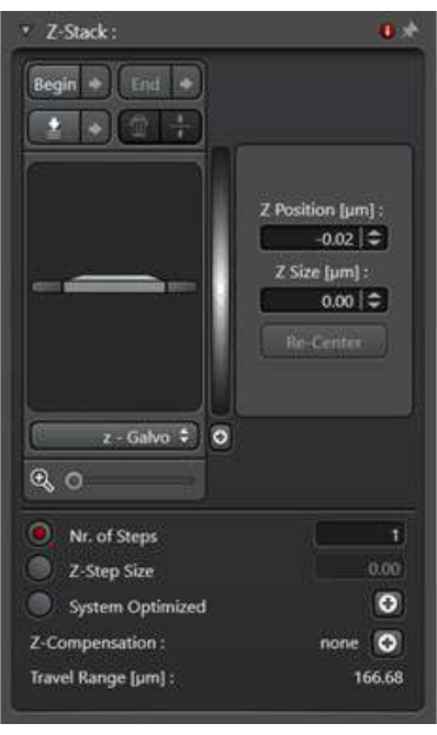
- Select the Z-wide option. Under the Scan Mode, define the top image and the bottom image of the Z-stack using the Begin and End buttons.
- Define the thickness of the z-step by clicking on z-step size. Alternatively, define how many images to take in order to cover the entire range of the Z-stack. To ensure consistency between the samples, define the z-step size (Figure 4).
- Image processing
- The maximum intensity projection of the z-stack images is generated by the confocal software (http://www.leica-microsystems.com). In the primary Process tab, first choose the interested z-stack file under the Open Projects tab on the far-left side. Then, switch to the middle tab named Process Tools, select the Projection function, choose Maximum in the dropdown panel of Method, and click on the Apply button. A z-stack projection image will be generated under the Open Projects tab.
- The video of the time series images is generated by Fiji (https://imagej.net/Fiji). In the File tab, use the Open function to open all the time series images in the correct order. In the Image tab, find the Stack function, and choose Images to stack to generate a video. Save the file in the .avi format with the desired frame rate (5 frames/s for Video 1).

Figure 2: The scan mode panel of the XY dimension. The scan mode panel is used to set up the image scan conditions. Please click here to view a larger version of this figure.

Figure 3: The time series utility under the xyt scan mode. The time series utility is used to set up the imaging conditions to consecutively collect a series of images. Please click here to view a larger version of this figure.

Figure 4: The z-stack utility under the xyz scan mode. The z-stack utility is used to set up the imaging conditions to collect a series of images on the z-axis. Please click here to view a larger version of this figure.
Results
A previous study indicated that ERL1 is an active receptor kinase that undergoes dynamic membrane trafficking events20. ERL1 is a transmembrane LRR-receptor kinase on the plasma membrane. Newly synthesized ERL1 in the endoplasmic reticulum is processed in the Golgi bodies and further transported to the plasma membrane. The ERL1 molecules on the plasma membrane can perceive EPF ligands using their extracellular LRR domain18. Upon activation by the inhibitory EPFs, including ...
Discussion
The endomembrane system separates the cytoplasm of a eukaryotic cell into different compartments, which enables the specialized biological function of these organelles. To deliver cargo proteins and macromolecules to their final destination at the right time, numerous vesicles are guided to shuttle between these organelles. Highly regulated membrane trafficking events play fundamental roles in the viability, development, and growth of cells. The mechanism regulating this crucial and complicated process is still poorly un...
Disclosures
The authors declare no conflicts of interest.
Acknowledgements
This work was supported by the National Science Foundation (IOS-2217757) (X.Q.) and the University of Arkansas for Medical Sciences (UAMS) Bronson Foundation Award (H.Z.).
Materials
| Name | Company | Catalog Number | Comments |
| 10 mL syringes | VWR | BD309695 | Vacuum samples |
| Brefeldin A (BFA) | Sigma | B7651 | membrane trafficking drug |
| Confocal Microscope | Leica | Lecia SP8 TCS with LAS-X software package | Imaging |
| Dissecting Forceps | VWR | 82027-402 | Genetic cross |
| Fiji | NIH | https://imagej.net/Fiji | Image processing |
| Leica LAS AF software | Leica | http://www.leica-microsystems.com | Image processing |
| transgenic seeds of ERL1-YFP | Qi, X. et al. The manifold actions of signaling peptides on subcellular dynamics of a receptor specify stomatal cell fate. Elife. 9, doi:10.7554/eLife.58097, (2020). | ||
| transgenic seeds of RFP-Ara7 | Ebine, K. et al. A membrane trafficking pathway regulated by the plant-specific RAB GTPase ARA6. Nat Cell Biol. 13 (7), 853-859, doi:10.1038/ncb2270, (2011). | ||
| Wortmannin (Wm) | Sigma | W1628 | membrane trafficking drug |
References
- Aniento, F., Sanchez de Medina Hernandez, V., Dagdas, Y., Rojas-Pierce, M., Russinova, E. Molecular mechanisms of endomembrane trafficking in plants. Plant Cell. 34 (1), 146-173 (2022).
- Sigismund, S., et al. Endocytosis and signaling: Cell logistics shape the eukaryotic cell plan. Physiological Reviews. 92 (1), 273-366 (2012).
- Lyu, Z., Genereux, J. C. Methodologies for measuring protein trafficking across cellular membranes. ChemPlusChem. 86 (10), 1397-1415 (2021).
- Rodriguez-Furlan, C., Raikhel, N. V., Hicks, G. R. Merging roads: Chemical tools and cell biology to study unconventional protein secretion. Journal of Experimental Botany. 69 (1), 39-46 (2017).
- Foissner, I., Sommer, A., Hoeftberger, M., Hoepflinger, M. C., Absolonova, M. Is wortmannin-induced reorganization of the trans-Golgi network the key to explain charasome formation. Frontiers in Plant Science. 7, 756 (2016).
- Qi, X., Torii, K. U. Hormonal and environmental signals guiding stomatal development. BMC Biology. 16 (1), 21 (2018).
- Han, S. K., Kwak, J. M., Qi, X. Stomatal lineage control by developmental program and environmental cues. Frontiers in Plant Science. 12, 751852 (2021).
- Bharath, P., Gahir, S., Raghavendra, A. S. Abscisic acid-induced stomatal closure: An important component of plant defense against abiotic and biotic stress. Frontiers in Plant Science. 12, 615114 (2021).
- Becklin, K. M., Ward, J. K., Way, D. A. . Photosynthesis, Respiration, and Climate Change., 1st edition. , (2021).
- Yang, M., Sack, F. D. The too many mouths and four lips mutations affect stomatal production in Arabidopsis. Plant Cell. 7 (12), 2227-2239 (1995).
- Hara, K., Kajita, R., Torii, K. U., Bergmann, D. C., Kakimoto, T. The secretory peptide gene EPF1 enforces the stomatal one-cell-spacing rule. Genes & Development. 21 (14), 1720-1725 (2007).
- Hara, K., et al. Epidermal cell density is autoregulated via a secretory peptide, EPIDERMAL PATTERNING FACTOR 2 in Arabidopsis leaves. Plant & Cell Physiology. 50 (6), 1019-1031 (2009).
- Hunt, L., Gray, J. E. The signaling peptide EPF2 controls asymmetric cell divisions during stomatal development. Current Biology. 19 (10), 864-869 (2009).
- Sugano, S. S., et al. Stomagen positively regulates stomatal density in Arabidopsis. Nature. 463 (7278), 241-244 (2010).
- Kondo, T., et al. Stomatal density is controlled by a mesophyll-derived signaling molecule. Plant & Cell Physiology. 51 (1), 1-8 (2010).
- Hunt, L., Bailey, K. J., Gray, J. E. The signalling peptide EPFL9 is a positive regulator of stomatal development. New Phytologist. 186 (3), 609-614 (2010).
- Shpak, E. D., McAbee, J. M., Pillitteri, L. J., Torii, K. U. Stomatal patterning and differentiation by synergistic interactions of receptor kinases. Science. 309 (5732), 290-293 (2005).
- Lin, G., et al. A receptor-like protein acts as a specificity switch for the regulation of stomatal development. Genes & Development. 31 (9), 927-938 (2017).
- Lee, J. S., et al. Direct interaction of ligand-receptor pairs specifying stomatal patterning. Genes & Development. 26 (2), 126-136 (2012).
- Qi, X., et al. The manifold actions of signaling peptides on subcellular dynamics of a receptor specify stomatal cell fate. Elife. 9, e58097 (2020).
- MacAlister, C. A., Ohashi-Ito, K., Bergmann, D. C. Transcription factor control of asymmetric cell divisions that establish the stomatal lineage. Nature. 445 (7127), 537-540 (2007).
- Pillitteri, L. J., Sloan, D. B., Bogenschutz, N. L., Torii, K. U. Termination of asymmetric cell division and differentiation of stomata. Nature. 445 (7127), 501-505 (2007).
- Ohashi-Ito, K., Bergmann, D. C. Arabidopsis FAMA controls the final proliferation/differentiation switch during stomatal development. Plant Cell. 18 (10), 2493-2505 (2006).
- Kanaoka, M. M., et al. SCREAM/ICE1 and SCREAM2 specify three cell-state transitional steps leading to Arabidopsis stomatal differentiation. Plant Cell. 20 (7), 1775-1785 (2008).
- Bergmann, D. C., Lukowitz, W., Somerville, C. R. Stomatal development and pattern controlled by a MAPKK kinase. Science. 304 (5676), 1494-1497 (2004).
- Wang, H., Ngwenyama, N., Liu, Y., Walker, J. C., Zhang, S. Stomatal development and patterning are regulated by environmentally responsive mitogen-activated protein kinases in Arabidopsis. Plant Cell. 19 (1), 63-73 (2007).
- Ho, C. M., Paciorek, T., Abrash, E., Bergmann, D. C. Modulators of stomatal lineage signal transduction alter membrane contact sites and reveal specialization among ERECTA kinases. Developmental Cell. 38 (4), 345-357 (2016).
- Le, J., et al. Auxin transport and activity regulate stomatal patterning and development. Nature Communications. 5, 3090 (2014).
- Geldner, N., et al. The Arabidopsis GNOM ARF-GEF mediates endosomal recycling, auxin transport, and auxin-dependent plant growth. Cell. 112 (2), 219-230 (2003).
- Qi, X., et al. Autocrine regulation of stomatal differentiation potential by EPF1 and ERECTA-LIKE1 ligand-receptor signaling. Elife. 6, 24102 (2017).
- Heilemann, M., et al. Subdiffraction-resolution fluorescence imaging with conventional fluorescent probes. Angewandte Chemie. 47 (33), 6172-6176 (2008).
- Leighton, R. E., Alperstein, A. M., Frontiera, R. R. Label-free super-resolution imaging techniques. Annual Review of Analytical Chemistry. 15 (1), 37-55 (2022).
- Oreopoulos, J., Berman, R., Browne, M. Spinning-disk confocal microscopy: Present technology and future trends. Methods in Cell Biology. 123, 153-175 (2014).
- Gao, R., et al. Cortical column and whole-brain imaging with molecular contrast and nanoscale resolution. Science. 363 (6424), (2019).
- Nwaneshiudu, A., et al. Introduction to confocal microscopy. Journal of Investigative Dermatology. 132 (12), (2012).
- Sanderson, J. Multi-photon microscopy. Current Protocols. 3 (1), 634 (2023).
Reprints and Permissions
Request permission to reuse the text or figures of this JoVE article
Request PermissionThis article has been published
Video Coming Soon
Copyright © 2025 MyJoVE Corporation. All rights reserved