Optimized Production and Analysis of Recombinant Protein-Filled Vesicles from E. coli
In This Article
Summary
The present protocol describes a detailed method for the bacterial production of recombinant proteins, including typically insoluble or disulfide-bond containing proteins, packaged inside extracellular membrane-bound vesicles. This has the potential to be applied to versatile areas of scientific research, including applied biotechnology and medicine.
Abstract
This innovative system, using a short peptide tag, that exports multiple recombinant proteins in membrane bound vesicles from E. coli, provides an effective solution to a range of problems associated with bacterial recombinant protein expression. These recombinant vesicles compartmentalise proteins within a micro-environment that facilitates the production of otherwise challenging, toxic, insoluble, or disulfide-bond containing proteins from bacteria. Protein yield is increased considerably when compared to typical bacterial expression in the absence of the vesicle-nucleating peptide tag. The release of vesicle-packaged proteins supports isolation from the culture medium and permits long-term active protein storage. This technology gives rise to increased yields of vesicle-packaged, functional proteins for simplified downstream processing for a diverse range of applications from applied biotechnology to discovery science and medicine. In the present article and the associated video, a detailed protocol of the method is provided, which highlights key steps in the methodology to maximize recombinant protein-filled vesicle production.
Introduction
The Gram-negative bacteria E. coli is an attractive system for recombinant protein production on both industrial and academic scale. It is not only cost-effective and straightforward to culture in batches to high densities, but a broad-spectrum of reagents, strains, tools, and promoters have been established to promote the generation of functional proteins in E. coli1. Additionally, synthetic biology techniques are now overcoming obstacles typically related to the application of post-translational modifications and folding of complex proteins2. The ability to target the secretion of recombinant proteins into culture media is attractive for improving yield and reducing manufacturing costs. Controlled packaging of user-defined proteins into membrane vesicles assists the development of products and technologies within the applied biotechnology and medical industries. Until now, there has been a lack of widely applicable methods for secreting recombinant proteins from E. coli 3.
Eastwood et al. have recently developed a peptide tagging-based method for producing and isolating recombinant protein-containing vesicles from E. coli1. This Vesicle Nucleating peptide (VNp) allows the production of extracellular bacterial membrane vesicles, into which the recombinant protein of choice can be targeted to simplify purification and storage of the target protein, and allows significantly higher yields than normally allowed from shaking flask cultures. Yields of close to 3 g of recombinant protein per liter of flask culture have been reported, with >100x higher yields than those obtained with equivalent proteins lacking the VNp tag. These recombinant protein-enriched vesicles can be rapidly purified and concentrated from the culture media and provide a stable environment for storage. This technology represents a major breakthrough in E. coli recombinant protein production. The vesicles compartmentalize toxic and disulfide-bond containing proteins in a soluble and functional form, and support the simple, efficient, and rapid purification of vesicle-packaged, functional proteins for long-term storage or direct processing1.
The major advantages this technology presents over current techniques are: (1) the applicability to a range of sizes (1 kDa to >100 kDa) and types of protein; (2) facilitating inter- and intra- protein disulfide-bond formation; (3) applicable to multiprotein complexes; (4) can be used with a range of promoters and standard lab E. coli strains; (5) the generation of yields of proteins from shaking flasks normally only seen with fermentation cultures; proteins are exported and packaged into membrane bound vesicles that (6) provide a stable environment for storage of the active soluble protein; and (7) simplifies downstream processing and protein purification. This simple and cost-effective recombinant protein tool is likely to have a positive impact on the biotechnology and medical industries, as well as discovery science.
Here, a detailed protocol, developed over several years, describes the optimal conditions to produce recombinant protein-filled vesicles from bacteria with the VNp technology. Example images of this system in practice are shown, with a fluorescent protein being expressed, allowing the presence of vesicles during different stages of the production, purification, and concentration to be visualized. Finally, guidance is provided on how to use live cell imaging to validate the production of VNp fusion-containing vesicles from the bacteria.
Protocol
The bacterial work undertaken follows the local, national, and international biosafety containment regulations befitting the particular biosafety hazard level of each strain.
1. Selection of different VNps
- Identify the VNp sequences.
NOTE: For the present study, three VNp sequences have been identified1 that result in maximal yield and vesicular export of the proteins examined to date: VNp2, VNp6, and VNp15. It is not currently clear why certain VNp variants perform more efficiently with some proteins than others; therefore, it is recommended that fusions are generated between a new protein of interest with each VNp variant (i.e., VNp2, 6, or 15).
VNp2: MDVFMKGLSKAKEGVVAAAEKTKQGVAEA
AGKTKEGVL
VNp6: MDVFKKGFSIADEGVVGAVEKTDQGVTEA
AEKTKEGVM
VNp15: MDVFKKGFSIADEGVVGAVE
Plasmids that allow expression of the protein of interest with different VNp amino terminal fusions have been made available commercially (see Table of Materials). - Design a cloning strategy to insert the gene of interest at the 3' end of the cDNA encoding for the VNp in one of these constructs, or adapt an existing plasmid by integrating synthesized VNp cDNA upstream of the first ATG codon of the gene encoding for the protein of interest. Use the methods as described in1.
- For toxic proteins, use a vector with a repressible expression promoter or a promotor with minimal uninduced expression noise.
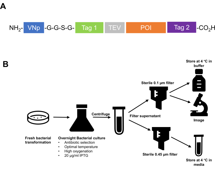
- Clone the VNp sequence tag at the amino-terminal of the fusion protein. Ensure that the affinity tags, protease cleavage sequences, etc., and the protein of interest are located on the carboxyl side of the VNp tag. It is recommended to separate the VNp from the downstream peptide with a flexible linker region, such as two or three repeats of a -G-G-S-G- polypeptide sequence (Figure 1).
NOTE: Use plasmids with an antibiotic selection that does not target peptidoglycan, which weakens the cell surface and reduces vesicle yield. Kanamycin and chloramphenicol (see Table of Materials) were the preferred antibiotics used for this study.
2. Bacterial cell culture and protein induction
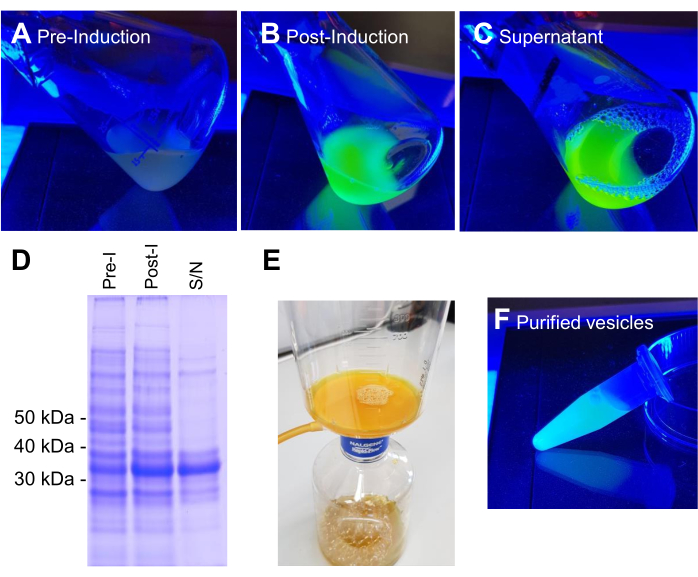
NOTE: Bacterial strains typically used in this protocol are either Escherichia coli BL21 (DE3) or W3110. E. coli cells are cultured in lysogeny broth (LB) (10 g/L tryptone; 10 g/L NaCl; 5 g/L yeast extract) or terrific broth (TB) (12 g/L tryptone; 24 g/L yeast extract; 4 mL/L 10% glycerol; 17 mM KH2PO4; 72 mM K2HPO4, salts autoclaved separately) media (see Table of Materials). Example images showing each step of the protein induction and subsequent isolation and purification process are shown in Figure 2.
- Culture 5 mL LB starters from fresh bacterial transformations at 37 °C to saturation and use them to inoculate 25 mL of TB in a 500 mL conical flask, all with appropriate antibiotic selection.
- The surface area:volume ratio is an important factor in optimizing this system. Use as large a volume flask as possible (e.g., 5 L flask containing 1 L of culture; for optimization runs, use 25 mL of media in a 500 mL flask).
- Incubate the larger shaking flask cultures in an incubator at 37 °C with shaking at 200 rpm (≥25 mm orbital throw) until a 600 nm optical density value (OD600) of 0.8-1.0 is reached.
NOTE: Vesiculation is optimal when cells are grown at 37 °C. However, some recombinant proteins require expression in lower temperatures. If this is the case for the protein of interest, the VNp6 tag must be used, as this allows high-yield vesicle export at temperatures down to 25 °C. - To induce recombinant protein expression from the T7 promoter, add isopropyl β-D-1-thiogalactopyranoside (IPTG) to a final concentration of up to 20 µg/mL (84 µM) (see Table of Materials). The induction of recombinant protein expression must occur at the late-log phase (i.e., typical OD600 of 0.8-1.0) for the production of vesicles.
NOTE: The length of the induction period may differ between proteins, with some reaching maximum production at 4 h and others overnight (18 h). To date, maximum vesicle export has been obtained in overnight cultures.
3. Recombinant vesicle isolation
- Pellet the cells by centrifugation at 3,000 x g (4 °C) for 20 min.
- To sterilize vesicle-containing media for long-term storage, pass the cleared culture media through a sterile and detergent-free 0.45 µm polyethersulfone (PES) filter (see Table of Materials).
NOTE: To test the exclusion of viable cells from the vesicle-containing filtrate, plate onto LB agar and incubate overnight at 37 °C. - To concentrate vesicles into a smaller volume, pass the sterile vesicle-containing media through a sterile and detergent-free 0.1 µm mixed cellulose esters (MCE) filter (see Table of Materials).
- Gently wash the membrane with 0.5-1 mL of sterile PBS using a cell scraper or plastic spreader to carefully remove vesicles from the membrane. Transfer to a fresh microfuge tube.
NOTE: Purified vesicles can be stored in sterile media or phosphate-buffered saline (PBS) at 4 °C. There are examples of recombinant proteins stored in these vesicles for 6 months, in this way, with no loss in enzymatic activity.
4. Soluble protein release from isolated vesicles
- Once protein-containing vesicles have been isolated into the sterile media/buffer, subject vesicular lipid membranes to sonication using an appropriate schedule for the apparatus (e.g., 6x 20 s on and off cycles) and centrifuge at 39,000 x g (4 °C) for 20 min to remove vesicle debris.
NOTE: Osmotic shock or detergent treatment can be used as an alternative to break open the vesicles, but consideration must be given to the impact upon protein functionality and/or downstream application. - If VNp fusion remains cytosolic and does not release into the media, isolate the protein using standard protocols (e.g., resuspend the cell pellets in 5 mL of an appropriate extraction buffer (20 mM tris, 500 mM NaCl), sonicate, and remove the cell debris by centrifugation).
5. Protein concentration determination
- Determine the concentration of proteins by gel densitometry analysis of triplicate samples1. Run alongside bovine serum albumin (BSA) loading standards on coomassie-stained sodium dodecyl-sulfate polyacrylamide gel electrophoresis (SDS-PAGE) gels. Scan and analyze the gels using appropriate software (e.g., Image J; see Table of Materials).
6. Visualization of vesicle formation and isolated vesicles by fluorescence microscopy
NOTE: If the cells contain fluorescently labeled VNp fusion or membrane markers, live cell imaging can be used to follow vesicle formation. Alternatively, fluorescent lipid dyes can be used to visualize vesicles to confirm production and purification.
- Cell mounting
- Induce VNp fusion expression for several hours before mounting onto the coverslip.
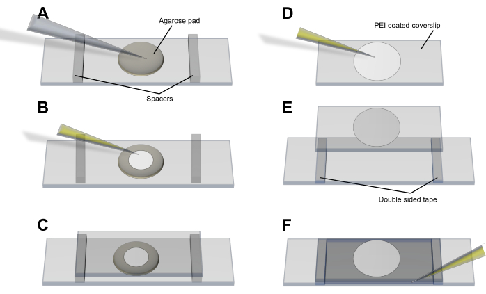
- Agarose pad method (Figure 3A-C): pipette the cells onto a thin (<1 mm), circular LB-agarose (2%) pad that has been allowed to form and set on a clean glass slide. Allow the cells to settle and equilibrate and place a 50 mm x 25 mm coverslip onto the pad and cells. Hold the coverslip in place with spacers and adhesive tape.
- Polyethyleneimine (PEI) method (Figure 3D-F): spread 20 µL of 0.05% PEI (in dH2O) onto a coverslip with a pipette tip and leave for 3-5 min to bind to glass without allowing to dry. Add 50 µL of cell culture and leave for 5-10 min to ensure bacteria have associated with the PEI-coated surface4. Wash the coverslip with 100 µL of media before placing it onto the slide and hold it in place with spacers and adhesive tape.
- Mounting vesicles
- Pipette purified vesicles onto a thin (<1 mm), circular LB-agarose (2%) pad that has been allowed to form and set on a clean glass slide. Once the liquid has dried, place a 50 mm x 25 mm coverslip onto the pad and vesicles. Hold the coverslip in place with spacers and adhesive tape.
- The fluorescent lipid dye FM4-64 is able to stain membranes5, and therefore can be used to visualize vesicles. Add FM4-64 (see Table of Materials) to purified vesicles at a final concentration of 2 µM (from a 2 mM stock dissolved in dimethyl sulfoxide [DMSO]) and image after a 10 min incubation. This is especially useful for identifying vesicles containing non-fluorescently labeled cargoes5.
- Rinse the coverslips with the same media used to culture the observed cells.
NOTE: Some complex media (e.g., TB) can exhibit autofluorescence, which may result in excess background signal.
- Imaging vesicles
NOTE: Example microscopy images of VNp recombinant vesicles can be seen in Figure 4.- Mount the slide onto an inverted microscope (see Table of Materials) using an oil immersion objective and leave for 2-3 min to allow the sample to settle and the temperature to equilibrate.
NOTE: All live cell imaging for each sample must be completed within 30 min of mounting cells onto coverslips to minimize the impact of phototoxicity and anaerobic stress. For this reason, single-plane images are preferred to z-stacks. - Use appropriate light sources (e.g., light emitting diode [LED] or halogen bulb; see Table of Materials) and filter combinations for the fluorescent protein(s)/dye(s) being used6.
- Use a high magnification (i.e., 100x or 150x) and high numerical aperture (i.e., NA ≥1.4) lens for imaging the microbial cells and vesicles.
- Determine the minimal light intensity required to visualize fluorescence signals from cells and / or vesicles. This may require some adjustment of the exposure and gain settings for the camera in use.
NOTE: Typical exposure times from current complementary metal-oxide semiconductor (CMOS) cameras vary between 50-200 ms depending upon the imaging system. - For single frame images, use three-image averaging to reduce hardware-dependent random background noise.
- For time-lapse imaging, allow 3-5 min between individual frames.
NOTE: Depending upon the microscope setup, the focus may need to be intermittently adjusted throughout longer time-lapse experiments.
- Mount the slide onto an inverted microscope (see Table of Materials) using an oil immersion objective and leave for 2-3 min to allow the sample to settle and the temperature to equilibrate.
Results
BL21 DE3 E. coli containing the VNp6-mNeongreen expression construct were grown to late-log phase (Figure 2A). VNp6-mNeongreen expression was induced by the addition of IPTG to the culture (20 µg/mL or 84 µM final concentration), which was subsequently left to grow overnight at 37 °C with vigorous shaking (200 rpm, ≥25 mm orbital throw). The following morning, the culture displayed mNeongreen fluorescence7 (Figure 2B), which remained visible in the media after the removal of bacterial cells by centrifugation (Figure 2C). The presence of VNp-mNeongreen within the culture and cleared culture media was confirmed by SDS-PAGE (Figure 2D). The mNeongreen-containing vesicles were isolated onto a 0.1 µm MCE filter (Figure 2E) and resuspended in PBS (Figure 2F). The purified vesicles were subsequently mounted on an agarose pad (Figure 3A-C) and imaged using widefield fluorescence microscopy (Figure 4A). The presence of vesicle membranes was confirmed using the lipophilic fluorescent dye FM4-64 (Figure 4B). The E. coli cells expressing the inner membrane protein CydB fused to mNeongreen (green) and VNp6-mCherry2 (magenta)8 show vesicle production and cargo insertion in live bacterial cells (Figure 4C). Figure 4A,B were captured using a widefield fluorescence microscope, while Figure 4C was acquired using structured illumination microscopy (SIM), using methods described previously9,10.

Figure 1: Summary of VNp technology from designing a cloning strategy to the purification and storage of extracellular vesicles. (A) Schematic of a typical VNp fusion protein. VNp at the NH2 terminus, followed by a flexible linker and an appropriate combination of affinity and fluorescence tags (Tag1, Tag 2, protease cleavage site [e.g., TEV]) and protein of interest. (B) Schematic diagram summarising the protocol for the expression and purification of recombinant protein-filled membrane vesicles from E. coli. Please click here to view a larger version of this figure.

Figure 2: Stages of production and purification of VNp6-mNg vesicles. Cultures of E. coli cells containing the VNp-mNeongreen expression construct in blue light either before (A) or after (B) IPTG-induced expression of the fusion protein. The cells from (B) were removed by centrifugation, leaving VNp-mNeongreen-filled vesicles in the media (C). (D) Equivalent samples from A, B, and C were analyzed by SDS-PAGE and coomassie staining. The vesicles were isolated onto a 0.1 µm filter (E) and subsequently washed off into an appropriate volume of buffer (F). Please click here to view a larger version of this figure.

Figure 3: Cell mounting procedure for imaging vesicles and vesicle production. (A-C) The agarose pad method and (D-F) the PEI method for mounting E. coli cells onto the coverslip. Please click here to view a larger version of this figure.

Figure 4: Microscopy images of VNp recombinant vesicles. Green (A) and red (B) emission images from different fields of FM4-64 labeled VNp6-mNeongreen-containing vesicles mounted on an agarose pad. (C) Imaging E. coli cells expressing the inner membrane protein CydB fused to mNeongreen (green) and VNp6-mCherry2 (magenta) shows vesicle production and cargo insertion in live bacterial cells. (A,B) were imaged using a widefield fluorescence microscope, while (C) was acquired using structured illumination microscopy (SIM). Scale bars: (A,B) = 10 µm; (C) = 1 µm. Please click here to view a larger version of this figure.
Discussion
The amino-terminal peptide-tagged method for the production of recombinant proteins described above is a simple process, which consistently yields large amounts of protein that can be efficiently isolated and/or stored for months.
It is important to highlight the key steps in the protocol that are required for optimal use of this system. Firstly, the VNp tag1 must be located at the N-terminus, followed by the protein of interest and any appropriate tags. It is also important to avoid using antibiotics that target the peptidoglycan layer, such as ampicillin.
In terms of growth conditions, rich media (e.g., LB or TB media) and a high surface area:volume ratio is necessary to maximize vesicle production. The optimal temperature for the production of extracellular vesicles is 37 °C, but the conditions typically required for expression of the protein of interest must be considered too. For lower induction temperatures, VNp6 must be used. Crucially, induction of the T7 promoter must be achieved using no greater than 20 µg/mL (84 µM) IPTG once the cells reach an OD600 of 0.8-1.0. Proteins expressed using the system reach maximum vesicle production at either 4 h or after overnight induction.
Despite the simplicity of this protocol, it requires optimization. VNp variant fusion, expression temperatures, and induction time periods may differ depending on the protein of interest. Furthermore, there is a need to optimize the purification and subsequent concentration of extracellular vesicles from the media. The current procedure is not scalable and can be time-consuming. These are the limitations of this methodology.
The VNp technology has many advantages over traditional methods2. It allows the vesicular export of diverse proteins, with the maximum size successfully expressed to date being 175 kDa for vesicles that remain internal and 85 kDa for those that are exported. Furthermore, this technology can significantly increase the yield of recombinant proteins with a range of physical properties and activities. Exported vesicles containing the protein of interest can be isolated by simple filtration from the precleared media and can subsequently be stored, in sterile culture media or buffer, at 4 °C for several months.
The applications for this system are diverse, from discovery science to applied biotechnology and medicine (e.g., through the production of functional therapeutics)3. The ease of production, downstream processing, and high yield are all attractive qualities in these areas and especially in industry.
Disclosures
The authors declare no competing financial interests or other conflicts of interest.
Acknowledgements
The authors thank diverse Twitter users who raised questions about the protocol presented in the paper describing the VNp technology. Figure 1A was generated using icons from flaticon.com. This work was supported by the University of Kent and funding from the Biotechnology and Biological Sciences Research Council (BB/T008/768/1 and BB/S005544/1).
Materials
| Name | Company | Catalog Number | Comments |
| Ampicillin | Melford | 69-52-3 | |
| Chloramphenicol | Acros Organics (Thermofisher Scientific) | 56-75-7 | |
| E. coli BL21 (DE3) | Lab Stock | N/A | |
| E. coli DH10β | Lab Stock | N/A | |
| Filters for microscope | Chroma | ||
| FM4-64 | Molecular Probes (Invitrogen) | T-3166 | Dissolved in DMSO, stock concentration 2 mM |
| ImageJ | Open Source | Downloaded from: https://imagej.net/ij/index.html | |
| Inverted microscope | Olympus | ||
| Isopropyl β-D-1-thiogalactopyranoside (IPTG) | Melford | 367-93-1 | |
| Kanamycin sulphate | Gibco (Thermofisher Scientific) | 11815-024 | |
| LED light source for micrscope | Cairn Research Ltd | ||
| Lysogeny Broth (LB) / LB agar | Lab Stock | N/A | 10 g/L Tryptone; 10 g/L NaCl; 5 g/L Yeast Extract (1.5 g/L agar) |
| Metamorph imaging software | Molecular Devices | ||
| MF-Millipore Membrane filter (0.1 µm, MCE) | Merck | VCWP04700 | |
| Millipore Express PLUS membrane filter (0.45 µm, PES) | Merck | HPWP04700 | |
| Phosphate buffered saline (PBS) | Lab Stock | N/A | |
| Plasmids allowing expression of protein of interest with different VNp amino terminal fusions | Addgene | https://www.addgene.org/Dan_Mulvihill/ | |
| Terrific Broth (TB) | Lab Stock | N/A | 12 g/L Tryptone; 24 g/L Yeast Extract; 4 ml/L 10% glycerol; 17 mM KH2PO4 72 mM K2HPO4 |
References
- Eastwood, T. A., et al. High-yield vesicle-packaged recombinant protein production from E. coli. Cell Reports Methods. 3 (2), 100396 (2023).
- Makino, T., Skretas, G., Georgiou, G. Strain engineering for improved expression of recombinant proteins in bacteria. Microbial Cell Factories. 10, 32 (2011).
- Peng, C., et al. Factors influencing recombinant protein secretion efficiency in gram-positive bacteria: Signal peptide and beyond. Frontiers in Bioengineering and Biotechnology. 7, 139 (2019).
- Lewis, K., Klibanov, A. M. Surpassing nature: rational design of sterile-surface materials. Trends in Biotechnology. 23 (7), 343-348 (2005).
- Betz, W. J., Mao, F., Smith, C. B. Imaging exocytosis and endocytosis. Current Opinion in Neurobiology. 6 (3), 365-371 (1996).
- Mulvihill, D. P. Live cell imaging in fission yeast. Cold Spring Harbor Protocols. 2017 (10), (2017).
- Shaner, N. C., et al. A bright monomeric green fluorescent protein derived from Branchiostoma lanceolatum. Nature Methods. 10 (5), 407-409 (2013).
- Shen, Y., Chen, Y., Wu, J., Shaner, N. C., Campbell, R. E. Engineering of mCherry variants with long Stokes shift, red-shifted fluorescence, and low cytotoxicity. PloS One. 12 (2), e0171257 (2017).
- Periz, J., et al. A highly dynamic F-actin network regulates transport and recycling of micronemes in Toxoplasma gondii vacuoles. Nature Communications. 10 (1), 4183 (2019).
- Qiu, H., et al. Uniform patchy and hollow rectangular platelet micelles from crystallizable polymer blends. Science. 352 (6286), 697-701 (2016).
Reprints and Permissions
Request permission to reuse the text or figures of this JoVE article
Request PermissionThis article has been published
Video Coming Soon
Copyright © 2025 MyJoVE Corporation. All rights reserved