
Bu içeriği görüntülemek için JoVE aboneliği gereklidir. Oturum açın veya ücretsiz deneme sürümünü başlatın.
Method Article
Ex Akciğer Metastaz ve Bunların mikroçevresinin Canlı Görüntüleme vivo
Bu Makalede
Özet
Farelerde floresan haberci kullanılarak, akciğer metastazı olan tümör hücre stromanın etkileşimleri ex vivo canlı görüntüleme için nispeten basit bir yöntem tarif eder. iplik disk konfokal mikroskopi kullanılarak, bu teknik, en az 4 saat boyunca canlı hücre görüntülenmesini sağlayarak ve diğer enflamatuar akciğer koşulları incelemek için adapte edilebilir.
Özet
Metastaz kanserle ilgili morbidite ve mortalite açısından önemli bir nedenidir. Metastaz çok aşamalı bir süreçtir ve karmaşıklığı nedeniyle, metastatik yayılması ve büyümesi yöneten tam hücresel ve moleküler süreçler hala sürüncemede bulunmaktadır. Canlı görüntüleme hücreleri ve bunların mikro dinamik ve mekansal etkileşimlerin görselleştirme sağlar. Solid tümörler genellikle akciğerlere metastaz. Ancak, akciğerlerde anatomik konumu intravital görüntüleme bir sorun teşkil etmektedir. Bu protokol, tümör hücreleri ve akciğer metastazı içindeki çevreleyen stroma arasındaki dinamik etkileşimin ex vivo canlı görüntüleme için nispeten basit ve hızlı bir yöntemdir. Bu yöntemi kullanarak, kendi mikro-kanser hücrelerinin stromal hücreleri arasında kanser hücrelerinin hareketliliği ve etkileşimler birkaç saat gerçek zamanlı olarak görülebilir. Transgenik flüoresan raportör fareler kullanılarak, bir flüoresan hücre çizgisi, enjekte edilebilir floresan etiketlimoleküller ve / veya antikorlar, akciğer mikro birden fazla bileşenler, kan damarları ve bağışıklık hücreleri gibi, görselleştirilebilir. farklı hücre tipleri görüntü için, hızlı, dört renkli görüntü elde uzun vadeli, sürekli görüntüleme sağlar dönen bir disk konfokal mikroskop kullanılmıştır. Birden fazla pozisyonları ve odak düzlemleri boyunca toplanan görüntülerden derlenen Time-lapse film en az 4 saat süreyle canlı metastatik ve bağışıklık hücreleri arasındaki etkileşimleri göstermektedir. Bu teknik, ayrıca, kemoterapi veya hedeflenen tedavi test etmek için de kullanılabilir. Dahası, bu yöntem, akciğer mikro-etkileyebilecek diğer akciğer ile ilgili patolojilerin çalışma için uyarlanabilir.
Giriş
The deadliest aspect of cancer is metastasis, which accounts for more than 90% of cancer-related morbidity and mortality1. Metastasis is a multistep process and due to its complexity, the exact cellular and molecular mechanisms that govern metastatic dissemination and growth are still elusive. To metastasize, tumor cells in the primary tumor must detach from their neighboring cells and basement membrane, cross through the extracellular matrix, intravasate, travel via blood or lymphatic vessels, extravasate at the secondary site, and finally, survive and establish secondary tumors. In addition to the properties of the tumor cells, the contribution from the microenvironment, which includes the adjacent stroma along with the normal counterparts of the cancer cells, is crucial for the seeding and establishment of metastatic lesions2.
Traditional methods to study metastatic seeding and growth examine static states, as tissues are excised and sectioned for histology. These data only generate a snapshot of this highly dynamic process. Although some useful information can be gained from these studies, the complicated process by which tumor and stromal cells interact during metastatic formation cannot be adequately assessed by these methods. Furthermore, it is not possible to gain insights into tumor or stromal cell migration patterns, which are important in establishing a colony at the distant site. In order to effectively study the metastatic process, it is essential to visualize various interactions between cancer cells and their microenvironment in a continuous manner and at real time.
The lung is a common site for metastases from solid tumors as breast, colorectal, pancreatic cancer, melanoma and sarcoma3. Intravital imaging was previously used to study cell-cell interaction in various primary tumor and metastatic models4,5. Methods of lung imaging in mice, including intravital imaging, lung section imaging, and an ex vivo pulmonary metastasis assay have been published6–9. Intravital imaging of mouse lungs utilizes a thoracic suction window to stabilize the lungs6. This method is used for time-lapse imaging of the lung microcirculation and alveolar spaces. The anatomical location of the lungs poses a challenge to intravital imaging. In order to access the lungs, the chest cavity must be opened which leads to loss of negative pressure and collapsed lungs. This method only allows the visualization of a small part of the lungs and is technically demanding; an unnecessary complication in studies that examine processes that are independent of blood flow. Moreover, this method also requires gating out movement caused by breathing. This is done either by collecting images between breaths or during post image acquisition analyses10. The alternative ex vivo lung section imaging provides stability and depth, and also prepares lung parenchyma for immunostaining7. However, the lengthy sectioning process leads to an extensive delay between the time of animal sacrifice and the start of the imaging session. Moreover, the process of sectioning a mouse lung causes considerable amount of cell death8, thus interfering with the quality and quantity of imaging samples and perhaps needlessly altering tumor-stroma interactions. In order to technically bridge between the methods of intravital imaging and lung section imaging, while exploiting the advantages of the two techniques, a relatively fast and easy method for ex vivo lung imaging was developed. This method was achieved by imaging of non-sectioned whole lung lobes. Using this method, the motility of cancer cells as well as interactions between cancer cells and stromal cells in their microenvironment can be visualized in real time for several hours.
Protokol
açıklanan tüm işlemler yerel Kurumsal Hayvan Bakımı ve Kullanımı Komitesi (IACUC) tarafından önceden onayı dahil olmak üzere, omurgalı hayvanların kullanımı için kurallara ve yönetmeliklere uygun olarak yapılmalıdır.
Ex Akciğer Metastazlarının 1. Nesil Canlı Görüntüleme vivo (transgenik veya Kuyruk Ven Enjeksiyon)
Not: Akciğer metastazı genetik olarak fare modelleri kullanılarak veya kanser hücrelerinin intravenöz (IV) enjeksiyonu vasıtasıyla üretilebilir.
- Örneğin, bir transgenik raportör fare içine genetik olarak tümörlü fare modeli geçerek görüntüleme için akciğer metastazı oluşturmak, ACTB-ECFP olarak meme kanseri fare modeli, fare meme tümör virüsü uzun terminal tekrarı-polioma orta T antijenini (MMTV PyMT) 11 arası fare modeli 12.
NOT: ACTB-ECFP modeli β-Yasası altında mavi floresan protein (ECFP) geliştirilmiş ifade ederpromoteri tüm hücreler mavi CFP kanal floresan şekilde. Bununla birlikte, kanser hücreleri tarafından bugüne kadar en önemli ve mikroskop altında ECFP-pozitif hücrelerin bir yığın olarak görünür. MMTV PyMT fare modeli meme tümör büyümesi, özellikle akciğerlere, çevresine kanser hücrelerinin yayılması ile ilişkili olduğu ilerleyici hastalık, geliştirir. FVB / n arka plan üzerinde MMTV PyMT farelerde, mikrometastazlar yaşı 10-11 hafta civarında görülebilir. Genellikle 13 yaş civarında 14 hafta makrometastazı bu ilerleme.
VEYA - birincil hücreler veya singeneik hücre hatları kullanılarak deneysel metastaz oluşturur. Iv enjeksiyonu 14 ve ardından nitro manipüle primer tümör hücreleri veya hücre çizgileri (örn., Transdüksiyon) kullanım.
- Kısaca, bu protokolde, flüoresan raportör farelerde (ACTB-ECFP) ya da vahşi tipli farelere yeşil floresan protein (GFP) -expressing (+) MMTV PyMT hücre hattı enjekte edilir. Daha sonra,yeşil, GFP kanalı kullanılarak VO-PyMT hücreleri 15 olarak anılacaktır bu hücreleri görselleştirmek.
NOT: Orijinal VO-PyMT hücre hattı Nashville, TN Vanderbilt Ortopedi de elde edilmiştir. VO Vanderbilt Ortopedi duruyor. - , Enjeksiyondan sonra birkaç saat hemen ve yukarı kanser hücresi ekstravazasyonu gözlemlemek (200 ul) 10 6 hücre enjeksiyonu takiben; enjeksiyondan sonra 1-3 hafta arasında mikrometastazlar gözlemlemek ve enjeksiyon 15 sonra makrometastazı 3 hafta algılar.
Not: daha az hücre metastatik büyümeye enjeksiyondan süresini uzatmak için enjekte edilebilir.
- Kısaca, bu protokolde, flüoresan raportör farelerde (ACTB-ECFP) ya da vahşi tipli farelere yeşil floresan protein (GFP) -expressing (+) MMTV PyMT hücre hattı enjekte edilir. Daha sonra,yeşil, GFP kanalı kullanılarak VO-PyMT hücreleri 15 olarak anılacaktır bu hücreleri görselleştirmek.
Metastatik mikroçevrede İlgi Bileşenlerinin 2. Etiketleme (Transgenik ve / veya Enjeksiyon)
Not: Etiketleme Transgenik farelerin ve / veya çeşitli enjekte ile elde edilebilir. Çeşitli hücre tiplerinin etiketlenmesi için farklı floresan renkler kullanmak emin olun.
- transgenik fareler kullanılarak metastatik mikroçevresinin etiket bileşenleri. Daha önce de belirtildiği fare tümör modeli çapraz ilgi stromal hücreler ECFP olmayan bir floresan protein, örneğin., C-fms EGFP etiketli olduğu bir transjenik fare modeli (örneğin., MMTV PyMT ACTB-ECFP x) 4,16.
Not: CFP kanal kanser hücrelerinin görselleştirme ek olarak, bu GFP kanalı 4 miyeloid hücrelerin görselleştirme sağlar.
VE / VEYA - Transgenik floresan muhabiri fareler ya da (floresan olmayan) wild tip farelere enjekte edilebilir kullanarak metastatik mikroçevresinin çeşitli bileşenleri etiketleyin.
Not: Çeşitli bileşikler, metastatik mikro, örneğin, bir AF647-konjuge Gr-1 antikoru nötrofil etiketlemek için kullanılan ve bazı monositler 13 ve farklı moleküler ağırlık dekstranlar kullanılır çeşitli bileşenleri etiketlemek için enjekte edilebilirAkciğer kılcal etiketlemek için. Bu enjektabl hazırlanması için adım 4'e bakın.
Diseksiyon önce Malzemelerin 3. Hazırlanması
- % 2 agaroz
- Agaroz 0.2 g tartılır ve 10 mi 1 x PBS ilave edin. agaroz çözmek için çözüm ısıtın. Agaroz oda sıcaklığında katılaşmaya, yani enflasyonu için kullanılana kadar bir 37 ° C su banyosu içinde muhafaza edecektir.
- CO2 ve sıcaklık kontrol
- Nemlendirme odasında GKD 2 O edin. gerektiğinde doldurun. Sıcaklık sahne plaka tutucu (iklim odası) içine yapılandırma plakasını yerleştirin. CO 2 kontrolör açın ve% 5 CO 2 olarak ayarlayın. hava akış oranı 0.4 Nl / dak ayarlanmış olduğundan emin olun.
- Hava ve CO 2 vanalarını açın. Sıcaklık kontrolörü açın. klima kabini sıcaklığı ve kapak 37 ° C'ye ayarlanmış olduğundan emin olun.
- CO 2 metrelik hava basıncını serbest bırakın. CO 2 artan edin, equilibration 30 dakika kadar sürebilir.
- İplik Disk konfokal mikroskop
Not: Mikroskop kurulumu ayrıntıları daha önce 4,17 tarif edilmiştir.- lazerler (488 nm uyarma ve solid-state 405 nm, 561 nm ve 640 nm lazerler için argon laser) açın. mikroskop, kamera, dönen disk kontrol ünitesi, AOTF, lazer kontrol ünitesi ve kamera kontrolörü açın.
- , Mikroskop deklanşör açın mikroskop çalıştıran bilgisayarı açın ve yazılımı açın.
- araçları ve diseksiyon platformunun hazırlanması.
- Sıcak boncuk sterilizatör açın ve 250 ° C ulaşmasını sağlar. su ve sabun ile cerrahi makas ve forseps temiz 2 çift. en az 30 saniye boyunca araçları sterilize edin. araçlar soğumasını bekleyin. diseksiyon platformu olarak bir polistiren kapağı kullanın. laboratuar nemlendiriciye bir parça ile örtün.
Enjeksiyonlar 4. hazırlanması
Not: yarı ömrü ve tercih edilen cevaba bağlı olarak, enjekte floresan etiketli antikorlar ve / veya floresan moleküller, ya hayvan kurban veya daha önce gün sonrası arasında, bir çift hemen önce.
- Görüntü Gr1 pozitif nötrofiller ve monositler için, başlık altında steril PBS 100 ul içinde hazır AF647-konjuge Gr-1 antikoru 7 ul (1 mg / ml) olan bir şırınga hazırlar. şırınga 27 G ½ iğne yerleştirin.
- Görüntü akciğer kılcallarına için, 100 ya da 70 kD rodamin kaplı dekstran (4 mg / mL) ul ya da 10 kD AF647-konjüge dekstran (4 mg / ml) ile, bir ikinci ve üçüncü şırınga hazırlar. şırınga üzerinde 27 G ½ iğne yerleştirin.
- Önceki akciğerlerin eksizyona AF647-konjuge antikor çözüm iv 5 saat enjekte edilir.
- iv hemen önce akciğerlerin eksizyona biri veya her ikisi dekstran çözümler enjekte edilir.
Ex Akciğerler 5. hazırlanması Canlı Görüntüleme vivo
NOT: akciğerler içinde bağışıklık hücrelerinin gereksiz sorunları önlemek için mümkün olduğunca steril ve dikkatli çalışmak için deneyin.
- IACUC onaylı protokol, hayvan, örn. Izin bir anestetik öldürücü bir dozu ile, fare karın içinden (ip) enjekte,% 2.5 Avertin 1 mi. nefes durdurmak ve ağrılı uyarılara (arka pençe tutam) tamamen sigara duyarlı olmaya fare için bekleyin.
Not: servikal dislokasyon ve karbondioksit ötanazi zarar verici akciğer hücre canlılığı etkileyebilir kaçınılmalıdır. - Bir diseksiyon gemide fare hareketsiz ve% 70 etanol ile fare sterilize edin.
- İlk periton aracılığıyla benzer bir kesi ardından deri yoluyla enine epigastrik kesi yapmak için cerrahi makas kullanın. , Dikey konumda diseksiyon kurulu tutun ve inen aorta kesip değil göğüs boşluğunda karın kan havuzları aşağı böylece.
- Svakumu boşaltmak için diyafram küçük bir açıklık nip. diyaframı tüketim ve akciğerlere görsel erişmek için 10 ve 12. kaburga boyunca kesilir.
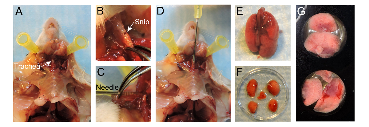
- göğüs kafesi üzerinden trakea cilt kesilmiş ama sağlam göğüs kafesi terk cerrahi makas kullanın. göğüs kafesi cilt ayırmak. Trakea kendisi (Şekil 1A) zarar vermemek için dikkatli olmak, çevredeki bağ dokusu kaldırarak trakea Açığa.
- Küçük bir açıklık mümkündür (Şekil 1B) halinde gırtlak yakın olarak kıkırdak halkalarının maruz trakea paralel çapı yaklaşık 1 mm, Kırpılmış. trakea yoluyla tamamen kesmek için dikkatli olun.
- 20 G iğne alın ve yavaşça herhangi bir karşı kuvvet (Şekil 1D) olmadan trakea içine iğne 4-5 mm yerleştirin. Iğne ucu trakea (Şekil 1C) ile görünür olmalıdır. trakea iğne stabilize etmek forseps kullanabilir. Alternatif olarak, bir dikiş aro bağlı olabiliriğneyi yerinde tutmak için trakea und.
NOT: Çok derin ekleyerek, karina travma olabilir ya da akciğerlerin sadece bir tarafı şişirilmiş olabilir. - (Sabit sıcaklık banyosuna doğrudan alınan) 37 ° C'de% 2 düşük erime sıcaklıklı agaroz 400 ul olan bir şırınga doldurun. diseksiyon tahtası ayakta emin olun ve yavaş yavaş akciğerlere iğneden sıcak agaroz aşılamak, akciğerleri şişirmek için ~ 400 ul kullanın.
NOT: göğüs kafesi içinde şişirme akciğerleri izleyin. Yarılmasına gibi akciğer aşırı şişirmek etmeyin. - Akciğerler şişirilmiş sonra, şırınga ayırmak ve sızıntı herhangi bir agaroz önlemek için trakea içine iğne tutmak, göğüs kafesinin ~ ⅔ dolum.
- ayarlamak ve katılaşmaya akciğer içindeki agaroz izin vermek için şişirilmiş akciğer boyunca 20 ° C PBS yaklaşık 50 ml dökün. Yavaş yavaş önlemek için forseps ile trakea iğneyi çıkarın ve kapatın olmayan katılaşmış agaroz sızmasını.
- Bir sternotomi gerçekleştirerek akciğerleri ortaya çıkarmak ve daha sonra akciğerleri tüketim. Tamamen trakea yoluyla kesim sırasında akciğerlerin eksizyonu için trakea üzerinde tutmak. Yavaşça akciğerler fare (Şekil 1E) ayrılana kadar göğüs boşluğunun dışında akciğerleri çekerken bağ dokusu ve yemek borusu kesip, trakea yukarı çekin.
- Aşırı kan yıkayın sıcak RPMI-1640 akciğerleri daldırın ve yavaşça hilus (Şekil 1F) de loblar ana kök bronşları kesmek için makas ve forseps kullanarak lobları ayırın.
- 24 oyuklu görüntüleme plaka (Şekil 1G) içindeki bir oyuk olarak, görüntüleme yüzeyinin en üst düzeye çıkarmak için aşağı düz yüzey ile, loblar yerleştirin. lob üstünde 37 ° C RPMI-1640 içinde 100 ul ekle. Kayan engellemek için lob üstüne birkaç 15 mm dairesel mikroskop kapak slaytlar yerleştirin.
- EVAP gelen RPMI-1640 medya önlemek için çevredeki kuyulara ılık PBS dökünparfüm içeren. Dengelenmiş klima kabini içine 24 oyuklu plaka ekleme ve hava ve% 5 CO2 ile 37 ° C 'de, akciğer lobları korumak. konfokal mikroskop sahnede iklim odasına yerleştirin.
Not: diğer gaz karışımları (örneğin, N2 içinde% 5 O2,% 5 CO2 hipoksi / düşük oksijen koşullarında hücre davranışını incelemek üzere) da düşünülebilir.

Canlı görüntüleme için akciğerlerin hazırlanması için Şekil 1. Protokol. Fare hazırlandıktan sonra trakea (A) Poz. Kıkırdak halkalar maruz trakea paralel olarak yapılan (B) Küçük kelepir. (C) 20 G iğne trakea içine 4-5 mm takılı. (D) agaroz akciğerlere 400 ul% 2 düşük erime sıcaklığı instilasyon. (E) enflamasyonATED akciğerler fare ayrılır. (F) lobları enflasyon sonra ayrılmış. (G) 24-iyi görüntüleme plaka iyi yerleştirilmiş lobları. Bu rakamın büyük halini görmek için lütfen buraya tıklayınız.
6. Toplama ve Görüntüler Analizi
NOT: Görüntüler çeşitli yazılım programları tarafından desteklenen bir disk konfokal mikroskoplar iplik çeşitli elde edilebilir. Imaris film düzenleme ve analizi için kullanılır ise, bu protokolde, piyasada bulunan dönen disk konfokal mikroskop ile bir ısmarlama dönen bir disk konfokal mikroskop ya da Zen ile ya μManager, görüntü elde etmek için kullanılır.
- μManager kullanarak görüntüleri elde edin. ΜManager yazılımını kullanarak görüntüleri edinimi için adım protokolü tarafından ayrıntılı bir adım daha önce 18 tarif edilmiştir.
VEYA - görüntüler eldeBöyle Zen olarak kullanarak görüntü analiz yazılımı (Şekil S1 bakınız).
- 'Bulun' sekmesine tıklayın ve 'Işık Yolu' aracı (Şekil S1A, kırmızı kutu) (10x veya 20x) objektif seçin. Mercek (Şekil S1A, mavi kutu) üzerinden CFP kanalına bakmak için - 'DAPI Gözler' Daha sonra, üzerine tıklayın. El ile mikroskop kullanılarak örnek yerelleştirilmesine. dokuda Off'after tüm görüş alanının merkezi 'seçeneğini tıklayın.
- görüntü elde etmek için tüm parametrelerini ayarlamak için 'Acquisition' sekmesine tıklayın.
- 'Kanallar' aracında, '+' tuşuna (Şekil S1B, kırmızı kutu) tıklayın. Bir pop-up menüsü görünür ve 'Boya Veritabanı' (Şekil S1 B) 'de numunede mevcut boya (ler) arayın. boya seçin ve 'Ekle' düğmesine tıklayın.
NOT: Program tüm filtreleri optimize edilmesi koyacaktır. Bir boya silinebilirseçerek o çöp tenekesi tuşuna (Şekil S1B, sarı kutu) tıklayarak izledi. - 'Edinim Modu' menüsünde, 5x5 'diyotları' olarak ayarlayın. seçmek için kanal menüsünde ECFP üzerine çift tıklayın. Örnek görüntü elde etmek için parametreleri kurarken ağartılmış olmayacak şekilde% 20 lazer gücünü azaltın.
- 'Deney Yöneticisi' bölümünde 'Karo Seramik'in kutusunu işaretleyin ve fayans aracı' Çok Boyutlu Toplama 'aracını grubunda (Şekil S1C) görünür. kameradan canlı görüntüyü görüntülemek için 'Gelişmiş Kurulum' düğmesine tıklayın. deney 4 ila 6 pozisyon eklemek için 'Positions' bölümünde 'Ekle' butonuna tıklayın. Bir konumu silmek için, o konumu seçin ve çöp kutusu düğmesine tıklayın.
- 'Edinim Parametre' aracını grubunda 'Absolut' Odak Stratejisi 'aracını açın ve Seçeneke açılan listeden Z-pozisyon 'düzeltildi.
- 'Deney Yöneticisi' bölümünde ve Z-Stack aracında Z-Yığın kutusunu işaretleyin 'Çok Boyutlu Acquisition' aracı grubunun (Şekil S1D) görünür. Pozisyonları 'bölümünde ve basın' Canlı 'pozisyonların birinde çift tıklayın. El ile ilk set ve görüntüleme alanının son konumunu ayarlamak. 4 mikron aralıklarla ayarlayın.
NOT: Program seçilen aralık ve aralık için dilim sayısını belirleyecektir. İdeal olarak, 5-7 dilim yeterli görselleştirme ve hızlı görüntü alımı sağlamak için uygundur. - 'Deney Yöneticisi' bölümünde 'Zaman Serisi' kutusunu işaretleyin. Set istenen 'Süre' ve 'Çok Boyutlu Toplama' aracını grubunda (Şekil S1E) ortaya 'Zaman Serisi' aracında 'Interval'ın' kez.
- 'Zinin içindeyon Modu 2x2 için 'diyotları' menüsünden, set '. kanallar menüsünde bir fluorofor çift tıklatın seçin ve% 100 lazer gücünü artırmak için. Basın 'Canlı' ve 'Pozlama Saati' ayarlayın. Her fluorofor için bu işlemi tekrarlayın.
- 'Enable Auto Kaydet' kutusunu işaretleyin. dosyanın adını bir klasör ve türünü seçin. Tüm edinilen görüntüler otomatik olarak bu klasöre kaydedilecektir.
- görüntü alımı başlatmak için 'Deney Yöneticisi' bölümünde 'Başlat Deney' üzerine tıklayın.
- Görüntü alındıktan sonra, Imaris yazılımında ham verileri derlemek. Dosyaları .ims görüntüleri dönüştürmek ve ayarlamalar yapılabilir. Dosyaların dönüştürülmesi, ayarlamalar yapmak ve Imaris kullanarak tasarruf filmler için adım protokolü tarafından ayrıntılı bir adım daha önce 18 tarif edilmiştir.
- Filmi kaydederken, ikinci (fps) başına 5 kare 'Frame Rate' set.
Sonuçlar
eğirme disk konfokal mikroskopi, çeşitli fare modeli sistemleri ve enjektabl kullanarak, metastatik mikroçevresinin görüntülendi ve zamanla izlenebilir. Bir MMTV PyMT kullanımı; ACTB-ECFP; c-FMS-EGFP üçlü transgenik fare modeli, farklı hücresel bileşenleri floresan (Şekil 2A, Film 1) etiketlenir. Akciğer parankimi tipik yapısı, tüm hücrelerin β-aktin organizatörü altında ECFP ifade beri CFP kanal görülebilir. Bu organize akciğer yapısı içindeki h?...
Tartışmalar
Bu yazıda metastaz taşıyan fare modellerinde akciğer metastazı ex vivo canlı görüntüleme için ayrıntılı bir yöntemi tarif etmektedir. Bu görüntüleme protokol akciğer mikroçevresinin içinde dinamik ve mekansal tümör hücre-stroma etkileşimleri doğrudan görselleştirme sağlar. En az 4 saat akciğer metastazı güvenilir bir görüntüleme sağlayan bir kolay ve hızlı bir yöntemdir. Bu deneylerden elde edilen filmler, hücre motilitesi ve hücresel etkileşimler gibi dinamik işlemleri...
Açıklamalar
The authors have no conflicts of interest to disclose. All animal experiments were conducted in accordance with IACUC approved protocols, UCSF.
Teşekkürler
We thank Nguyen H. Nguyen for her technical help and Audrey O’Neill for support with the Zeiss Cell Observer spinning-disk confocal microscope. This work was supported by a Department of Defense postdoctoral fellowship (W81XWH-11-01-0139) and the Weizmann Institute of Science-National Postdoctoral Award Program for Advancing Women in Science (to V.P.).
Malzemeler
| Name | Company | Catalog Number | Comments |
| MMTV-PyMT/FVB mice | Jackson Laboratory | 2374 | Female mice |
| ACTB-ECFP/FVB mice | UCSF Werb lab | Female mice | |
| c-fms-EGFP/FVB mice | UCSF Werb lab | Female mice | |
| FVB mice | Jackson Laboratory | 1800 | Female mice |
| GFP+ VO-PyMT cells | UCSF Werb lab | ||
| 70,000 kDa Dextran, rhodamine-conjugated | Invitrogen | D1818 | Dilute to 4mg/ml in 1 x PBS and store at -20 °C. Use 0.4 mg per animal. |
| 10,000 kDa Dextran, Alexa Fluor 647 conjugated | Invitrogen | D22914 | Dilute to 4mg/ml in 1 x PBS and store at -20 °C. Use 0.4 mg per animal. |
| Anti-mouse Gr-1 antibody Alexa Fluor 647 | UCSF Monoclonal antibody core | Stock 1mg/ml. Use 7 ug per animal. | |
| Anesthetic | Anesthesia approved by IACUC, used for anesthesia and/or euthanesia | ||
| 1X PBS | UCSF cell culture facility | ||
| PBS, USP sterile | Amresco INC | K813-500ML | Ultra pure grade for i.v. injection |
| Styrofoam platform | Will be used as dissection board | ||
| Fine scissors sharp | Fine Science Tools | 14060-11 | |
| Forceps | Roboz Surgical Store | RS-5135 | |
| Hot bead sterilizer | Fine Science Tools | 18000-45 | Turn ON 30min before use |
| Air | UCSF | ||
| Oxygen | UCSF | ||
| Carbon dioxide | UCSF | ||
| 1 mL syringe without needle | BD | 309659 | |
| 27 G x 1/2 needle | BD | 305109 | for i.v. injection |
| 20 G x 1 needle, short bevel | BD | 305178 | |
| Low-melting-temperature agarose | Lonza | 50111 | To make 10 ml of solution, weigh 0.2 g of agarose, add to 10 ml 1 x PBS, and heat to dissolve. Agarose will solidify at room temperature, so maintain in a 37 °C water bath until used for inflation. |
| RPMI-1640 medium without phenol red | Life Technologies | 11835-030 | |
| 24 well Imaging plate | E&K scientific | EK-42892 | |
| Glass cover slides, 15 mm | Fisher Scientific | 22-031-144 | |
| Digital CO2 and temperature controller | Okolab | DGTCO2BX | http://www.oko-lab.com |
| Climate chamber | Okolab | http://www.oko-lab.com | |
| Cell Observer spinning disk confocal microscope | Zeiss | ||
| Zen software | Zeiss | ||
| Inverted microscope | Carl Zeiss Inc | Zeiss Axiovert 200M | |
| ICCD camera | Stanford Photonics | XR-Mega-10EX S-30 | |
| Spinning disk confocal scan-head | Yokogawa Corporation | CSU-10b | |
| Imaris | Bitplane | ||
| mManager | Vale lab, UCSF | Open-source software |
Referanslar
- Chaffer, C. L., Weinberg, R. A. A perspective on cancer cell metastasis. Science. 331 (6024), 1559-1564 (2011).
- Plaks, V., Kong, N., Werb, Z. The cancer stem cell niche: how essential is the niche in regulating stemness of tumor cells. Cell stem cell. 16 (3), 225-238 (2015).
- Nguyen, D. X., Bos, P. D., Massague, J. Metastasis: from dissemination to organ-specific colonization. Nat Rev Cancer. 9 (4), 274-284 (2009).
- Egeblad, M., Ewald, A. J., et al. Visualizing stromal cell dynamics in different tumor microenvironments by spinning disk confocal microscopy. Dis Model Mech. 1 (2-3), 155-167 (2008).
- Ellenbroek, S. I. J., van Rheenen, J. Imaging hallmarks of cancer in living mice. Nat Rev Cancer. 14 (6), 406-418 (2014).
- Looney, M. R., Thornton, E. E., Sen, D., Lamm, W. J., Glenny, R. W., Krummel, M. F. Stabilized imaging of immune surveillance in the mouse lung. Nat Methods. 8 (1), 91-96 (2011).
- Thornton, E. E., Krummel, M. F., Looney, M. R. Live imaging of the lung. Cur Protoc Cytom. , (2012).
- Thornton, E. E., Looney, M. R., et al. Spatiotemporally separated antigen uptake by alveolar dendritic cells and airway presentation to T cells in the lung. J Exp Med. 209 (6), 1183-1199 (2012).
- Mendoza, A., Hong, S. -. H., et al. Modeling metastasis biology and therapy in real time in the mouse lung. J Clin Invest. 120 (8), 2979-2988 (2010).
- Lelkes, E., Headley, M. B., Thornton, E. E., Looney, M. R., Krummel, M. F. The spatiotemporal cellular dynamics of lung immunity. Trends Immunol. 35 (8), 379-386 (2014).
- Guy, C. T., Cardiff, R. D., Muller, W. J. Induction of mammary tumors by expression of polyomavirus middle T oncogene: a transgenic mouse model for metastatic disease. Mol Cell Biol. 12 (3), 954-961 (1992).
- Hadjantonakis, A. -. K., Macmaster, S., Nagy, A. Embryonic stem cells and mice expressing different GFP variants for multiple non-invasive reporter usage within a single animal. BMC Biotechnol. 2, (2002).
- Casbon, A. -. J., Reynaud, D., et al. Invasive breast cancer reprograms early myeloid differentiation in the bone marrow to generate immunosuppressive neutrophils. Proc Natl Acad Sci USA. 112 (6), 566-575 (2015).
- Donovan, J., Brown, P. Parenteral injections. Curr Protoc Immunol. , (2006).
- Halpern, J., Lynch, C. C., et al. The application of a murine bone bioreactor as a model of tumor: bone interaction. Clin Exp Metastas. 23 (7-8), 345-356 (2006).
- Sasmono, R. T., Oceandy, D., et al. A macrophage colony-stimulating factor receptor-green fluorescent protein transgene is expressed throughout the mononuclear phagocyte system of the mouse. Blood. 101 (3), 1155-1163 (2003).
- Ewald, A. J., Werb, Z., Egeblad, M. Dynamic, long-term in vivo imaging of tumor-stroma interactions in mouse models of breast cancer using spinning-disk confocal microscopy. Cold Spring Harb Protoc. (2), (2011).
- Bonnans, C., Lohela, M., Werb, Z. Real-time imaging of myeloid cells dynamics in ApcMin/+ intestinal tumors by spinning disk confocal microscopy. J Vis Exp. (92), (2014).
- Nakasone, E. S., Askautrud, H. A., et al. Imaging tumor-stroma interactions during chemotherapy reveals contributions of the microenvironment to resistance. Cancer cell. 21 (4), (2012).
- Cheng, N., Lambert, D. L. Mammary transplantation of stromal cells and carcinoma cells in C57BL/6J mice. J Vis Exp. (54), (2011).
- Al-Mehdi, A. B., Tozawa, K., Fisher, A. B., Shientag, L., Lee, A., Muschel, R. J. Intravascular origin of metastasis from the proliferation of endothelium-attached tumor cells: a new model for metastasis. Nat Med. 6 (1), 100-102 (2000).
- Wong, C. W., Song, C., et al. Intravascular location of breast cancer cells after spontaneous metastasis to the lung. Am J Pathol. 161 (3), 749-753 (2002).
- Liang, C. -. C., Park, A. Y., Guan, J. -. L. In vitro scratch assay: a convenient and inexpensive method for analysis of cell migration in vitro. Nat protoc. 2 (2), 329-333 (2007).
- Nelson, K., Bobba, C., Ghadiali, S., Hayes, D. J., Black, S. M., Whitson, B. A. Animal models of ex vivo lung perfusion as a platform for transplantation research. World J Exp Med. 4 (2), (2014).
- Magness, S. T., Bataller, R., Yang, L., Brenner, D. A. A dual reporter gene transgenic mouse demonstrates heterogeneity in hepatic fibrogenic cell populations. Hepatology. 40 (5), 1151-1159 (2004).
- Motoike, T., Loughna, S., et al. Universal GFP reporter for the study of vascular development. Genesis. 28 (2), (2000).
- Srivastava, M. K., Andersson, A., et al. Myeloid suppressor cells and immune modulation in lung cancer. Immunotherapy. 4 (3), (2012).
- Craig, A., Mai, J., Cai, S., Jeyaseelan, S. Neutrophil recruitment to the lungs during bacterial pneumonia. Infect Immun. 77 (2), 568-575 (2009).
- Kreisel, D., Nava, R. G., et al. In vivo two-photon imaging reveals monocyte-dependent neutrophil extravasation during pulmonary inflammation. Proc Natl Acad Sci USA. 107 (42), 18073-18078 (2010).
Yeniden Basımlar ve İzinler
Bu JoVE makalesinin metnini veya resimlerini yeniden kullanma izni talebi
Izin talebiThis article has been published
Video Coming Soon
JoVE Hakkında
Telif Hakkı © 2020 MyJove Corporation. Tüm hakları saklıdır