Thyroid Exam
Source: Richard Glickman-Simon, MD, Assistant Professor, Department of Public Health and Community Medicine, Tufts University School of Medicine, MA
The thyroid gland is located in the neck anterior trachea between the cricoid cartilage (above) and the suprasternal notch (below) (Figure 1). It consists of a right and left lobe connected by an isthmus. The isthmus covers the second, third, and fourth tracheal rings, and the lobes curve posteriorly around the sides of the trachea and esophagus. The normal gland, weighing 10 - 25 g, is usually invisible on inspection and often difficult to palpate. A goiter is an enlarged thyroid from any cause. In addition to assessing its size, it is important to palpate the thyroid for its shape, mobility, consistency, and tenderness. A normal thyroid is soft, smooth, symmetrical, and non-tender, and it slides upward slightly when swallowing. Symmetrical enlargement of a soft, smooth thyroid suggests endemic hypothyroidism due to iodine deficiency or one of two prevalent autoimmune disorders: Graves' disease or Hashimoto's thyroiditis. Thyroid nodules are common and usually incidental; however, 10% of thyroid nodules turn out to be malignant. They may be single or multiple, and are most often firm and non-tender. A tender, symmetrical goiter typically indicates thyroiditis.
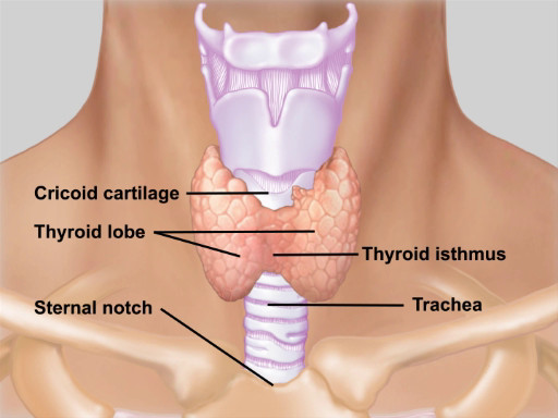
Figure 1. Anatomy of the thyroid gland. Illustration of the location and anatomy of the thyroid gland with respect to the neck structures.
Thyroid disease rarely manifests as a palpable goiter in isolation. Thyroid hormones serve to maintain homeostasis primarily by stimulating cellular metabolism throughout the body. Thus, hypo and hyperthyroidism are associated with a range of symptoms and physical findings (Table 1). It is important to note that goiters may be euthyroid (normal thyroid hormone levels), hyperthyroid, or hypothyroid. Headaches or visual disturbances may suggest a secondary thyroid disorder due to a pituitary adenoma
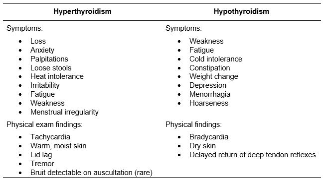
Table 1. Symptoms and physical findings for hypo- and hyper-thyroidism.
1. Inspection
- Tip the patient's head slightly back, and carefully inspect the anterior neck. If visible, the thyroid appears between the cricoid cartilage and suprasternal notch. Check for symmetry, diffuse swelling, and obvious masses.
- Have the patient swallow, and observe as the cricoid cartilage, thyroid cartilage, and thyroid gland move up and down.
2. Palpate
Although the thyroid can be palpated from either anterior or posterior positions, the latter
An enlarged thyroid gland, or goiter, is most often associated with normal thyroid gland function (euthyroid), but may be associated with hyper- or hypothyroid conditions. Therefore, thyroid abnormality found on physical examination should prompt a careful evaluation for the systemic signs and symptoms associated with both high and low thyroid hormone levels. A normal thyroid can be difficult to palpate, particularly in patients with large necks. However, its location can be precisely determined by identifying the bony a
ABOUT JoVE
Copyright © 2024 MyJoVE Corporation. All rights reserved