
Finding Your Blind Spot and Perceptual Filling-in
Source: Laboratory of Jonathan Flombaum—Johns Hopkins University
In the back of everyone's eye is a small piece of neural tissue called the retina. The retina has photosensitive cells that respond to stimulation by light. The responses of these cells are sent into the brain through the optic nerve, a bundle of neural fibers. In each retina there is a place somewhere in the periphery where the outputs from retinal cells collect and the bundled optic nerve exits to the brain. At that location, there is no photosensitivity-whatever light reflects from the world and lands in that position does not produce a signal in the brain. As a result, humans have a blind spot, a place in the visual field for which they don't process incoming stimuli.
However, people are not aware that they have blind spots; there is not an empty hole in the visual images in front of the eyes. So what do people see in their blind spots? The brain actually fills-in missing input based on the surroundings.
This video demonstrates how to find a person's blind spot, and how to investigate the mechanisms of perceptual filling-in.
1. Stimulus Design
- Open a blank white page in a slide editor (PowerPoint or Keynote will suffice).
- Near the right edge of the page, make a circle about as large as penny, when the slide is printed. This will be the fixation point.
- Near the left edge of the page, make a small star (or any other shape), so that when you print the slide, the shape is about the size of a penny This slide is the left eye blind spot stimulus.
- Duplicate the slide, and switch the positions of th
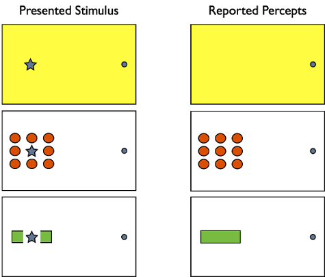
Figure 3. Perceptual reports: What participants report seeing (shown in the right column) when the star in the left column images disappears in the blind spot. The reports reveal several principles of how the brain creates perceptual experience. Most broadly, in all cases, the brain fills-in the blind spot as the most likely content given the local context.
Log in or to access full content. Learn more about your institution’s access to JoVE content here
Because the brain fills-in perception within the blind spot, one application involves studies that seek to identify the brain areas involved in producing conscious experience. For example, for a long time, researchers were uncertain whether the earliest part of human visual cortex, called V1, was involved directly in the production of conscious experience. To address this issue, researchers used fMRI (Functional Magnetic Resonance Imaging) to measure neural responses in the monocular regions of V1 that mapped to the blin
- Tong, F., & Engel, S. A. (2001). Interocular rivalry revealed in the human cortical blind-spot representation. Nature, 411(6834), 195-199.
Skip to...
ABOUT JoVE
Copyright © 2024 MyJoVE Corporation. All rights reserved