Immunofluorescence Microscopy: Immunofluorescence Staining of Paraffin-Embedded Tissue Sections
Source: Thomas Chaffee1, Thomas S. Griffith2,3,4, and Kathryn L. Schwertfeger1,3,4
1 Department of Lab Medicine and Pathology, University of Minnesota, Minneapolis, MN 55455
2 Department of Urology, University of Minnesota, Minneapolis, MN 55455
3 Masonic Cancer Center, University of Minnesota, Minneapolis, MN 55455
4 Center for Immunology, University of Minnesota, Minneapolis, MN 55455
Pathologic analyses of tissue sections can be used to obtain a better understanding of normal tissue structure and contribute to our understanding of mechanisms of disease. Tissue biopsies, either from patients or from experimental in vivo models, are often preserved by fixing in formalin or paraformaldehyde and embedding in paraffin wax. This allows for long-term storage and for the tissues to be sectioned. Tissues are cut into thin (5 µm) sections using a microtome and the sections are adhered to glass slides. The tissues sections can be stained with antibodies, which allow for the detection of specific proteins within the tissue sections. Staining with antibodies conjugated to fluorophores (also known as fluorochromes) - compounds that emit light at specific wavelengths when excited by a laser - is known as immunofluorescence. The ability to detect proteins within a section can provide information such as cell type heterogeneity within the tissue, activation of specific signaling pathways, and expression of biomarkers. Depending on the fluorophores used and type of microscope available for analysis, multiple colors can be used, which allows for multiplexed analysis of targets.
The following protocol outlines the basic steps involved in immunofluorescence staining of paraffin embedded tissue sections. It is important to note that this protocol will not include any details on the fixation of tissue, process of paraffin embedding, or sectioning of the tissues. Once tissues have been sectioned and placed on glass slides, they are rehydrated through a series of graded ethanol (EtOH) incubations. The sections are incubated with a blocking reagent to reduce non-specific binding of antibody to the tissue section. The sections are then incubated with a primary antibody that may or may not be directly labeled with a fluorophore. If the primary antibody is not directly labeled, the sections are then incubated with a secondary antibody labeled with a fluorophore. Different antibodies may require different staining conditions, thus suggestions for optimization of antibodies are included. Following washing to remove all unbound antibody, the slides are mounted with media containing DAPI to fluorescently label the nucleus. Once the mounting media has dried, the slides can be imaged using a microscope with lasers that can detect the different fluorophores.
1. Set-up
- The typical staining protocol involves the following steps:
- Re-hydrating the tissue sections on the slides using a series of graded ethanols.
- Incubating the tissue sections with a blocking buffer, which will help to block non-specific binding of antibody to the tissue and reduce background fluorescence.
- Removing the blocking buffer and incubating the section in primary antibody, at which time the antibody will bind its peptide target.
- Rem
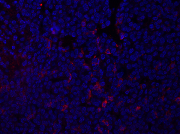
Figure 1: F4/80 staining of a mammary tumor section. Following fixation, a mouse mammary tumor was sectioned and stained with anti-F4/80 and mounted using a DAPI-containing mounting media. Staining is shown by cell surface F4/80 staining in red. Please click here to view a larger vers
Immunofluorescence allows for the investigation of protein expression and localization within the context of a tissue section. This technique can be used to understand how tissues change in the context of disease by examining protein localization or cell number in normal and diseased tissues. Changes in localization or in expression patterns can be determined and linked to specific attributes of the samples.
- Im K, Mareninov S., Diaz MFP, Yong WH. An Introduction to Performing Immunofluorescence Staining. Yong W. (eds) Biobanking. Methods in Molecular Biology. 1897, Humana Press, New York, NY (2019)
- Ramos-Vara JA. Principles and Methods of Immunohistochemistry. Gautier JC. (eds) Drug Safety Evaluation. Methods in Molecular Biology. 1641, Humana Press, New York, NY (2017)
- Donaldson JG. Immunofluorescence Staining. Current protocols in Cell Biology. 69 (1):1 4.3.1-4.3.7. (2015)
Skip to...
ABOUT JoVE
Copyright © 2024 MyJoVE Corporation. All rights reserved