
Visualizing Dengue Virus through Alexa Fluor Labeling
July 9th, 2011
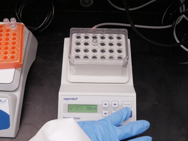
•Taking advantage of the advancements in fluorophore development and imaging technology, a simple method of Alexa Fluor labeling of dengue virus was devised to visualize the early interactions between virus and cell.
Tags
Related Videos

Neutrophil Isolation and Analysis to Determine their Role in Lymphoma Cell Sensitivity to Therapeutic Agents

Evaluation of the Efficacy And Toxicity of RNAs Targeting HIV-1 Production for Use in Gene or Drug Therapy

Rescue and Characterization of Recombinant Virus from a New World Zika Virus Infectious Clone

Visualizing Macrophage Extracellular Traps Using Confocal Microscopy
A Simple Flow Cytometry Based Assay to Determine In Vitro Antibody Dependent Enhancement of Dengue Virus Using Zika Virus Convalescent Serum

Rapid, Safe, and Simple Manual Bedside Nucleic Acid Extraction for the Detection of Virus in Whole Blood Samples

Measuring Dengue Virus RNA in the Culture Supernatant of Infected Cells by Real-time Quantitative Polymerase Chain Reaction

Identification of Coding and Non-coding RNA Classes Expressed in Swine Whole Blood

A Murine Model of Dengue Virus-induced Acute Viral Encephalitis-like Disease

In Vitro ELISA Test to Evaluate Rabies Vaccine Potency
ABOUT JoVE
Copyright © 2024 MyJoVE Corporation. All rights reserved