
A TIRF Microscopy Technique for Real-time, Simultaneous Imaging of the TCR and its Associated Signaling Proteins
March 22nd, 2012
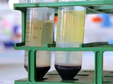
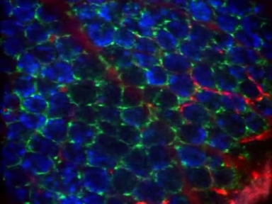
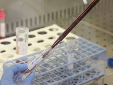
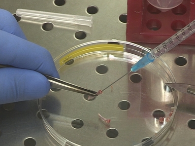
•The compartmentalization of proteins either within the plasma membrane or into intracellular locations is one regulatory mechanism that can greatly influence signaling outcomes; hence, to understand signaling it is important to study the spatial and temporal behavior of the proteins involved. We describe here a TIRF microscopy based system to study signal transduction in T cells, but is broadly applicable.
Tags
Related Videos

Real-time Imaging of Leukotriene B4 Mediated Cell Migration and BLT1 Interactions with β-arrestin

Real-time Live Imaging of T-cell Signaling Complex Formation
Real-time Imaging of Endothelial Cell-cell Junctions During Neutrophil Transmigration Under Physiological Flow
Real-time Imaging of Myeloid Cells Dynamics in ApcMin/+ Intestinal Tumors by Spinning Disk Confocal Microscopy
Simultaneous Quantification of T-Cell Receptor Excision Circles (TRECs) and K-Deleting Recombination Excision Circles (KRECs) by Real-time PCR

Analysis of Yersinia enterocolitica Effector Translocation into Host Cells Using Beta-lactamase Effector Fusions
Imaging Neutrophils and Monocytes in Mesenteric Veins by Intravital Microscopy on Anaesthetized Mice in Real Time

Purification of Viral DNA for the Identification of Associated Viral and Cellular Proteins

Real-time Imaging and Quantification of Fungal Biofilm Development Using a Two-Phase Recirculating Flow System

Pan-lyssavirus Real Time RT-PCR for Rabies Diagnosis
ABOUT JoVE
Copyright © 2024 MyJoVE Corporation. All rights reserved