A Microscopic Phenotypic Assay for the Quantification of Intracellular Mycobacteria Adapted for High-throughput/High-content Screening
January 17th, 2014
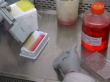
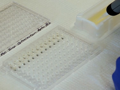
•Here, we describe a phenotypic assay applicable to the High-throughput/High-content screens of small-interfering synthetic RNA (siRNA), chemical compound, and Mycobacterium tuberculosis mutant libraries. This method relies on the detection of fluorescently labeled Mycobacterium tuberculosis within fluorescently labeled host cell using automated confocal microscopy.
Tags
Related Videos

A Functional Whole Blood Assay to Measure Viability of Mycobacteria, using Reporter-Gene Tagged BCG or M.Tb (BCG lux/M.Tb lux)

A Parasite Rescue and Transformation Assay for Antileishmanial Screening Against Intracellular Leishmania donovani Amastigotes in THP1 Human Acute Monocytic Leukemia Cell Line

A New Screening Method for the Directed Evolution of Thermostable Bacteriolytic Enzymes

Generation and Multi-phenotypic High-content Screening of Coxiella burnetii Transposon Mutants

Visualization of the Charcoal Agar Resazurin Assay for Semi-quantitative, Medium-throughput Enumeration of Mycobacteria

A Simple Fluorescence Assay for Quantification of Canine Neutrophil Extracellular Trap Release

A Fluorescence-based Lymphocyte Assay Suitable for High-throughput Screening of Small Molecules

System for Efficacy and Cytotoxicity Screening of Inhibitors Targeting Intracellular Mycobacterium tuberculosis

Quantification of Intracellular Growth Inside Macrophages is a Fast and Reliable Method for Assessing the Virulence of Leishmania Parasites

A High-throughput, High-content, Liquid-based C. elegans Pathosystem
ABOUT JoVE
Copyright © 2024 MyJoVE Corporation. All rights reserved