
Super-resolution Imaging of the Natural Killer Cell Immunological Synapse on a Glass-supported Planar Lipid Bilayer Video (Video) | JoVE
February 11th, 2015
•We describe here a combination of the glass-supported lipid bilayer technique of forming immunological synapses with the super-resolution imaging technique of stimulated emission depletion (STED) microscopy. The goal of this protocol is to provide users with the instructions necessary to successfully carry out these two techniques.
Related Videos

Facilitating the Analysis of Immunological Data with Visual Analytic Techniques (Video) | JoVE
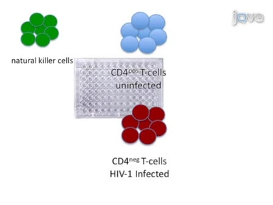
Preparation and Use of HIV-1 Infected Primary CD4+ T-Cells as Target Cells in Natural Killer Cell Cytotoxic Assays (Video) | JoVE

Imaging of HIV-1 Envelope-induced Virological Synapse and Signaling on Synthetic Lipid Bilayers (Video) | JoVE

Artificial Antigen Presenting Cell AAPC Mediated Activation and Expansion of Natural Killer T Cells Video (Video) | JoVE

Visualization of the Immunological Synapse by Dual Color Time-gated Stimulated Emission Depletion STED Nanoscopy Video (Video) | JoVE
An Optimized Method for Isolating and Expanding Invariant Natural Killer T Cells from Mouse Spleen Video (Video) | JoVE

An Endothelial Planar Cell Model for Imaging Immunological Synapse Dynamics Video (Video) | JoVE

Flow Cytometric Analysis of Natural Killer Cell Lytic Activity in Human Whole Blood Video (Video) | JoVE

Highly Multiplexed, Super-resolution Imaging of T Cells Using madSTORM Video (Video) | JoVE
A Novel Feeder-free System for Mass Production of Murine Natural Killer Cells In Vitro Video (Video) | JoVE
ABOUT JoVE
Copyright © 2024 MyJoVE Corporation. All rights reserved