Long Term Intravital Multiphoton Microscopy Imaging of Immune Cells in Healthy and Diseased Liver Using CXCR6.Gfp Reporter Mice
March 24th, 2015
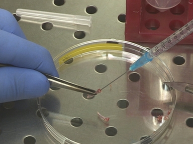
•Stable intravital high-resolution imaging of immune cells in the liver is challenging. Here we provide a highly sensitive and reliable method to study migration and cell-cell-interactions of immune cells in mouse liver over long periods (about 6 hours) by intravital multiphoton laser scanning microscopy in combination with intensive care monitoring.
Tags
Related Videos

Seven Steps to Stellate Cells

Intravital Imaging of the Mouse Thymus using 2-Photon Microscopy

Long Term Chronic Pseudomonas aeruginosa Airway Infection in Mice

Highly Resolved Intravital Striped-illumination Microscopy of Germinal Centers

Intravital Microscopy Imaging of the Liver following Leishmania Infection: An Assessment of Hepatic Hemodynamics

Intravital Microscopy of Leukocyte-endothelial and Platelet-leukocyte Interactions in Mesenterial Veins in Mice

Analysis of Yersinia enterocolitica Effector Translocation into Host Cells Using Beta-lactamase Effector Fusions

Imaging Neutrophils and Monocytes in Mesenteric Veins by Intravital Microscopy on Anaesthetized Mice in Real Time

Intravital Imaging of Neutrophil Priming Using IL-1β Promoter-driven DsRed Reporter Mice

Imaging Mycobacterium tuberculosis in Mice with Reporter Enzyme Fluorescence
ABOUT JoVE
Copyright © 2024 MyJoVE Corporation. All rights reserved