
Precision-cut Mouse Lung Slices to Visualize Live Pulmonary Dendritic Cells
April 5th, 2017
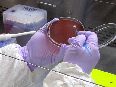
•We describe a method for generating Precision-cut Lung Slices (PCLS) and immunostaining them to visualize the localization of various immune cell types in the lung. Our protocol can be extended to visualize the location and function of many different cell types under a variety of conditions.
Tags
Related Videos

Isolation of Mouse Lung Dendritic Cells

Analysis of Pulmonary Dendritic Cell Maturation and Migration during Allergic Airway Inflammation

Isolation and Characterization of Dendritic Cells and Macrophages from the Mouse Intestine

Right Ventricular Systolic Pressure Measurements in Combination with Harvest of Lung and Immune Tissue Samples in Mice
Pseudomonas aeruginosa Induced Lung Injury Model

Influenza Virus Propagation in Embryonated Chicken Eggs

Legionella pneumophila Outer Membrane Vesicles: Isolation and Analysis of Their Pro-inflammatory Potential on Macrophages

Human Lung Dendritic Cells: Spatial Distribution and Phenotypic Identification in Endobronchial Biopsies Using Immunohistochemistry and Flow Cytometry

Assessment of the Cytotoxic and Immunomodulatory Effects of Substances in Human Precision-cut Lung Slices

Development and Validation of an Ultrasensitive Single Molecule Array Digital Enzyme-linked Immunosorbent Assay for Human Interferon-α
ABOUT JoVE
Copyright © 2024 MyJoVE Corporation. All rights reserved