
Methods for Detecting Cytotoxic Amyloids Following Infection of Pulmonary Endothelial Cells by Pseudomonas aeruginosa
July 12th, 2018
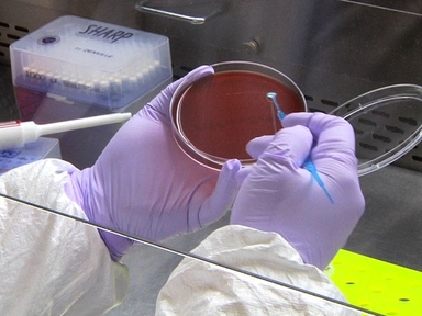
•Simple methods are described for demonstrating the production of cytotoxic amyloids following infection of pulmonary endothelium by Pseudomonas aeruginosa.
Tags
Related Videos

Co-culture Models of Pseudomonas aeruginosa Biofilms Grown on Live Human Airway Cells

Pseudomonas aeruginosa and Saccharomyces cerevisiae Biofilm in Flow Cells

Methods to Assess Beta Cell Death Mediated by Cytotoxic T Lymphocytes

Selection of Plasmodium falciparum Parasites for Cytoadhesion to Human Brain Endothelial Cells

RNA Isolation of Pseudomonas aeruginosa Colonizing the Murine Gastrointestinal Tract

Following Cell-fate in E. coli After Infection by Phage Lambda

Long Term Chronic Pseudomonas aeruginosa Airway Infection in Mice
Pseudomonas aeruginosa Induced Lung Injury Model

Pneumococcus Infection of Primary Human Endothelial Cells in Constant Flow

Culture of Small Colony Variant of Pseudomonas aeruginosa and Quantitation of its Alginate
ABOUT JoVE
Copyright © 2024 MyJoVE Corporation. All rights reserved